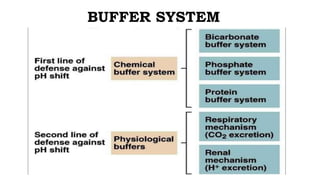
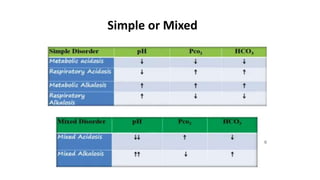
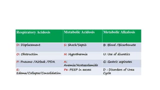
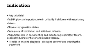
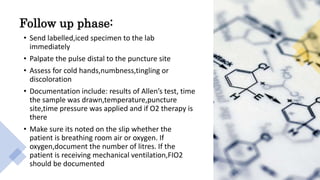
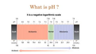
Dr Venkata Sai kumar
P.G.-J.R.2
Dept. of Pediatrics
Hind Institute of Medical
Sciences, BBK
ABG INTERPRETATION
Dr PRAGATI SISODIA
ASSISTANT PROFESSOR
Dept. of Pediatrics
Hind Institute of Medical
Sciences, BBK
Learning objectives
what
Indication
who
Doctors,staff Nurse & Trained
Medical Persons
How & Why
How To Do
Clinical Significane And How To
Interpret
Learning objectives
Definition
Indication
 Any sick child
✓ABGA plays an important role in critically ill children with respiratory
distress.
✓Reveals oxygenation status,
✓Adequacy of ventilation and acid-base balance.
✓Significant role in documenting and monitoring respiratory failure,
especially during ventilator and oxygen therapy.
• IT helps in making diagnosis , assessing severity and titrating the
treatment
EQUIPMENT
Blood gas kit
• 1ml/2ml syringe
• 23-26 gauge needle
• Stopper or cap
• Alcohol swab
• Disposible gloves
• Plastic bag& crushed ice
• Lidocaine(optional)
• Vial of heparin(1:1000)
• Bar code or label
PREPARATORY PHASE:
• Record patient inspired oxygen concentration
• Check patient temperature
• Explain the procedure to the patient
• Provide privacy for client
• If not using heparinised syringe,heparanize the needle
• Perform allen’s test
• Wait at least 20 minutes before drawing blood for ABG after
initiating,changing,or discontinuing oxygen therapy,or
settings of mechanical ventilation,after suctioning the
patient or after extubation
ALLEN’S TEST
• It is a test done to determine that collateral circulation is present
from the ulnar artery in case thrombosis occur in the radial
Sites for obtaining ABG
• Radial artery(most common)
• Brachial artery
• Femoral artery
Radial is the most preferable site used because:
• It is easy to access
• It is not a deep artery which facilitate
palpation,stabilization and puncturing
• The artery has a collateral blood circulation
•OBSE
PERFORMANCE PHASE
• Wash hands
• Put on gloves
• Palpate the artery for maximum pulsation
• If radial,perform Allen’s test
• Place a small towel roll under the patient wrist
• Instruct the patient to breath normally durning the test and warn him that
he may feel brief cramping or throbbing pain at the puncture site
• Clean with alcohol swab in circular motion
• Skin and subcutaneous tissue may be infiltrated with local anesthetic agent
if needed
• Insert needle at 45 radial, 60 brachial and 90
femoral
• Withdraw the needle and apply digital pressure
• Check bubbles in syringe
• Place the capped syringe in the container of ice
immediately
• Maintain firm pressure on the puncture site for 5
minutes, if patient has coagulation abnormalities
apply pressure for 10-15 minutes
Follow up phase:
• Send labelled,iced specimen to the lab
immediately
• Palpate the pulse distal to the puncture site
• Assess for cold hands,numbness,tingling or
discoloration
• Documentation include: results of Allen’s test, time
the sample was drawn,temperature,puncture
site,time pressure was applied and if O2 therapy is
there
• Make sure its noted on the slip whether the
patient is breathing room air or oxygen. If
oxygen,document the number of litres. If the
patient is receiving mechanical ventilation,FIO2
should be documented
Errors in ABG
Excessive heparin
•Dilution Effect on Results decrease in HCO3 - & PaCO2
•Only .05 ml Heparin required for 1ml blood
•So syringe be emptied of heparin after flushing or
only dead space volume is sufficient or dry heparin
should be used
AIR BUBBLES
1. PO2 150 mmHg & PCO2 0mm Hg in air bubble(R.A)
2. Mixing with sample,lead to increase PaO2 & decrease in PaCO2
To avoid air bubble,sample drawn very slowly and preferabily in glass
syringe
Steady State:
Sampling should be done during steady state after change in oxygen
therapy or ventilator parameter
Steady state is achieved usually within 3-10 minutes
Dilution of blood sample with IV fluids
• Dilution of blood sample with IV fluid will typically lower the PaCO2
and increase the basedeficit without affecting the pH
• This is probably because of the diffusion of CO2 from blood onto the
IV fluid,which contains no CO2.
• Because of the buffering capacityof the blood,the pH changes
little,despite the decrease in PaCO2,giving the appearance of a
combined metabolic acidosis and respiratory alkalosis.
• Dilution of a blood gas sample with a liquid emulsion does not appear
to have any affect on the blood gas measurements. Dry heparin also
does not affect blood gas results.
Complications
•Arteriospasm
•Hematoma
•Hemorrhage
•Distal ischemia
•Infection
•numbness
WHAT IS pH
It is a negative logarithmic scale
What is pH ?
Henderson-Hasselbalch Equation
 Correlates metabolic & respiratoryregulations
HCO -
pH = pK + log ----------------
.03 x [PaCO2]
Simplified
HCO3 -
pH ~ ---------
PaCO2
CheckT
able
H+ ion conc. Correspondingto pH
pH H+ (nmol/L)
6.8 160
7.0 100
7.2 65
7.4 40
7.6 25
7.8 15
Why do we need pH?
Facilitaes cellular processes to
occcur effectively
Failure inefficient cellular function
and cell death
BUFFER SYSTEM
1. pH
2.
Respiratory/metabolic
3.Stage of
compensation
4. Oxygenation status
5. Simple
disorder/mixed
6. acute/chronic
7.Laboratory error
ABG Interpretation- Step by Step approach
Things to be look for/What You Get
Approach to ABG
• ABG should never be interpreted alone
• Previous blood gases
• Patient clinical status
• X-ray
• Other investigations e.g. Sepsis screening
• You have to treat the patient not the blood gas go with the clinical
picture
AbnormalValues
pH < 7.35
• Acidosis(metabolic and/or
respiratory)
paCO2 < 35 mmHg
• Respiratory alkalosis (alveolar
hyperventilation)
HCO3 -< 22meq/L
• Metabolic acidosis
pH > 7.45
Alkalosis(metabolic and/or
respiratory)
paCO2 > 45 mmHg
• Respiratory acidosis (alveolar
hypoventilation)
HCO3 -> 26meq/L
• Metabolic alkalosis
Define the cause?
Step 2:
 Study paco2 & HCO3
 Respiratory if PaCO2 abnormal & HCO3 normal
 Metabolic if HCO3 abnormal & PaCO2 normal
SAMPLE 2
pH: 7.21
Pco2:55
HCO3:27
ACIDOSIS
RESPIRATORY
PH: 7.51
PCO2:50
HCO3: 38
ALKALOSIS
METABOLIC
STEP 3: COMPENSATION
Ability to resist a change in pH
COMPENSATION
Respiratory compensation
• Respiratory compensation
• Starts within minutes
• complete within 12-24 days
Metabolic compensation
• Starts within Hours
• complete within 2-5 days
• renal base excretion faster
Compensation by Respiratory Response to
Metabolic Acidosis
✓Predicted Change in PCO2 = (1.5 x HCO3 + 8)± 2
✓If patient’s PCO2 is roughly this value, his or her response is
appropriate
✓If patient’s PCO2 is higher than this value, they are failing to
compensate adequately
Metabolic Alkalosis
✓Every 10 mmHCO3 increase PaCO2 7
In respiratory acidosis, for every 10mm increase in
PaCO2 , HCO3 is increased by 1 in acute condition
and by 3.5 in chronic condition
In respiratory alkalosis, for every 10mmHg
decrease in PaCO2 , HCO3 is decreased by 2 in
acute and by 4 in chronic condition
Compensation by Metabolic Response to
Examples
Metabolic acidosis
paCO2=37 : N
HCO3=21 : ↓
pH =7.25 : ↓
Respiratory Alkalosis
• pH = 7.56 : ↑
• paCO2=28 : ↓
• HCO3=22 : N
Partially compensated Metabolic Alkalosis
• pH = 7.50 : ↑
• paCO2=51 : ↑ (Slightly ,35-45)
• HCO3=40 : ↑( more ,22-26)
Metabolic Alkalosis with compensated respiratory acidosis
• pH = 7.44 :↑
• paCO2=48 :↑
• HCO3=35 : ↑
Oxygenation Status
o Depends upon age & inspired oxygen concentration (FiO2)
o Normal value of PaO2 is 80-100 mm of Hg in children and adults
o Newborn it ranges from 40-70 mm of Hg
o On oxygen therapy the value of PaO2 will high (PaO2 = FiO2 × 5)
o Oxygen supplementation should below 60% to avoid toxicity
o Anemic patient may have normal PaO2 and SaO2 but final delivery of
O2 to the tissues
will be compromised
o Generally oxygen saturation measured by pulse oximetry (SpO2)
lower than arterial oxygen saturation (SaO2) measured directly by
blood gas analysis.
Simple or Mixed
Simple or Mixed Disorder
• In simple acid base disorder, both PaCO2 and HCO3– change in the
same direction
• If they do not follow this trend, the possibility of mixed disorder is
likely
• The expected compensation for simple acid base disorder is
predetermined and
• if they deviate from the norms there is the possibility of mixed
disorder
Anion gap
• Difference of unmeasured cations & Anions
• Anion gap(AG)= Na+k-(Cl+HCO3)
• Normal AG Acidosis: RTA, Diarrhoea
• Increase AG acidosis: lactic acidosis,ketoacidosis,uremia
• Normal 8-12
• If >12= high anion gas acidosis
• If <12= non anion gas acidosis
Q
• A 6 y male child presented in emergency with c/c of fever and
abnormal body movement
• for 2 days , ABGA was showing following
• pH =7.39
• PaCO2=40.2q
• PO2= 74.5
• HCO3= 22.1
• BE=-0.8
• What is your diagnosis ?
Q
• 6-year-old child came with fever for 5 days and rapid breathing since today morning. You
did ABGA which reveals the following results:
what is your interpretation
PaO2: 68.2 mmHg (82.5 – 97.5 mmHg)
pH: 7.30 (7.35 – 7.45)
PaCO2: 63 mmHg (35.2 – 45 mmHg)
HCO3-: 29 (22 – 26 mEq/L)
BE: +4 (-2 to +2)
i. Metabolic Acidosis
ii. Metabolic Acidosis with Respiratory Compensation
iii. Respiratory Acidosis
iv. Respiratory Acidosis with metabolic compensation
THANK YOU

ABG INTERPRETATION.pptx

  • 1.
    Dr Venkata Saikumar P.G.-J.R.2 Dept. of Pediatrics Hind Institute of Medical Sciences, BBK ABG INTERPRETATION Dr PRAGATI SISODIA ASSISTANT PROFESSOR Dept. of Pediatrics Hind Institute of Medical Sciences, BBK
  • 2.
    Learning objectives what Indication who Doctors,staff Nurse& Trained Medical Persons How & Why How To Do Clinical Significane And How To Interpret Learning objectives
  • 3.
  • 4.
    Indication  Any sickchild ✓ABGA plays an important role in critically ill children with respiratory distress. ✓Reveals oxygenation status, ✓Adequacy of ventilation and acid-base balance. ✓Significant role in documenting and monitoring respiratory failure, especially during ventilator and oxygen therapy. • IT helps in making diagnosis , assessing severity and titrating the treatment
  • 5.
    EQUIPMENT Blood gas kit •1ml/2ml syringe • 23-26 gauge needle • Stopper or cap • Alcohol swab • Disposible gloves • Plastic bag& crushed ice • Lidocaine(optional) • Vial of heparin(1:1000) • Bar code or label
  • 6.
    PREPARATORY PHASE: • Recordpatient inspired oxygen concentration • Check patient temperature • Explain the procedure to the patient • Provide privacy for client • If not using heparinised syringe,heparanize the needle • Perform allen’s test • Wait at least 20 minutes before drawing blood for ABG after initiating,changing,or discontinuing oxygen therapy,or settings of mechanical ventilation,after suctioning the patient or after extubation
  • 7.
    ALLEN’S TEST • Itis a test done to determine that collateral circulation is present from the ulnar artery in case thrombosis occur in the radial
  • 8.
    Sites for obtainingABG • Radial artery(most common) • Brachial artery • Femoral artery Radial is the most preferable site used because: • It is easy to access • It is not a deep artery which facilitate palpation,stabilization and puncturing • The artery has a collateral blood circulation
  • 9.
  • 10.
    PERFORMANCE PHASE • Washhands • Put on gloves • Palpate the artery for maximum pulsation • If radial,perform Allen’s test • Place a small towel roll under the patient wrist • Instruct the patient to breath normally durning the test and warn him that he may feel brief cramping or throbbing pain at the puncture site • Clean with alcohol swab in circular motion • Skin and subcutaneous tissue may be infiltrated with local anesthetic agent if needed
  • 11.
    • Insert needleat 45 radial, 60 brachial and 90 femoral • Withdraw the needle and apply digital pressure • Check bubbles in syringe • Place the capped syringe in the container of ice immediately • Maintain firm pressure on the puncture site for 5 minutes, if patient has coagulation abnormalities apply pressure for 10-15 minutes
  • 12.
    Follow up phase: •Send labelled,iced specimen to the lab immediately • Palpate the pulse distal to the puncture site • Assess for cold hands,numbness,tingling or discoloration • Documentation include: results of Allen’s test, time the sample was drawn,temperature,puncture site,time pressure was applied and if O2 therapy is there • Make sure its noted on the slip whether the patient is breathing room air or oxygen. If oxygen,document the number of litres. If the patient is receiving mechanical ventilation,FIO2 should be documented
  • 13.
    Errors in ABG Excessiveheparin •Dilution Effect on Results decrease in HCO3 - & PaCO2 •Only .05 ml Heparin required for 1ml blood •So syringe be emptied of heparin after flushing or only dead space volume is sufficient or dry heparin should be used
  • 14.
    AIR BUBBLES 1. PO2150 mmHg & PCO2 0mm Hg in air bubble(R.A) 2. Mixing with sample,lead to increase PaO2 & decrease in PaCO2 To avoid air bubble,sample drawn very slowly and preferabily in glass syringe Steady State: Sampling should be done during steady state after change in oxygen therapy or ventilator parameter Steady state is achieved usually within 3-10 minutes
  • 15.
    Dilution of bloodsample with IV fluids • Dilution of blood sample with IV fluid will typically lower the PaCO2 and increase the basedeficit without affecting the pH • This is probably because of the diffusion of CO2 from blood onto the IV fluid,which contains no CO2. • Because of the buffering capacityof the blood,the pH changes little,despite the decrease in PaCO2,giving the appearance of a combined metabolic acidosis and respiratory alkalosis. • Dilution of a blood gas sample with a liquid emulsion does not appear to have any affect on the blood gas measurements. Dry heparin also does not affect blood gas results.
  • 16.
  • 17.
    WHAT IS pH Itis a negative logarithmic scale What is pH ?
  • 18.
    Henderson-Hasselbalch Equation  Correlatesmetabolic & respiratoryregulations HCO - pH = pK + log ---------------- .03 x [PaCO2] Simplified HCO3 - pH ~ --------- PaCO2
  • 19.
    CheckT able H+ ion conc.Correspondingto pH pH H+ (nmol/L) 6.8 160 7.0 100 7.2 65 7.4 40 7.6 25 7.8 15
  • 20.
    Why do weneed pH? Facilitaes cellular processes to occcur effectively Failure inefficient cellular function and cell death
  • 21.
  • 22.
    1. pH 2. Respiratory/metabolic 3.Stage of compensation 4.Oxygenation status 5. Simple disorder/mixed 6. acute/chronic 7.Laboratory error ABG Interpretation- Step by Step approach
  • 23.
    Things to belook for/What You Get
  • 24.
    Approach to ABG •ABG should never be interpreted alone • Previous blood gases • Patient clinical status • X-ray • Other investigations e.g. Sepsis screening • You have to treat the patient not the blood gas go with the clinical picture
  • 25.
    AbnormalValues pH < 7.35 •Acidosis(metabolic and/or respiratory) paCO2 < 35 mmHg • Respiratory alkalosis (alveolar hyperventilation) HCO3 -< 22meq/L • Metabolic acidosis pH > 7.45 Alkalosis(metabolic and/or respiratory) paCO2 > 45 mmHg • Respiratory acidosis (alveolar hypoventilation) HCO3 -> 26meq/L • Metabolic alkalosis
  • 26.
    Define the cause? Step2:  Study paco2 & HCO3  Respiratory if PaCO2 abnormal & HCO3 normal  Metabolic if HCO3 abnormal & PaCO2 normal
  • 27.
    SAMPLE 2 pH: 7.21 Pco2:55 HCO3:27 ACIDOSIS RESPIRATORY PH:7.51 PCO2:50 HCO3: 38 ALKALOSIS METABOLIC
  • 29.
    STEP 3: COMPENSATION Abilityto resist a change in pH
  • 30.
    COMPENSATION Respiratory compensation • Respiratorycompensation • Starts within minutes • complete within 12-24 days Metabolic compensation • Starts within Hours • complete within 2-5 days • renal base excretion faster
  • 32.
    Compensation by RespiratoryResponse to Metabolic Acidosis ✓Predicted Change in PCO2 = (1.5 x HCO3 + 8)± 2 ✓If patient’s PCO2 is roughly this value, his or her response is appropriate ✓If patient’s PCO2 is higher than this value, they are failing to compensate adequately Metabolic Alkalosis ✓Every 10 mmHCO3 increase PaCO2 7
  • 33.
    In respiratory acidosis,for every 10mm increase in PaCO2 , HCO3 is increased by 1 in acute condition and by 3.5 in chronic condition In respiratory alkalosis, for every 10mmHg decrease in PaCO2 , HCO3 is decreased by 2 in acute and by 4 in chronic condition Compensation by Metabolic Response to
  • 34.
    Examples Metabolic acidosis paCO2=37 :N HCO3=21 : ↓ pH =7.25 : ↓ Respiratory Alkalosis • pH = 7.56 : ↑ • paCO2=28 : ↓ • HCO3=22 : N
  • 35.
    Partially compensated MetabolicAlkalosis • pH = 7.50 : ↑ • paCO2=51 : ↑ (Slightly ,35-45) • HCO3=40 : ↑( more ,22-26)
  • 36.
    Metabolic Alkalosis withcompensated respiratory acidosis • pH = 7.44 :↑ • paCO2=48 :↑ • HCO3=35 : ↑
  • 37.
    Oxygenation Status o Dependsupon age & inspired oxygen concentration (FiO2) o Normal value of PaO2 is 80-100 mm of Hg in children and adults o Newborn it ranges from 40-70 mm of Hg o On oxygen therapy the value of PaO2 will high (PaO2 = FiO2 × 5) o Oxygen supplementation should below 60% to avoid toxicity o Anemic patient may have normal PaO2 and SaO2 but final delivery of O2 to the tissues will be compromised o Generally oxygen saturation measured by pulse oximetry (SpO2) lower than arterial oxygen saturation (SaO2) measured directly by blood gas analysis.
  • 38.
  • 39.
    Simple or MixedDisorder • In simple acid base disorder, both PaCO2 and HCO3– change in the same direction • If they do not follow this trend, the possibility of mixed disorder is likely • The expected compensation for simple acid base disorder is predetermined and • if they deviate from the norms there is the possibility of mixed disorder
  • 40.
    Anion gap • Differenceof unmeasured cations & Anions • Anion gap(AG)= Na+k-(Cl+HCO3) • Normal AG Acidosis: RTA, Diarrhoea • Increase AG acidosis: lactic acidosis,ketoacidosis,uremia • Normal 8-12 • If >12= high anion gas acidosis • If <12= non anion gas acidosis
  • 43.
    Q • A 6y male child presented in emergency with c/c of fever and abnormal body movement • for 2 days , ABGA was showing following • pH =7.39 • PaCO2=40.2q • PO2= 74.5 • HCO3= 22.1 • BE=-0.8 • What is your diagnosis ?
  • 44.
    Q • 6-year-old childcame with fever for 5 days and rapid breathing since today morning. You did ABGA which reveals the following results: what is your interpretation PaO2: 68.2 mmHg (82.5 – 97.5 mmHg) pH: 7.30 (7.35 – 7.45) PaCO2: 63 mmHg (35.2 – 45 mmHg) HCO3-: 29 (22 – 26 mEq/L) BE: +4 (-2 to +2) i. Metabolic Acidosis ii. Metabolic Acidosis with Respiratory Compensation iii. Respiratory Acidosis iv. Respiratory Acidosis with metabolic compensation
  • 46.