Definition
• An ulceris a break in the continuity of the
covering epithelium, either skin or mucous
membrane due to molecular death.
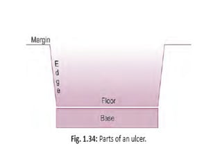
Parts of an Ulcer
a. Margin: It may be regular or irregular. It may
be rounded or oval.
b. Edge: Edge is the one which connects floor of
the ulcer to the margin.
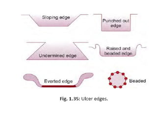
3.
Different edges are:
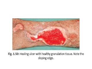
•Sloping edge. It is seen in a healing ulcer.
• Its inner part is red because of red, healthy
granulation tissue.
• Its outer part is white due to scar/fibrous
tissue.
• Its middle part is blue due to epithelial
proliferation.
4.
• Undermined edgeis seen in a tuberculous ulcer
• Punched out edge is seen in a gummatous (syphilitic)
ulcer and trophic ulcer.
It is due to endarteritis.
• Raised and beaded edge (pearly white) is seen in a
rodent ulcer (BCC).
Beads are due to proliferating active cells.
• Everted edge (rolled out edge): It is seen in a
carcinomatous ulcer due to spill of the proliferating
malignant tissues over the normal skin.
5.
c. Floor:
• Itis the one which is seen. Floor may contain
discharge, granulation tissue or slough.
d. Base:
Base is the one on which ulcer rests. It may be bone or
soft tissue.
8.
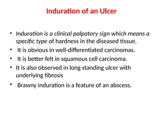
Induration of anUlcer
• Induration is a clinical palpatory sign which means a
specific type of hardness in the diseased tissue.
• It is obvious in well-differentiated carcinomas.
• It is better felt in squamous cell carcinoma.
• It is also observed in long standing ulcer with
underlying fibrosis
• Brawny induration is a feature of an abscess.
9.
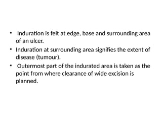
• Induration isfelt at edge, base and surrounding area
of an ulcer.
• Induration at surrounding area signifies the extent of
disease (tumour).
• Outermost part of the indurated area is taken as the
point from where clearance of wide excision is
planned.
10.
Classifications
Classification I (Clinical)
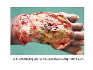
1.Spreading ulcer:
• Here edge is inflamed, irregular and oedematous.
• It is an acute painful ulcer; floor does not contain
healthy granulation tissue (or granulation tissue is
absent) but with profuse purulent discharge and
slough; surrounding area is red and edematous.
12.
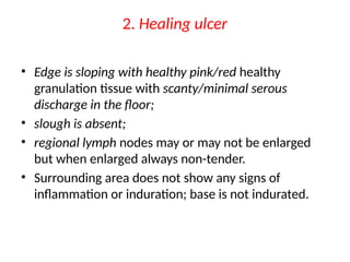
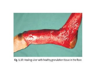
2. Healing ulcer
•Edge is sloping with healthy pink/red healthy
granulation tissue with scanty/minimal serous
discharge in the floor;
• slough is absent;
• regional lymph nodes may or may not be enlarged
but when enlarged always non-tender.
• Surrounding area does not show any signs of
inflammation or induration; base is not indurated.
13.
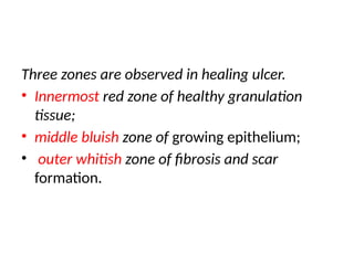
Three zones areobserved in healing ulcer.
• Innermost red zone of healthy granulation
tissue;
• middle bluish zone of growing epithelium;
• outer whitish zone of fibrosis and scar
formation.
15.
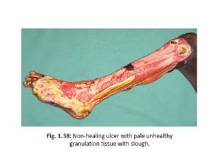
3. Non-healing ulcer
•It may be a chronic ulcer depending on the cause of
the ulcer; here edge will be depending on the cause—
punched out (trophic), undermined (tuberculous),
rolled out (carcinomatous ulcer), beaded (rodent
ulcer);
• floor contains unhealthy granulation tissue and slough,
and serosanguineous/purulent/bloody discharge;
• Regional draining lymph nodes may be enlarged but
non-tender.
17.
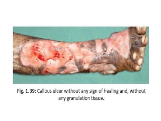
4. Callous (stationary)ulcer
• It is also a chronic non-healing ulcer; floor contains
pale unhealthy, flabby, whitish yellow granulation
tissue and thin scanty serous discharge or often with
copious serosanguinous discharge, with indurated
nontender edge; base is indurated, nontender and
often fixed.
• Ulcer does not show any tendency to heal.
• It lasts for many months to years.
• Tissue destruction is more with absence of or only
minimal regeneration.
18.
• Induration andpigmentation may be seen in the
surrounding area.
• There is no/less discharge.
• Regional lymph nodes may be enlarged; are firm/
hard and nontender.
• It is callousness towards healing; wordcallous means
—insensitive and cruel; and also it means— hard
skinned.
20.
Classification II (Basedon Duration)
1. Acute ulcer
duration is less than 2 weeks.
2. Chronic ulcer
duration is more than 2 weeks (long).
21.
Classification III (Pathological)
1.Specific ulcers:
• Tuberculous ulcer.
• Syphilitic ulcer: It is punched out, deep, with “wash-
leather” slough in the floor and with indurated base.
• Actinomycosis.
• Meleney’s ulcer.
22.
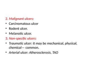
2. Malignant ulcers:
•Carcinomatous ulcer
• Rodent ulcer.
• Melanotic ulcer.
3. Non-specific ulcers:
• Traumatic ulcer: It may be mechanical, physical,
chemical— common.
• Arterial ulcer: Atherosclerosis, TAO
23.
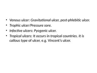
• Venous ulcer:Gravitational ulcer, post-phlebitic ulcer.
• Trophic ulcer/Pressure sore.
• Infective ulcers: Pyogenic ulcer.
• Tropical ulcers: It occurs in tropical countries. It is
callous type of ulcer, e.g. Vincent’s ulcer.
24.
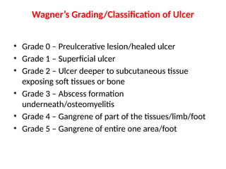
Wagner’s Grading/Classification ofUlcer
• Grade 0 – Preulcerative lesion/healed ulcer
• Grade 1 – Superficial ulcer
• Grade 2 – Ulcer deeper to subcutaneous tissue
exposing soft tissues or bone
• Grade 3 – Abscess formation
underneath/osteomyelitis
• Grade 4 – Gangrene of part of the tissues/limb/foot
• Grade 5 – Gangrene of entire one area/foot
26.
GRANULATION TISSUE
• Itis proliferation of new capillaries and
fibroblasts intermingled with red blood cells
and white blood cells with thin fibrin cover
over it.
28.
Unhealthy granulation tissue
•It is pale with purulent discharge.
• Its floor is covered with slough.
• Its edge is inflamed and oedematous.
• It is a spreading ulcer.
• Unhealthy, pale, flat granulation tissue: It is seen in
chronic nonhealing ulcer (callous ulcer).
29.
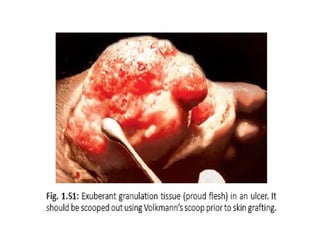
Exuberant granulation tissue(Proud flesh)
• It occurs in a sinus or ulcer wherein granulation
tissue protrudes out of the sinus opening or ulcer
bed like a proliferating mass.
• It is commonly associated with a retained foreign
body in the sinus cavity.
31.
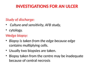
INVESTIGATIONS FOR ANULCER
Study of discharge:
• Culture and sensitivity, AFB study,
• cytology.
Wedge biopsy:
• Biopsy is taken from the edge because edge
contains multiplying cells.
• Usually two biopsies are taken.
• Biopsy taken from the centre may be inadequate
because of central necrosis
32.
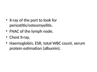
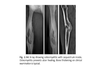
• X-ray ofthe part to look for
periostitis/osteomyelitis.
• FNAC of the lymph node.
• Chest X-ray,
• Haemoglobin, ESR, total WBC count, serum
protein estimation (albumin).
34.
MANAGEMENT OF ANULCER
• Cause should be found and treated.
• Correction of the anaemia, deficiencies like of
protein and vitamins.
• Proper investigation as needed.
• Transfusion of the blood if required.
• Control the pain and infection.
• Rest, immobilization, elevation, avoidance of
repeated trauma.
35.
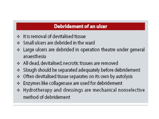
• Care ofthe ulcer by debridement, ulcer cleaning and
dressing.
• Desloughing is done either mechanically or
chemically. Mechanically it is done using scissor by
excising the slough.
• Hydrogen peroxide which releases nascent oxygen is
used as chemical agent.
36.
• Eusol (EdinburghUniversity Solution) which contains
sodium hypochlorite releases nascent chlorine which
forms a water soluble complex with slough to
dissolve it.
• Use of povidone iodine in ulcer cleaning is
controversial (open wound is not suitable; it is mainly
for cleaning the surgical field prior to incision).
37.
• Maggots ifpresent in the wound will cause crawling
sensation and are removed using turpentine
solution.
• Removal of the exuberant granulation tissue is also
required when present.
• Ulcer cleaning and dressing is done daily or twice
daily or once in 2–3 days depending on the type of
ulcer and type of dressing used.
38.
• Normal salineis ideal for ulcer cleaning.
• Various dressings are available.
• Films (opsite/semipermeable polyurethane),
hydrocolloids (duoderm), hydrogels (polyethylene
oxide with water), hydroactives (nonpectin-based
polyurethane matrix), foams.
42.
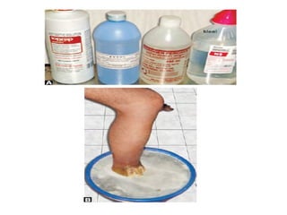
EUSOL bath.
• DiluteEUSOL solution in a basin is used wherein ulcer
foot is dipped and kept in place for 20–30 minutes.
• EUSOL removes the slough and cleans the ulcer bed.
• Hydrogen peroxide releases nascent oxygen and
helps in removing necrotic material.
• Povidone iodine is not used for open wound; it is only
a surface antiseptic
43.
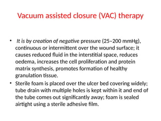
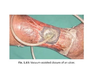
Vacuum assisted closure(VAC) therapy
• It is by creation of negative pressure (25–200 mmHg),
continuous or intermittent over the wound surface; it
causes reduced fluid in the interstitial space, reduces
oedema, increases the cell proliferation and protein
matrix synthesis, promotes formation of healthy
granulation tissue.
• Sterile foam is placed over the ulcer bed covering widely;
tube drain with multiple holes is kept within it and end of
the tube comes out significantly away; foam is sealed
airtight using a sterile adhesive film.
44.
• Tube isconnected to suction system.
• Suction is maintained initially continuously
later intermittently.
• Redressing is done only after 4–7 days.
• Therapy using infrared/short wave/ultraviolet
rays to decrease the ulcer size is often used
but their benefits are not proved.
46.
Maggot debridement therapy
•It is used as biotherapy (but not commonly) by placing
cultured live disinfected maggots.
• Maggots are larvae of the green bottle fly, also known
as the green blowfly (Lucilia sericata).
• They act by dissolving and engulfing dead necrotic
tissues; they may reduce the bacterial content in the
wound.
• They can inhibit many bacteria including MRSA
(methicillin resistant bacteria), anaerobic and aerobic
bacteria.
47.
• They secreteproteolytic enzymes to have mechanical
effects; secretion of ammonia alters the pH in the
ulcer bed which inhibits bacterial growth.
• They increase the granulation tissue formation also.
• Once ulcer granulates, defect is closed with
secondary suturing, skin graft or flaps
48.
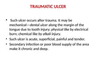
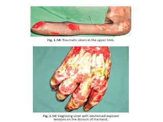
TRAUMATIC ULCER
• Suchulcer occurs after trauma. It may be
mechanical—dental ulcer along the margin of the
tongue due to tooth injury; physical like by electrical
burn; chemical like by alkali injury.
• Such ulcer is acute, superficial, painful and tender.
• Secondary infection or poor blood supply of the area
make it chronic and deep.
50.
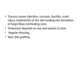
• Trauma causesinfection, necrosis, fasciitis, crush
injury, endarteritis of the skin leading into formation
of large/deep nonhealing ulcer.
• Treatment depends on size and extent of ulcer.
• Regular dressing,
• later skin grafting .
51.
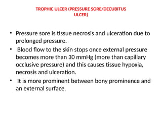
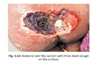
TROPHIC ULCER (PRESSURESORE/DECUBITUS
ULCER)
• Pressure sore is tissue necrosis and ulceration due to
prolonged pressure.
• Blood flow to the skin stops once external pressure
becomes more than 30 mmHg (more than capillary
occlusive pressure) and this causes tissue hypoxia,
necrosis and ulceration.
• It is more prominent between bony prominence and
an external surface.
54.
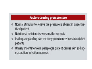
It is dueto:
• Impaired nutrition.
• Defective blood supply.
• Neurological deficit.
55.
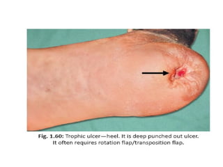
Sites
• Over theischial tuberosity.
• Sacrum.
• In the heel.
• In relation to heads of metatarsals.
• Buttocks.
• Over the shoulder.
• Occiput.
56.
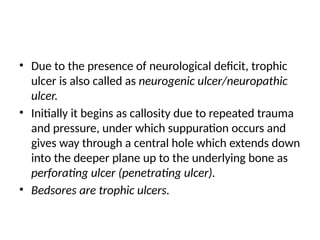
• Due tothe presence of neurological deficit, trophic
ulcer is also called as neurogenic ulcer/neuropathic
ulcer.
• Initially it begins as callosity due to repeated trauma
and pressure, under which suppuration occurs and
gives way through a central hole which extends down
into the deeper plane up to the underlying bone as
perforating ulcer (penetrating ulcer).
• Bedsores are trophic ulcers.
58.
Clinical Features
• Occursin 5% of all hospitalised patients.
• Painless ulcer which is punched out.
• Ulcer is non-mobile with base formed by bone.
59.
Investigations
• Study ofdischarge, blood sugar, biopsy from the
edge, X-ray of the part, X-ray spine
60.
Treatment
• Cause shouldbe treated.
• Nutritional supplementation.
• Rest, antibiotics, slough excision, regular dressings.
• Vacuum-assisted closure (VAC): It is the creation of
intermittent negative pressure of minus 125 mmHg
to promote formation of healthy granulation tissue.
61.
• Proper care:Change in position once in 2 hours;
lifting the limb upwards for 10 seconds once in 10
minutes; nutrition; use of water bed/air bed/air-fluid
floatation bed and pressure dispersion cushions to
the affected area; urinary and faecal care; hygiene;
psychological counselling.
• Regular skin observation; keeping skin clean and dry
(using regular use of talcum powder); oil massaging
of the skin and soft tissues using clean, absorbent
porous clothing; control and prevention of sepsis
helps in the management.
62.
ULCER DUE TOFROSTBITE
• It is due to exposure of a part to wet cold
below the freezing point (cold wind).
• There is arteriolar spasm, denaturation of
proteins and cell destruction.
• It leads to gangrene of the part.
• Ulcers here are always deep.


![Presentation dr rahul seminar (2)[1387]](https://cdn.slidesharecdn.com/ss_thumbnails/presentationdrrahulseminar21387-210324042322-thumbnail.jpg?width=640&height=640&fit=bounds)


