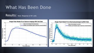
The Heterogeneity of intrinsically photosensitive
Retinal Ganglion Cells (ipRGCs)
Ben Loreto & Kris Cronise
Outline
ipRGCs
Function
Heterogeneity of ipRGCs
Anatomy and Physiology
Model
Future Goals
References and Acknowledgements
ipRGCs
Classical
Photoreceptors
(rods and cones)
Horizontal Cells,
Bipolar Cells,
And Amacrine
Cells
Ganglion Cells
ipRGCs = intrinically
photosensitive retinal
ganglion cells
Source: http://www.skidmore.edu/~hfoley/images/Retina.jpg
Melanopsin
Amination here
Heterogeneity of ipRGCs
5 major classes of ipRGCs
Each classified by certain
differences in anatomy
and physiology
Adapted from: http://www.nature.com/nature/journal/v415/n6871/images/415493a-
f1.2.jpg
Soma Size
Soma size = Size of cell body
Both figures modified from: Schmidt et al. (2011)
Dendritic Stratification and Field
Dendritic Stratification = Where signal is received in the retina
Dendritic field = Amount of area covered by dendrites
Both figures modified from: Ecker. (2011)
Molecular Identity
Molecular Identity = Level of Gene Expression
Melanopsin gene = e.g. Opn4
Both figures modified from: Schmidt et al. (2011)
Intrinsic Light Response
Evaluation of response to light
Traits include sensitivity based
on both size of peak and
duration
Modified from: Schmidt et al
(2011)
Role in Vision
Rods and Cones = IF
Vision
ipRGCs = NIF Vision
Adapted from:
http://www.photobiology.info/Sengupta.html
What Has Been Done
Methods:
•Chemical reactions based on hypothesized pathway
Example:
•Conversion of chemical reactions to differential equations using mass action (and Michaelis-Menten
kinetics)
Example:
•System of differential equations was solved in Matlab and the solution for the concentration of open
channels was plotted
•Parameters (rate constants) were adjusted to fit experimental data with the help of MATLAB function
fminunc.
What Has Been Done
Results: Note: Response of M1 cells
afff
Electrophysiological data obtained from Jess and Abby Electrophysiological data obtained from
R.L. Brown
What We’re Doing
Further Directions
Complete literature review of non-M1 ipRGCs
Compile differences among different ipRGC classes
Incorporate those differences into the model
References
References:
Schmidt. "Melanopsin-Positive Intrinsically Photosensitive Retinal Ganglion Cells: From Form to Function." The Journal of
Neuroscience, 9 Nov. 2011. Web. 20 Oct. 2014.
Wong. "Photoresponse Diversity among the Five Types of Intrinsically Photosensitive Retinal Ganglion Cells." The Journal of
Physiology, 3 Feb. 2014. Web. 20 Oct. 2014.
Hattar et al. "Melanopsin-Expressing Retinal Ganglion-Cell Photoreceptors: Cellular Diversity and Role in Pattern Vision."
Neuron (2010): 49-60. Print.
Acknowledgements
We would like to thank:
UBM @ UMBC
Drs. Phyllis Robinson Kathleen Hoffman, and Hye-Won Kang
Abigail Jackson and Jessica Ortega
Previous Cohorts
The Robinson Lab and its collaborators
NSF

UBM Presentation

  • 1.
    The Heterogeneity ofintrinsically photosensitive Retinal Ganglion Cells (ipRGCs) Ben Loreto & Kris Cronise
  • 2.
    Outline ipRGCs Function Heterogeneity of ipRGCs Anatomyand Physiology Model Future Goals References and Acknowledgements
  • 3.
    ipRGCs Classical Photoreceptors (rods and cones) HorizontalCells, Bipolar Cells, And Amacrine Cells Ganglion Cells ipRGCs = intrinically photosensitive retinal ganglion cells Source: http://www.skidmore.edu/~hfoley/images/Retina.jpg
  • 4.
  • 5.
    Heterogeneity of ipRGCs 5major classes of ipRGCs Each classified by certain differences in anatomy and physiology Adapted from: http://www.nature.com/nature/journal/v415/n6871/images/415493a- f1.2.jpg
  • 6.
    Soma Size Soma size= Size of cell body Both figures modified from: Schmidt et al. (2011)
  • 7.
    Dendritic Stratification andField Dendritic Stratification = Where signal is received in the retina Dendritic field = Amount of area covered by dendrites Both figures modified from: Ecker. (2011)
  • 8.
    Molecular Identity Molecular Identity= Level of Gene Expression Melanopsin gene = e.g. Opn4 Both figures modified from: Schmidt et al. (2011)
  • 9.
    Intrinsic Light Response Evaluationof response to light Traits include sensitivity based on both size of peak and duration Modified from: Schmidt et al (2011)
  • 10.
    Role in Vision Rodsand Cones = IF Vision ipRGCs = NIF Vision Adapted from: http://www.photobiology.info/Sengupta.html
  • 11.
    What Has BeenDone Methods: •Chemical reactions based on hypothesized pathway Example: •Conversion of chemical reactions to differential equations using mass action (and Michaelis-Menten kinetics) Example: •System of differential equations was solved in Matlab and the solution for the concentration of open channels was plotted •Parameters (rate constants) were adjusted to fit experimental data with the help of MATLAB function fminunc.
  • 12.
    What Has BeenDone Results: Note: Response of M1 cells afff Electrophysiological data obtained from Jess and Abby Electrophysiological data obtained from R.L. Brown
  • 13.
  • 14.
    Further Directions Complete literaturereview of non-M1 ipRGCs Compile differences among different ipRGC classes Incorporate those differences into the model
  • 15.
    References References: Schmidt. "Melanopsin-Positive IntrinsicallyPhotosensitive Retinal Ganglion Cells: From Form to Function." The Journal of Neuroscience, 9 Nov. 2011. Web. 20 Oct. 2014. Wong. "Photoresponse Diversity among the Five Types of Intrinsically Photosensitive Retinal Ganglion Cells." The Journal of Physiology, 3 Feb. 2014. Web. 20 Oct. 2014. Hattar et al. "Melanopsin-Expressing Retinal Ganglion-Cell Photoreceptors: Cellular Diversity and Role in Pattern Vision." Neuron (2010): 49-60. Print.
  • 16.
    Acknowledgements We would liketo thank: UBM @ UMBC Drs. Phyllis Robinson Kathleen Hoffman, and Hye-Won Kang Abigail Jackson and Jessica Ortega Previous Cohorts The Robinson Lab and its collaborators NSF

Editor's Notes

  • #3 SIMPLIFY
  • #4 pathway = LIGHT → PHOTORECEPTORS → HORIZONTAL → BIPOLAR → AMACRINE → GANGLION→ OPTIC NERVE Photoreceptors at the back of the eye 2 types: the rods and cones. Their outer segment disks are the site of phototransduction. LIGHT HITS THIS FIRST Bipolar Cells transfer all signals between photoreceptors and ganglions Horizontal Cells their processes enable lateral interactions between photoreceptors and bipolar cells that maintain the visual system’s sensitivity to luminance (AKA contrast) Amacrine cells processes are postsynaptic to bipolar cell terminals and presynaptic to dendrites of ganglion cells Ganglion Cells their signals feed into the optic nerve Some of the ganglion cells mentioned are intrinsically photosensitive, and thus make up a third class of photoreceptors - known as ipRGCs
  • #5 Like rods and cones which express their own photopigment, rhodopsin, ipRGCs use the photopigment melanopsin Activation Light converts 11-cis retinal in melanopsin to trans configuration this allows for binding of a g protein (GP), in its inactive form bound to GDP it then exchanges GDP for GTP, thus activating the g protein activated g protein dissociates its alpha subunit from its beta and gamma subunits, and is then free to interact with phospholipase c (PLC) phospholipase c is then able to cleave pip2 into a second messenger this second messenger is then able bind to calcium channels and allow them to open, resulting in an influx of extracellular calcium ions which depolarize the cell Deactivation In response to a rise in calcium concentration, a kinase phosphorylates the melanopsin tail, thus allowing arrestin to bind with arrestin bound, the inactive g protein is unable to bind to melanopsin, thus stopping the cascade
  • #6 Ask about how diversity among iprgcs discovered This is a “photoreceptive net” made up of ipRGCs, obtained with immunoflouresence using antibodies labeled against melanopsin Back when Melanopsin was first discovered, it was believed that only one type of ipRGC existed, so this picture was believed to compose of only one type But as scientists have made more and more discoveries. It is now believed that this is a Mosaic of just 2 of the 5 types (M1 and M2) of Melanopsin containing ipRGCs each type of ipRGC is classified by a few of its differences in its anatomy and physiology (structure and function
  • #7 The comparative sizes of each class. M1=M5 is the smallest cell body, M4 is the largest. M2 & M3 intermediate On the left is a picture of some examples of ipRGC’s, m1, m2 and m4, captured using an Intracellular dye GFP,
  • #8 Looking at this graph, we can tell the morphological differences between each melanopsin class: RGCs with dendrites stratifying in the outer sublamina of the IPL receive excitatory synaptic inputs from bipolar cells that respond to light decrements (OFF) whereas RGCs with dendrites stratifying in the inner sublamina of the IPL receive excitatory synaptic inputs from bipolar cells that respond to light increments (ON) Where each ipRGC class ramifies its dendrites. M1 stratify in the outermost sublamina of the IPL (OFF), M2 in the innermost sublamina (ON) M3 stratifies in both the inner and outer sublamina (OFF/ON) M4 and M5 stratify in the innermost sublamina (ON) What’s interesting is that M1 receives majority of ON signals, even when stratified in the off sublamina
  • #9 level of OPN 4 expression → amount of melanopsin expression Strong Opn4+ Opntau-lacZ+ OpnCre+ Brn3b+/- Weak Opn4+ Opntau-lacZ- OpnCre+ Brn3b+ Weak Opn4+ Opntau-lacZ+ OpnCre+ Brn3b+ Opn4- Opntau-lacZ- OpnCre+ Brn3b+ Opn4- Opntau-lacZ- OpnCre+ Brn3b+ because of this M1 is the most well studied, because it expresses the most melanopsin
  • #10 corresponding whole cell voltage clamp recordings below, measured in pico amps, It measures 1 flash, for 20 seconds after flash. the logs correspond to intensity of logs, with 0 log having the highest intensity M1 cells have largest, most sensitive intrinsic light-evoked responses whereas M2 cells have significantly smaller light responses that are at least one logarithmic unit less sensitive to light M1 cells have higher input resistance, a more depolarized resting membrane potential, and spike at lower frequencies than M2 cells In-depth electrophysiological analyses of the rare M3 cells demonstrated that these cells are remarkably invariable, which is surprising given the dendritic variability within this subtype M4 and M5 have the smallest and least sensitive intrinsic light responses of the ipRGCs they could be considered insensitive
  • #11 IF vision = colors, shapes, textures, distance NIF vision = brightness, and darkness M1 ipRGCs innervate only the shell of the OPN, Non-M1 cells supply signals to the core of the OPN for PLR Non-M1 cells also contribute substantial synaptic input to the dorsal lateral geniculate nucleus (dLGN), a structure involved in image-forming vision, and the superior colliculus (SC), also involved in image forming vision
  • #12 Based on the hypothesized melanopsin signaling pathway, Abbey and Jess came up with a list of chemical reactions They then converted these chemical reactions into differential equations using the law of mass action, and Michaelis Menten kinetics Solved the system of differential equations using Matlab, and plotted the solution for the concentration of open channels They then adjusted the parameters (or rate constants and initial conditions) to fit the experimental data Law of Mass Action in chemical kinetics states that the rate at which a chemical is produced is proportional to the concentration of the reactants
  • #13 (Left) Single flash HEK cells Human embryonic kidney (HEK) cells were transfected with mouse melanopsin gene. A calcium-sensitive dye was added to the cells and they were exposed to a flash of light, and they measured fluorescence model parameters (rate constants) then adjusted to fit this data (Right) Single flash ipRGCs Electrophysiological data obtained from collaborator R. L. Brown Measured current across cell membrane of an ipRGC Results: Model fits well with WT experimental data for a single flash. The same parameters are sensitive in both models
  • #14 Single flash data we obtained for HEK cells transfected with the murine (mouse) melanopsin gene we have yet to fit a model to our data