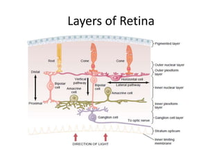
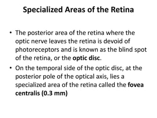
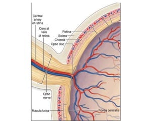
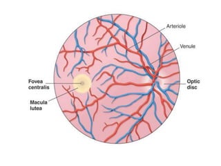
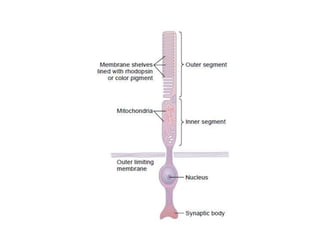
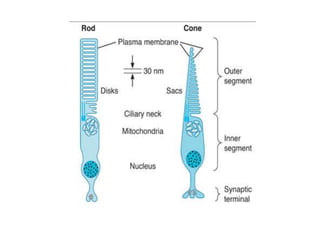
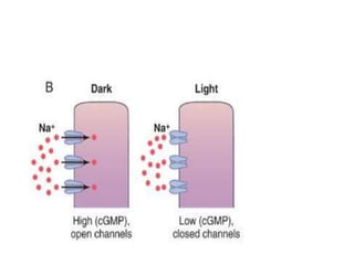
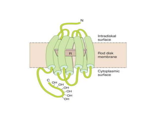
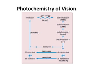
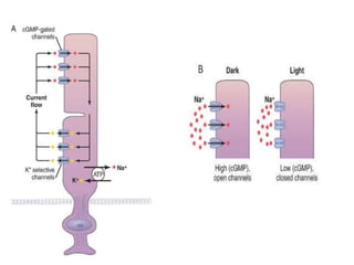
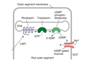
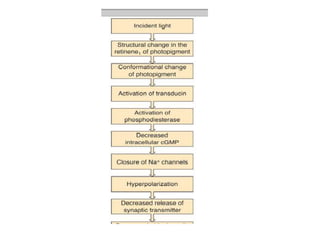
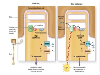
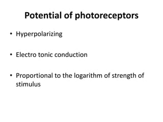
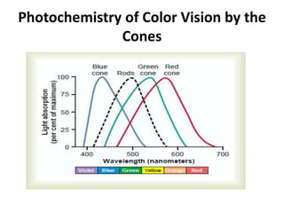
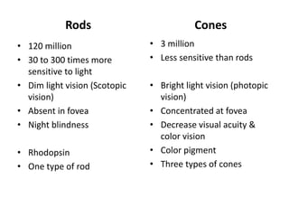
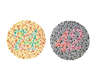
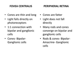
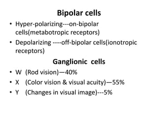
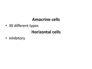
RECEPTOR AND NEURAL
FUNCTION OF RETINA
Layers of Retina
Specialized Areas of the Retina
• The posterior area of the retina where the
optic nerve leaves the retina is devoid of
photoreceptors and is known as the blind spot
of the retina, or the optic disc.
• On the temporal side of the optic disc, at the
posterior pole of the optical axis, lies a
specialized area of the retina called the fovea
centralis (0.3 mm)
• Pigment layer of retina (Importance)
• Albinos have low visual acuity
• Night blindness
• Retinal detachment & consequences
Photochemistry of Vision
Potential of photoreceptors
• Hyperpolarizing
• Electro tonic conduction
• Proportional to the logarithm of strength of
stimulus
Photochemistry of Color Vision by the
Cones
Rods
• 120 million
• 30 to 300 times more
sensitive to light
• Dim light vision (Scotopic
vision)
• Absent in fovea
• Night blindness
• Rhodopsin
• One type of rod
Cones
• 3 million
• Less sensitive than rods
• Bright light vision (photopic
vision)
• Concentrated at fovea
• Decrease visual acuity &
color vision
• Color pigment
• Three types of cones
LIGHT AND DARK ADAPTATION
LIGHT ADAPTATION
• If a person has been in bright light for hours,
large portions of the photochemicals in both
the rods and the cones will have been reduced
to retinal and opsins. The sensitivity of the eye
to light is correspondingly reduced. This is
called light adaptation.
• Pupillary constriction
• Neural adaptation
DARK ADAPTATION
• If a person remains in darkness for a long
time, the retinal and opsins in the rods and
cones are converted back into the light-
sensitive pigments. Sensitivity to light
increases. This is called dark adaptation.
• Pupillary dilation
• Adaptation at level of ganaglion and bipolar
cells
Automatic Regulation of
Retinal Sensitivity—Light and
Dark Adaptation
Color vision and color deficiency
Color Vision
• The Young–Helmholtz theory of color vision in
humans postulates the existence of three
kinds of cones
• Each containing a different photo pigment and
that are maximally sensitive to one of the
three primary colors (blue, green, red)
• The sensation of any given color being
determined by the relative frequency of the
impulses from each of these cone systems.
• R: G: B
• Blue 445 nm (0:0:97)
• Green 535 nm (31:67:36)
• Red 570 nm
• Orange (580) 99:42:0
• Yellow 83:83:0
R:G:B
• White light?
How colors are perceived
• Three types of cones
• Specific ganglionic cells (X or parvocellular
cells)
• Specific areas of LGB and visual cortex
• Color blobs of visual cortex
Color Blindness
• Protanope (Red cones deficient)
• Deuteranope (Green cones deficient)
• Tritanope (Blue cones deficient)
Color Blindness
• Red-Green Color blindness
• A person with loss of red cones is called a
protanope;
• the overall visual spectrum is noticeably
shortened at the long wavelength end because of
a lack of the red cones.
• A color-blind person who lacks green cones is
called a deuteranope;
this person has a perfectly normal visual spectral
width because red cones are available to detect
the long wavelength red
• Red-Green Blindness almost always occur in
males
• Its gene is on X chromosome
• Blue Weakness. Only rarely are blue cones
missing, although sometimes they are
underrepresented, which is a genetically
inherited state giving rise to the phenomenon
called blue weakness.
Color weakness
• Ishihara chart
• Edridge-Green lantern
• Holmgren colored wool
FOVEA CENTRALIS
• Cones are thin and long
• Light falls directly on
photoreceptors
• 1:1 connection with
bipolar and ganglionic
cells
• Cones- Bipolar-
Ganglionic cells
PERIPHERAL RETINA
• Cones are fatter
• Light does not fall
directly
• Many rods and cones
converge on bipolar and
ganglionic cells
• Rods & cones- Bipolar-
Amacrine- Ganglionic
cells
Bipolar cells
• Hyper-polarizing---on-bipolar
cells(metabotropic receptors)
• Depolarizing ----off-bipolar cells(ionotropic
receptors)
Ganglionic cells
• W (Rod vision)—40%
• X (Color vision & visual acuity)—55%
• Y (Changes in visual image)---5%
Amacrine cells
• 30 different types
Horizontal cells
• inhibitory
Nodal point (Point at the center of lens
where light rays pass un deviated)

EYE 2.pptx

  • 1.
  • 2.
  • 3.
    Specialized Areas ofthe Retina • The posterior area of the retina where the optic nerve leaves the retina is devoid of photoreceptors and is known as the blind spot of the retina, or the optic disc. • On the temporal side of the optic disc, at the posterior pole of the optical axis, lies a specialized area of the retina called the fovea centralis (0.3 mm)
  • 10.
    • Pigment layerof retina (Importance) • Albinos have low visual acuity • Night blindness • Retinal detachment & consequences
  • 11.
  • 16.
    Potential of photoreceptors •Hyperpolarizing • Electro tonic conduction • Proportional to the logarithm of strength of stimulus
  • 17.
    Photochemistry of ColorVision by the Cones
  • 18.
    Rods • 120 million •30 to 300 times more sensitive to light • Dim light vision (Scotopic vision) • Absent in fovea • Night blindness • Rhodopsin • One type of rod Cones • 3 million • Less sensitive than rods • Bright light vision (photopic vision) • Concentrated at fovea • Decrease visual acuity & color vision • Color pigment • Three types of cones
  • 19.
    LIGHT AND DARKADAPTATION
  • 20.
    LIGHT ADAPTATION • Ifa person has been in bright light for hours, large portions of the photochemicals in both the rods and the cones will have been reduced to retinal and opsins. The sensitivity of the eye to light is correspondingly reduced. This is called light adaptation. • Pupillary constriction • Neural adaptation
  • 21.
    DARK ADAPTATION • Ifa person remains in darkness for a long time, the retinal and opsins in the rods and cones are converted back into the light- sensitive pigments. Sensitivity to light increases. This is called dark adaptation. • Pupillary dilation • Adaptation at level of ganaglion and bipolar cells
  • 22.
    Automatic Regulation of RetinalSensitivity—Light and Dark Adaptation
  • 23.
    Color vision andcolor deficiency
  • 24.
    Color Vision • TheYoung–Helmholtz theory of color vision in humans postulates the existence of three kinds of cones • Each containing a different photo pigment and that are maximally sensitive to one of the three primary colors (blue, green, red) • The sensation of any given color being determined by the relative frequency of the impulses from each of these cone systems.
  • 25.
    • R: G:B • Blue 445 nm (0:0:97) • Green 535 nm (31:67:36) • Red 570 nm • Orange (580) 99:42:0 • Yellow 83:83:0 R:G:B • White light?
  • 27.
    How colors areperceived • Three types of cones • Specific ganglionic cells (X or parvocellular cells) • Specific areas of LGB and visual cortex • Color blobs of visual cortex
  • 28.
    Color Blindness • Protanope(Red cones deficient) • Deuteranope (Green cones deficient) • Tritanope (Blue cones deficient)
  • 29.
    Color Blindness • Red-GreenColor blindness • A person with loss of red cones is called a protanope; • the overall visual spectrum is noticeably shortened at the long wavelength end because of a lack of the red cones. • A color-blind person who lacks green cones is called a deuteranope; this person has a perfectly normal visual spectral width because red cones are available to detect the long wavelength red
  • 30.
    • Red-Green Blindnessalmost always occur in males • Its gene is on X chromosome
  • 31.
    • Blue Weakness.Only rarely are blue cones missing, although sometimes they are underrepresented, which is a genetically inherited state giving rise to the phenomenon called blue weakness.
  • 32.
    Color weakness • Ishiharachart • Edridge-Green lantern • Holmgren colored wool
  • 34.
    FOVEA CENTRALIS • Conesare thin and long • Light falls directly on photoreceptors • 1:1 connection with bipolar and ganglionic cells • Cones- Bipolar- Ganglionic cells PERIPHERAL RETINA • Cones are fatter • Light does not fall directly • Many rods and cones converge on bipolar and ganglionic cells • Rods & cones- Bipolar- Amacrine- Ganglionic cells
  • 35.
    Bipolar cells • Hyper-polarizing---on-bipolar cells(metabotropicreceptors) • Depolarizing ----off-bipolar cells(ionotropic receptors) Ganglionic cells • W (Rod vision)—40% • X (Color vision & visual acuity)—55% • Y (Changes in visual image)---5%
  • 36.
    Amacrine cells • 30different types Horizontal cells • inhibitory
  • 37.
    Nodal point (Pointat the center of lens where light rays pass un deviated)