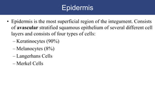
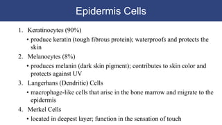
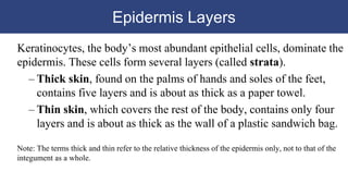
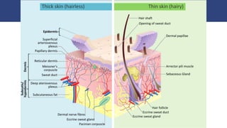
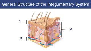
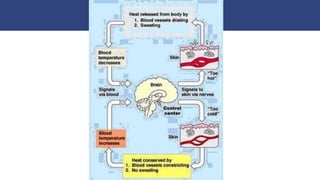
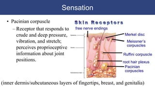
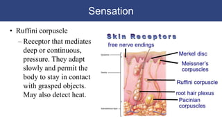
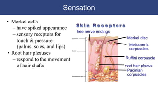
Unit
3
The
Integumentary
System
Chapter 5 in Textbook
3.2 Skin and its Tissues
Epidermis
• Epidermis is the most superficial region of the integument. Consists
of avascular stratified squamous epithelium of several different cell
layers and consists of four types of cells:
– Keratinocytes (90%)
– Melanocytes (8%)
– Langerhans Cells
– Merkel Cells
Epidermis Cells
1. Keratinocytes (90%)
• produce keratin (tough fibrous protein); waterproofs and protects the
skin
2. Melanocytes (8%)
• produces melanin (dark skin pigment); contributes to skin color and
protects against UV
3. Langerhans (Dendritic) Cells
• macrophage-like cells that arise in the bone marrow and migrate to the
epidermis
4. Merkel Cells
• located in deepest layer; function in the sensation of touch
Epidermis Layers
Keratinocytes, the body’s most abundant epithelial cells, dominate the
epidermis. These cells form several layers (called strata).
– Thick skin, found on the palms of hands and soles of the feet,
contains five layers and is about as thick as a paper towel.
– Thin skin, which covers the rest of the body, contains only four
layers and is about as thick as the wall of a plastic sandwich bag.
Note: The terms thick and thin refer to the relative thickness of the epidermis only, not to that of the
integument as a whole.
Epidermis Layers
The five strata of epidermis from basement
membrane to apical surface:
1. Stratum basale or germinativum
– single row of cells attached to dermis; youngest cells; through
cell division it generates all of the other layers.
2. Stratum spinosum (spiny layer)
– 8-10 rows of polyhedral (many sided) cells covered with
prickly spines; participate in the immune response.
3. Stratum granulosum (grainy layer)
– 3-5 rows of flattened cells; produces keratin.
4. Stratum lucidum (clear layer)
– thick skin only; 3-5 rows of clear, flat, dead cells.
5. Stratum corneum
• 15-30 rows of flat dead cells; completely filled with keratin.
Structure of the Epidermis
Epidermis
• Keratinization
– The formation of a layer of dead, protective cells filled with
keratin
– Occurs on all exposed skin surfaces except eyes
– Skin life cycle takes 2 to 4 weeks for a cell to move from stratum
germinativum to stratum corneum
Skin Color
The color of your skin is caused by the interaction between (1)
epidermal pigmentation and (2) the dermal blood supply.
Three pigments play a role in skin color:
1. Carotene:
• Orange-yellow pigment
• Mostly in the stratum corneum and fatty areas of the dermis
• Precursor of Vitamin A (synthesizes vision pigments)
• Accounts for the yellowish hue of Asian skin
• Fun Fact: also found in orange vegetables (carrots)
Skin Color
Three pigments play a role in skin color:
2. Melanin:
• Mostly in the epidermis; provides protection against sun damage
• Yellow brown-black pigment produced by melanocytes in stratum basale
• Number of melanocytes about the same in all races, amount of melanin
differs
• Most plentiful in the mucous membranes, penis, nipples, areola (around
nipples), face, and extremities
• Freckles (clusters of melanin), Albinism (no melanin produced), Vitiligo
(partial or complete loss of melanin; irregular white spots)
Skin Color
Three pigments play a role in skin color:
3. Hemoglobin:
• Carries oxygen in the blood contributing to the pink of light skin
• Oxygenated blood contributes to changes in skin color
–Blood vessels dilate from heat, skin reddens
–Blood flow decreases, skin pales
• Erythma:
– Redness in skin due to engorgement of blood vessels (blushing)
• Cyanosis:
– Bluish skin tint
– Caused by severe reduction in blood flow or oxygenation
Fingerprints, Palmprints, Footprints
• The stratum basale forms epidermal ridges, which extend into the
dermis, increasing the area of contact between the two regions.
• The contours of skin surface follow the ridge patterns, which vary from
small conical pegs (in thin skin) to the complex whorls on the thick skin
of the palms and soles.
• Epidermal ridges increases friction and acts like tiny suction cups.
• Ridge contours are genetically determined; those of each person are
unique and do not change over the course of a lifetime.
• Fingerprints are ridge patterns on the tips of fingers!
The Dermis
• The Dermis
– Is located between epidermis and subcutaneous layer; plays critical role
in temperature regulation and nourishment to the epidermis.
– Anchors epidermal accessory structures (hair follicles, sweat glands).
– Very thick in palms and soles. Very thin in eyelids, penis, and scrotum.
– Has two components
• Outer papillary layer
• Deep reticular layer
The Dermis
• The Papillary Layer
– Superficial layer; consists of areolar tissue
– Contains capillaries and nerves supplying the surface of the skin
– Has dermal papillae projecting between epidermal ridges
• The Reticular Layer
– Deep layer; consists of dense irregular connective tissue
– Contains larger blood vessels, lymph vessels, nerve fibers, and glands
– Contains collagen and elastic fibers that allows the skin to stretch and
return to original shape
The Hypodermis
• The subcutaneous layer or hypodermis
– Lies below the skin (not a TRUE layer of the integument)
– Stabilizes the skin, allows for independent movement
– Is made of elastic areolar and adipose tissues
– Is connected to the reticular layer of integument by connective tissue fibers
– Contains no vital organs and few capillaries, which makes subcutaneous
injections a useful method for administering drugs using a hypodermic needle
– Deposits of subcutaneous fat have distribution patterns determined by hormones
General Structure of the Integumentary System
Body Temperature
• Normal body temperature ranges from 97 ºF to 100 ºF (average is
98.6 ºF)
• Body temperature fluctuates throughout the day
– lower in the morning
– higher in the afternoon
• Core temperature (cranial, thoracic and abdominal cavities)
• Shell temperature (skin and mouth)
Body Temperature
• Body temperature is maintained by balancing heat production and
heat loss (remember homeostasis?)
– Heat is produced by the millions of chemical reactions occurring
in the body and is affected by food consumption, hormone
secretion, and physical activity
– 80% of heat is lost through the skin, the rest through the
respiratory tract and excretion of wastes
• This balance is known as thermoregulation
• Excessive changes in body temperature can be severe, even fatal
(hyperthermia vs hypothermia)
Body Temperature
• The skin helps to regulate the homeostasis of body temperature
through a negative feedback system
• Perspiration from sweat glands and dilation of the superficial blood
vessels help to remove excess heat from the body
• Constriction of the blood vessels in the skin aids in conserving heat
when body temperature drops; also, hair stands up, trapping
insulating air
• YOU can help thermoregulate by stretching out when hot and
curling up when cold!
Body Temperature
Body Temperature
• Thermoregulation is difficult for newborns
– Large surface area increases heat loss
– Thin layer of subcutaneous fat
– Neonates cannot shiver
• Neonates have brown adipose tissue (BAT) which produces a large
amount of heat when metabolized; this is called non-shivering
thermogenesis
Sensation
• The skin acts as a sensory organ. There are receptors located
throughout the dermis and the epidermis.
• Free nerve endings found in the dermal papillae and possibly in the
stratum basale and stratum spinosum detect itch, pain, hot, and cold.
Sensation
• Touch is a skin sensation that results from an active or passive
contact between a person's skin and an object. Pressure applied onto
the skin is the primary stimulus for the sense of touch. Another
stimulus, vibration, emerges when there is a rapid and regular
change in pressure.
• Fine (discriminative) touch
Touch that is subtle but easily located
• Crude (non-discriminative) touch
Touch that is more easily identified, but more difficult to
locate (tends to spread)
Sensation
• Meissner corpuscle
(AKA: tactile corpuscle)
– Receptor that mediates
sensations of
discriminative touch
such as light versus
deep pressure, as well
as low-frequency
vibration.
(dermal papillae of the fingertips, lips, eyelids, nipples, and genitalia)
Sensation
• Pacinian corpuscle
– Receptor that responds to
crude and deep pressure,
vibration, and stretch;
perceives proprioceptive
information about joint
positions.
(inner dermis/subcutaneous layers of fingertips, breast, and genitalia)
Sensation
• Ruffini corpuscle
– Receptor that mediates
deep or continuous,
pressure. They adapt
slowly and permit the
body to stay in contact
with grasped objects.
May also detect heat.
Sensation
• Merkel cells
– have spiked appearance
– sensory receptors for
touch & pressure
(palms, soles, and lips)
• Root hair plexuses
– respond to the movement
of hair shafts

U3.2 Integumentary System

  • 1.
    Unit 3 The Integumentary System Chapter 5 inTextbook 3.2 Skin and its Tissues
  • 2.
    Epidermis • Epidermis isthe most superficial region of the integument. Consists of avascular stratified squamous epithelium of several different cell layers and consists of four types of cells: – Keratinocytes (90%) – Melanocytes (8%) – Langerhans Cells – Merkel Cells
  • 3.
    Epidermis Cells 1. Keratinocytes(90%) • produce keratin (tough fibrous protein); waterproofs and protects the skin 2. Melanocytes (8%) • produces melanin (dark skin pigment); contributes to skin color and protects against UV 3. Langerhans (Dendritic) Cells • macrophage-like cells that arise in the bone marrow and migrate to the epidermis 4. Merkel Cells • located in deepest layer; function in the sensation of touch
  • 4.
    Epidermis Layers Keratinocytes, thebody’s most abundant epithelial cells, dominate the epidermis. These cells form several layers (called strata). – Thick skin, found on the palms of hands and soles of the feet, contains five layers and is about as thick as a paper towel. – Thin skin, which covers the rest of the body, contains only four layers and is about as thick as the wall of a plastic sandwich bag. Note: The terms thick and thin refer to the relative thickness of the epidermis only, not to that of the integument as a whole.
  • 6.
    Epidermis Layers The fivestrata of epidermis from basement membrane to apical surface: 1. Stratum basale or germinativum – single row of cells attached to dermis; youngest cells; through cell division it generates all of the other layers. 2. Stratum spinosum (spiny layer) – 8-10 rows of polyhedral (many sided) cells covered with prickly spines; participate in the immune response. 3. Stratum granulosum (grainy layer) – 3-5 rows of flattened cells; produces keratin. 4. Stratum lucidum (clear layer) – thick skin only; 3-5 rows of clear, flat, dead cells. 5. Stratum corneum • 15-30 rows of flat dead cells; completely filled with keratin.
  • 7.
  • 8.
    Epidermis • Keratinization – Theformation of a layer of dead, protective cells filled with keratin – Occurs on all exposed skin surfaces except eyes – Skin life cycle takes 2 to 4 weeks for a cell to move from stratum germinativum to stratum corneum
  • 9.
    Skin Color The colorof your skin is caused by the interaction between (1) epidermal pigmentation and (2) the dermal blood supply. Three pigments play a role in skin color: 1. Carotene: • Orange-yellow pigment • Mostly in the stratum corneum and fatty areas of the dermis • Precursor of Vitamin A (synthesizes vision pigments) • Accounts for the yellowish hue of Asian skin • Fun Fact: also found in orange vegetables (carrots)
  • 10.
    Skin Color Three pigmentsplay a role in skin color: 2. Melanin: • Mostly in the epidermis; provides protection against sun damage • Yellow brown-black pigment produced by melanocytes in stratum basale • Number of melanocytes about the same in all races, amount of melanin differs • Most plentiful in the mucous membranes, penis, nipples, areola (around nipples), face, and extremities • Freckles (clusters of melanin), Albinism (no melanin produced), Vitiligo (partial or complete loss of melanin; irregular white spots)
  • 11.
    Skin Color Three pigmentsplay a role in skin color: 3. Hemoglobin: • Carries oxygen in the blood contributing to the pink of light skin • Oxygenated blood contributes to changes in skin color –Blood vessels dilate from heat, skin reddens –Blood flow decreases, skin pales • Erythma: – Redness in skin due to engorgement of blood vessels (blushing) • Cyanosis: – Bluish skin tint – Caused by severe reduction in blood flow or oxygenation
  • 12.
    Fingerprints, Palmprints, Footprints •The stratum basale forms epidermal ridges, which extend into the dermis, increasing the area of contact between the two regions. • The contours of skin surface follow the ridge patterns, which vary from small conical pegs (in thin skin) to the complex whorls on the thick skin of the palms and soles. • Epidermal ridges increases friction and acts like tiny suction cups. • Ridge contours are genetically determined; those of each person are unique and do not change over the course of a lifetime. • Fingerprints are ridge patterns on the tips of fingers!
  • 13.
    The Dermis • TheDermis – Is located between epidermis and subcutaneous layer; plays critical role in temperature regulation and nourishment to the epidermis. – Anchors epidermal accessory structures (hair follicles, sweat glands). – Very thick in palms and soles. Very thin in eyelids, penis, and scrotum. – Has two components • Outer papillary layer • Deep reticular layer
  • 14.
    The Dermis • ThePapillary Layer – Superficial layer; consists of areolar tissue – Contains capillaries and nerves supplying the surface of the skin – Has dermal papillae projecting between epidermal ridges • The Reticular Layer – Deep layer; consists of dense irregular connective tissue – Contains larger blood vessels, lymph vessels, nerve fibers, and glands – Contains collagen and elastic fibers that allows the skin to stretch and return to original shape
  • 15.
    The Hypodermis • Thesubcutaneous layer or hypodermis – Lies below the skin (not a TRUE layer of the integument) – Stabilizes the skin, allows for independent movement – Is made of elastic areolar and adipose tissues – Is connected to the reticular layer of integument by connective tissue fibers – Contains no vital organs and few capillaries, which makes subcutaneous injections a useful method for administering drugs using a hypodermic needle – Deposits of subcutaneous fat have distribution patterns determined by hormones
  • 16.
    General Structure ofthe Integumentary System
  • 17.
    Body Temperature • Normalbody temperature ranges from 97 ºF to 100 ºF (average is 98.6 ºF) • Body temperature fluctuates throughout the day – lower in the morning – higher in the afternoon • Core temperature (cranial, thoracic and abdominal cavities) • Shell temperature (skin and mouth)
  • 18.
    Body Temperature • Bodytemperature is maintained by balancing heat production and heat loss (remember homeostasis?) – Heat is produced by the millions of chemical reactions occurring in the body and is affected by food consumption, hormone secretion, and physical activity – 80% of heat is lost through the skin, the rest through the respiratory tract and excretion of wastes • This balance is known as thermoregulation • Excessive changes in body temperature can be severe, even fatal (hyperthermia vs hypothermia)
  • 19.
    Body Temperature • Theskin helps to regulate the homeostasis of body temperature through a negative feedback system • Perspiration from sweat glands and dilation of the superficial blood vessels help to remove excess heat from the body • Constriction of the blood vessels in the skin aids in conserving heat when body temperature drops; also, hair stands up, trapping insulating air • YOU can help thermoregulate by stretching out when hot and curling up when cold!
  • 20.
  • 21.
    Body Temperature • Thermoregulationis difficult for newborns – Large surface area increases heat loss – Thin layer of subcutaneous fat – Neonates cannot shiver • Neonates have brown adipose tissue (BAT) which produces a large amount of heat when metabolized; this is called non-shivering thermogenesis
  • 22.
    Sensation • The skinacts as a sensory organ. There are receptors located throughout the dermis and the epidermis. • Free nerve endings found in the dermal papillae and possibly in the stratum basale and stratum spinosum detect itch, pain, hot, and cold.
  • 23.
    Sensation • Touch isa skin sensation that results from an active or passive contact between a person's skin and an object. Pressure applied onto the skin is the primary stimulus for the sense of touch. Another stimulus, vibration, emerges when there is a rapid and regular change in pressure. • Fine (discriminative) touch Touch that is subtle but easily located • Crude (non-discriminative) touch Touch that is more easily identified, but more difficult to locate (tends to spread)
  • 24.
    Sensation • Meissner corpuscle (AKA:tactile corpuscle) – Receptor that mediates sensations of discriminative touch such as light versus deep pressure, as well as low-frequency vibration. (dermal papillae of the fingertips, lips, eyelids, nipples, and genitalia)
  • 25.
    Sensation • Pacinian corpuscle –Receptor that responds to crude and deep pressure, vibration, and stretch; perceives proprioceptive information about joint positions. (inner dermis/subcutaneous layers of fingertips, breast, and genitalia)
  • 26.
    Sensation • Ruffini corpuscle –Receptor that mediates deep or continuous, pressure. They adapt slowly and permit the body to stay in contact with grasped objects. May also detect heat.
  • 27.
    Sensation • Merkel cells –have spiked appearance – sensory receptors for touch & pressure (palms, soles, and lips) • Root hair plexuses – respond to the movement of hair shafts