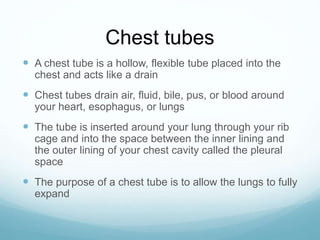
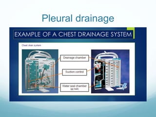
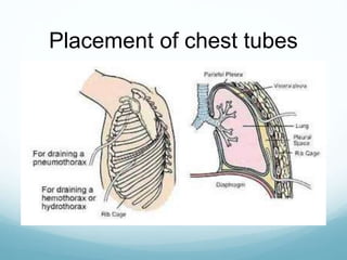
Chest Tubes
Nursing Clinical III
Chest tubes
 A chest tube is a hollow, flexible tube placed into the
chest and acts like a drain
 Chest tubes drain air, fluid, bile, pus, or blood around
your heart, esophagus, or lungs
 The tube is inserted around your lung through your rib
cage and into the space between the inner lining and
the outer lining of your chest cavity called the pleural
space
 The purpose of a chest tube is to allow the lungs to fully
expand
Types of Chest tubes
 Large (36F to 40F) tubes used to drain blood
 Medium (24F to 36F) tubes used to drain fluid
 Small (12F to 24F) tubes used to drain air
 Pigtail tubes are very small (10F to 14F) with a curly
end designed to keep them in place
Pleural drainage
Chest drainage unit
 First compartment (collection chamber) receives fluid and air
from the pleural space
 Second compartment (water-seal chamber) contains 2 cm of
water, which acts as a one-way valve
 Tidaling – normal flunctuation of the water within the water-seal
chamber and reflects respirations on inspiration and expiration
 The air enters from the collection chamber and bubbles up
through the water
 Third compartment (suction control chamber) applies
suction to the chest drainage system
 Two types: water and dry
Chest tube indications
 Pneumothorax
 Hemothorax
 Empyema
 Pleural Effusion
 Hemopneumothorax
 Hydrothorax
 Chylothorax
 Patients with penetrating wall injury who are intubated or
about to be intubated
Chest tube insertion
 Make sure patient is aware of the procedure and
informed consent is obtained
 Gather equipment
 Thoracotomy tray
 Chest drainage unit (CDU)
 Chest tube
 Bottle of sterile water
 1% lidocaine
 Suction tubing and collection container
 Occlusive dressing
Chest tube insertion (cont..)
 Prepare CDU
 Wet suction: add sterile water to 2-cm mark in water-seal
chamber and to 20-cm mark (or as ordered) in suction
control chamber
 Dry suction: add sterile water to the fill line of the air leak
meter. Attach suction tubing and increase suction until the
bellows-like float moves across the display window
 Position and support the patient to minimize movement
during procedure
Placement of chest tubes
Risks and complications
 Bleeding or infection where the tube is inserted
 Improper placement of the tube (into the tissues,
abdomen, or too far into the chest)
 Injury to the lung
 Injury to organs near the tube (spleen, liver, stomach,
or diaphragm)
Nursing considerations
 Assess vital signs, lung sounds and pain
 Assess for manifestations of reaccumulation of air and fluid in the chest (decrease or absent breath
sounds)
 Assess for significant bleeding (100 mL/hr)
 Assess for chest drainage site infection (drainage, erythema, fever, increase WBC) or poor wound
healing
 Encourage patient to cough and breath deeply
 Make sure connections are taped securely
 Keep collection below the level of the patients chest
 Check water seal and suction control chambers
 Measure drainage every 8 hours or more often depending on patients condition
 Document assessment of drainage system and how patient is responding
Care of chest tube and
drainage unit
 Mark the time of measurement and the fluid level on the drainage unit according to
the unit standards
 Keep all tubing loosely coiled below the clients level
 Observe for air flunctuations (tidaling) and bubbling in the water-seal chamber
 Keep the suction control chamber at the appropriate water level by adding sterile
water as needed
 If no bubbling is seen in the suction control chamber, there is no suction, suction is
not high enough, or the pleural air leak is so large that suction is not high enough to
evacuate it
Removing the chest tube
 Chest tubes are removed when:
 The lungs are reexpanded and fluid drainage has ceased
or is minimal
 The suction is discontinued and the chest drain is on
gravity drainage for 24 hours before removed
Chest tube removal
Procedure chest tube removal
 The physician or an advanced practice nurse in most settings
will remove the tube
 Give patient pain medications about 30 minutes before
 Gather dressings and supplies and petroleum jelly
 Explain procedure
 The suture is cut, and a sterile airtight petroleum jelly gauze
dressing is prepared
 With the patient holding his or her breath or bearing down
(Valsalva) the tube is removed
 The site is covered with airtight dressing
 A chest x-ray is done to evaluate pneumothorax or
reaccumulation of fluid
 Assess wound for drainage and patient for respiratory distress
References
 Tube Thoracostomy. (2017,August 18). Retrieved November
22, 2017, from https://emedicine.medscape.com/article/80678-
overview#a9
 Chest tube insertion. (n.d) Retrieved Novemeber 22, 2017, from
https://medlineplus.gov/article/002947.htm
 A novel technique for chest drain removal using a two layer
method with triclosan-coated sutures. (n.d.) Retrieved
November 23, 2017, from
https://jtd.amegroups.com/article/view/11552/html
 Dirksen, S.M., Heitkemper, M.M, Bucher, L., & Lewis, S.M.
(2014). Clinical companion to Medical-surgical nursing:
assessment and management of clinical problems. St. Louis,
MO: Elsevier.

Chesttubes benoy

  • 1.
  • 2.
    Chest tubes  Achest tube is a hollow, flexible tube placed into the chest and acts like a drain  Chest tubes drain air, fluid, bile, pus, or blood around your heart, esophagus, or lungs  The tube is inserted around your lung through your rib cage and into the space between the inner lining and the outer lining of your chest cavity called the pleural space  The purpose of a chest tube is to allow the lungs to fully expand
  • 3.
    Types of Chesttubes  Large (36F to 40F) tubes used to drain blood  Medium (24F to 36F) tubes used to drain fluid  Small (12F to 24F) tubes used to drain air  Pigtail tubes are very small (10F to 14F) with a curly end designed to keep them in place
  • 4.
  • 5.
    Chest drainage unit First compartment (collection chamber) receives fluid and air from the pleural space  Second compartment (water-seal chamber) contains 2 cm of water, which acts as a one-way valve  Tidaling – normal flunctuation of the water within the water-seal chamber and reflects respirations on inspiration and expiration  The air enters from the collection chamber and bubbles up through the water  Third compartment (suction control chamber) applies suction to the chest drainage system  Two types: water and dry
  • 6.
    Chest tube indications Pneumothorax  Hemothorax  Empyema  Pleural Effusion  Hemopneumothorax  Hydrothorax  Chylothorax  Patients with penetrating wall injury who are intubated or about to be intubated
  • 7.
    Chest tube insertion Make sure patient is aware of the procedure and informed consent is obtained  Gather equipment  Thoracotomy tray  Chest drainage unit (CDU)  Chest tube  Bottle of sterile water  1% lidocaine  Suction tubing and collection container  Occlusive dressing
  • 8.
    Chest tube insertion(cont..)  Prepare CDU  Wet suction: add sterile water to 2-cm mark in water-seal chamber and to 20-cm mark (or as ordered) in suction control chamber  Dry suction: add sterile water to the fill line of the air leak meter. Attach suction tubing and increase suction until the bellows-like float moves across the display window  Position and support the patient to minimize movement during procedure
  • 9.
  • 10.
    Risks and complications Bleeding or infection where the tube is inserted  Improper placement of the tube (into the tissues, abdomen, or too far into the chest)  Injury to the lung  Injury to organs near the tube (spleen, liver, stomach, or diaphragm)
  • 11.
    Nursing considerations  Assessvital signs, lung sounds and pain  Assess for manifestations of reaccumulation of air and fluid in the chest (decrease or absent breath sounds)  Assess for significant bleeding (100 mL/hr)  Assess for chest drainage site infection (drainage, erythema, fever, increase WBC) or poor wound healing  Encourage patient to cough and breath deeply  Make sure connections are taped securely  Keep collection below the level of the patients chest  Check water seal and suction control chambers  Measure drainage every 8 hours or more often depending on patients condition  Document assessment of drainage system and how patient is responding
  • 12.
    Care of chesttube and drainage unit  Mark the time of measurement and the fluid level on the drainage unit according to the unit standards  Keep all tubing loosely coiled below the clients level  Observe for air flunctuations (tidaling) and bubbling in the water-seal chamber  Keep the suction control chamber at the appropriate water level by adding sterile water as needed  If no bubbling is seen in the suction control chamber, there is no suction, suction is not high enough, or the pleural air leak is so large that suction is not high enough to evacuate it
  • 13.
    Removing the chesttube  Chest tubes are removed when:  The lungs are reexpanded and fluid drainage has ceased or is minimal  The suction is discontinued and the chest drain is on gravity drainage for 24 hours before removed
  • 14.
  • 15.
    Procedure chest tuberemoval  The physician or an advanced practice nurse in most settings will remove the tube  Give patient pain medications about 30 minutes before  Gather dressings and supplies and petroleum jelly  Explain procedure  The suture is cut, and a sterile airtight petroleum jelly gauze dressing is prepared  With the patient holding his or her breath or bearing down (Valsalva) the tube is removed  The site is covered with airtight dressing  A chest x-ray is done to evaluate pneumothorax or reaccumulation of fluid  Assess wound for drainage and patient for respiratory distress
  • 16.
    References  Tube Thoracostomy.(2017,August 18). Retrieved November 22, 2017, from https://emedicine.medscape.com/article/80678- overview#a9  Chest tube insertion. (n.d) Retrieved Novemeber 22, 2017, from https://medlineplus.gov/article/002947.htm  A novel technique for chest drain removal using a two layer method with triclosan-coated sutures. (n.d.) Retrieved November 23, 2017, from https://jtd.amegroups.com/article/view/11552/html  Dirksen, S.M., Heitkemper, M.M, Bucher, L., & Lewis, S.M. (2014). Clinical companion to Medical-surgical nursing: assessment and management of clinical problems. St. Louis, MO: Elsevier.