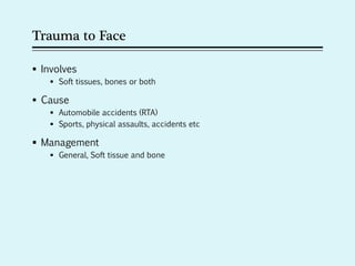
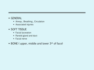
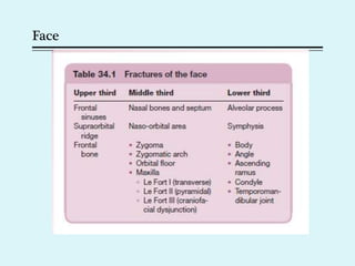
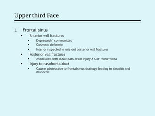
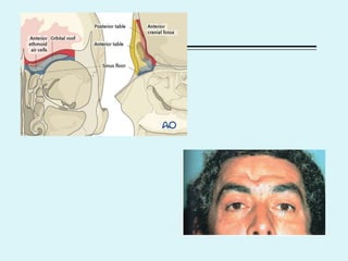
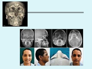
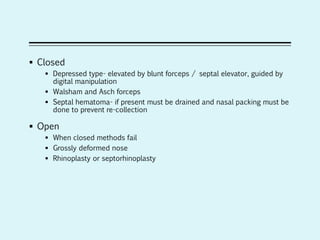
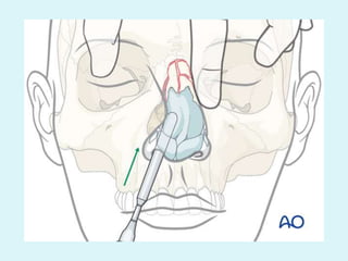
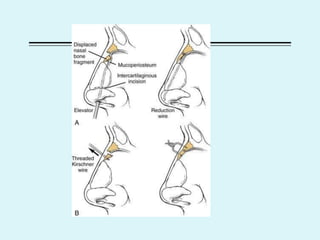
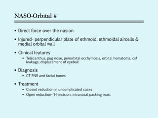
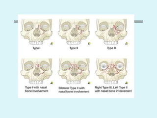
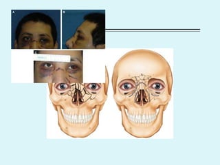
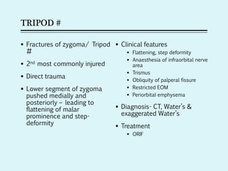
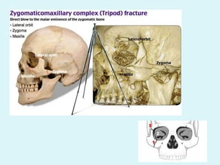
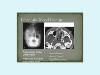
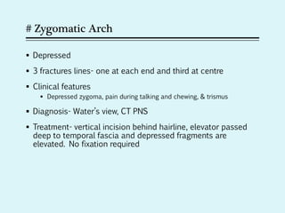
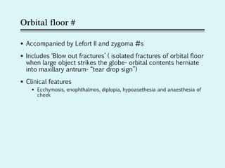
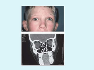
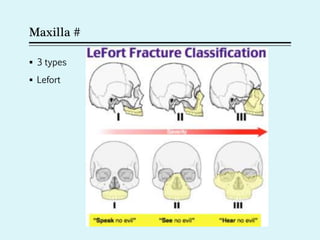
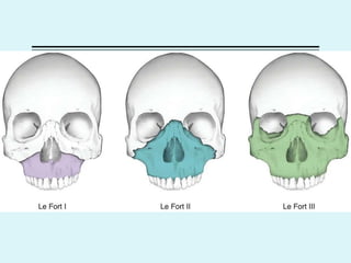
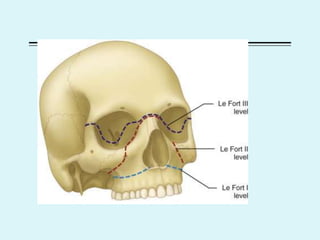
TRAUMA TO FACE
Dr.R.Malarvizhi MS(ENT), DLO
Senior Resident
Department of Otorhinolaryngology
Trauma to Face
 Involves
 Soft tissues, bones or both
 Cause
 Automobile accidents (RTA)
 Sports, physical assaults, accidents etc
 Management
 General, Soft tissue and bone
 GENERAL
 Airway , Breathing , Circulation
 Associated injuries
 SOFT TISSUE
 Facial laceration
 Parotid gland and duct
 Facial nerve
 BONE ( upper, middle and lower 3rd of face)
Face
Upper third Face
1. Frontal sinus
 Anterior wall fractures
 Depressed/ communitted
 Cosmetic deformity
 Interior inspected to rule out posterior wall fractures
 Posterior wall fractures
 Associated with dural tears, brain injury & CSF rhinorrhoea
 Injury to nasofrontal duct
 Causes obstruction to frontal sinus drainage leading to sinusitis and
mucocele
2. Supraorbital ridge
 Cause periorbital ecchymosis, flattening of eyebrow, proptosis,
 Bone fragments may be pushed into orbit
3. Fractures of Frontal bone
 Depressed/ linear with or without separation
 Extend into orbit
 Associated with brain injury and/ or cerebral oedema
Middle third Face
1. Nasal bone and septum
2. Naso-orbital fractures
3. Fractures of Zygoma
4. Fractures of Zygomatic arch
5. Fractures of Orbital floor
6. Fractures of Maxilla
Fractures of Nasal bone and Septum
 Most common
 Magnitude of force will determine depth of injury
 Types
 Depressed: due to frontal blow. Lower thinner part gives away. If force
greater- open book type can occur (nasal septum collapses and nasal
bones are splayed)
 Angulated : due to lateral blow
 Clinical features
 Swelling
 Periorbital echymosis
 Tenderness & crepitus
 Nasal deformity
 Epistaxis
 Nasal obstruction
 Lacerations of nasal skin
 Diagnosis
 PHYSICAL examination
 Xray – lateral views and Water’s view
 CT PNS with 3D reconstruction of facial bones
 Treatement
- SIMPLE # without displacement – NO intervention
- If displaced and presenting within 6 hours easy to reduce
- Best time to reduce – before edema sets in (i.e, within 6 hours of injury)
and 5-7 days later (i.e, after edema reduces)
- Difficult to reduce after 14 days
 Closed
 Depressed type- elevated by blunt forceps / septal elevator, guided by
digital manipulation
 Walsham and Asch forceps
 Septal hematoma- if present must be drained and nasal packing must be
done to prevent re-collection
 Open
 When closed methods fail
 Grossly deformed nose
 Rhinoplasty or septorhinoplasty
NASO-Orbital #
 Direct force over the nasion
 Injured- perpendicular plate of ethmoid, ethmoidal aircells &
medial orbital wall
 Clinical features
 Telecanthus, pug nose, periorbital ecchymosis, orbital hematoma, csf
leakage, displacement of eyeball
 Diagnosis
 CT PNS and facial bones
 Treatment
 Closed reduction in uncomplicated cases
 Open reduction- ‘H’ incision, intranasal packing must
TRIPOD #
 Fractures of zygoma/ Tripod
#
 2nd most commonly injured
 Direct trauma
 Lower segment of zygoma
pushed medially and
posteriorly – leading to
flattening of malar
prominence and step-
deformity
 Clinical features
 Flattening, step deformity
 Anaesthesia of infraorbital nerve
area
 Trismus
 Obliquity of palperal fissure
 Restricted EOM
 Periorbital emphysema
 Diagnosis- CT, Water’s &
exaggerated Water’s
 Treatment
 ORIF
# Zygomatic Arch
 Depressed
 3 fractures lines- one at each end and third at centre
 Clinical features
 Depressed zygoma, pain during talking and chewing, & trismus
 Diagnosis- Water’s view, CT PNS
 Treatment- vertical incision behind hairline, elevator passed
deep to temporal fascia and depressed fragments are
elevated. No fixation required
Orbital floor #
 Accompanied by Lefort II and zygoma #s
 Includes ‘Blow out fractures’ ( isolated fractures of orbital floor
when large object strikes the globe- orbital contents herniate
into maxillary antrum- “tear drop sign”)
 Clinical features
 Ecchymosis, enophthalmos, diplopia, hypoasethesia and anaesthesia of
cheek
 Diagnosis
 CT PNS, Water’s
 Traction test- nerve entrapment
 Treatment
 Indication – enophthalmos & persistent diplopia
 Transantal approach
 Infraorbital approach
 Reforcement of orbital floor
TEAR DROP SIGN
Maxilla #
 3 types
 Lefort
 Clinical features
 Malocclusion of teeth, elongation of midface, mobility of maxilla, csf
rhinorrhoea( II & III)
 Diagnosis
 CT PNS, Water’s , postero-anterior, lateral
 Treatment
 Immediate
 Airway
 Hemorrhage- maxillary artery br
 Fixation (interdental wiring, intermaxillary wiring- arch bars, Open
reduction with intraosseous wiring & wire slings)
Fractures of lower third
 MANDIBULAR fractures
 Classified based on location
 CONDYLE> angle> body> symphysis
 Uncommon – ramus, coronoid and alveolar processes
 Multiple – more common
 Direct trauma
 Displacement determined by
 Pull of muscles attached to fragments
 Direction of # line
 Bevel of #
 Clinical features- tenderness and crepitus at the fracture site
 Condyle
 Pain, trismus (when not displaced)
 When displaced associated with malocclusion of teeth and deviation of jaw to
opp side on opening mouth
 Angle, body & symphysis
 intra- & extra- oral palpation
 Step deformity, malocclusion of teeth, ecchymoss of mucosa,
 Diagnosis
 OPG, Water’s , CT PNS with 3D reconstruction of facial bones
 Treatment
 Closed: interdental wiring, intermaxillary fixation. External pin fixation
 Open : # site exposed and direct wiring ( figure of 8) aided with
compression plates
 CAUTION- Immobilasation beyond 3 weeks causes ankylosis. Therefore
jaw exercises are started as soon as wires are removed after a week.
OROANTRAL FISTULA
 Communication between maxillary antrum and oral cavity
 Aetio
 Dental extraction
 Failure of sublabial incision to close
 Carcinoma
 Fractures of maxilla
 Osteitis – syphilis, malignant granuloma
 Clinical features
 Food regurgitation- into nose
 Antral discharge- foul smelling from infected antral mucosa
 Inability to build positive or negative pressure in the mouth ( holding air
within mouth or drinking from straw)
 Diagnosis
 Probe test
 CT PNS
 Treatment
 Recent – if no infection – suturing of margins and antibiotics
 Chronic – palatal or buccal flap, dental obturators, removing fistulous
tract
 CALDWELL LUC surgery is both the cause and treatment of oroantral
fistula
Trauma to face
Trauma to face

Trauma to face

  • 1.
    TRAUMA TO FACE Dr.R.MalarvizhiMS(ENT), DLO Senior Resident Department of Otorhinolaryngology
  • 2.
    Trauma to Face Involves  Soft tissues, bones or both  Cause  Automobile accidents (RTA)  Sports, physical assaults, accidents etc  Management  General, Soft tissue and bone
  • 3.
     GENERAL  Airway, Breathing , Circulation  Associated injuries  SOFT TISSUE  Facial laceration  Parotid gland and duct  Facial nerve  BONE ( upper, middle and lower 3rd of face)
  • 4.
  • 5.
    Upper third Face 1.Frontal sinus  Anterior wall fractures  Depressed/ communitted  Cosmetic deformity  Interior inspected to rule out posterior wall fractures  Posterior wall fractures  Associated with dural tears, brain injury & CSF rhinorrhoea  Injury to nasofrontal duct  Causes obstruction to frontal sinus drainage leading to sinusitis and mucocele
  • 8.
    2. Supraorbital ridge Cause periorbital ecchymosis, flattening of eyebrow, proptosis,  Bone fragments may be pushed into orbit 3. Fractures of Frontal bone  Depressed/ linear with or without separation  Extend into orbit  Associated with brain injury and/ or cerebral oedema
  • 9.
    Middle third Face 1.Nasal bone and septum 2. Naso-orbital fractures 3. Fractures of Zygoma 4. Fractures of Zygomatic arch 5. Fractures of Orbital floor 6. Fractures of Maxilla
  • 10.
    Fractures of Nasalbone and Septum  Most common  Magnitude of force will determine depth of injury  Types  Depressed: due to frontal blow. Lower thinner part gives away. If force greater- open book type can occur (nasal septum collapses and nasal bones are splayed)  Angulated : due to lateral blow
  • 14.
     Clinical features Swelling  Periorbital echymosis  Tenderness & crepitus  Nasal deformity  Epistaxis  Nasal obstruction  Lacerations of nasal skin
  • 15.
     Diagnosis  PHYSICALexamination  Xray – lateral views and Water’s view  CT PNS with 3D reconstruction of facial bones
  • 16.
     Treatement - SIMPLE# without displacement – NO intervention - If displaced and presenting within 6 hours easy to reduce - Best time to reduce – before edema sets in (i.e, within 6 hours of injury) and 5-7 days later (i.e, after edema reduces) - Difficult to reduce after 14 days
  • 17.
     Closed  Depressedtype- elevated by blunt forceps / septal elevator, guided by digital manipulation  Walsham and Asch forceps  Septal hematoma- if present must be drained and nasal packing must be done to prevent re-collection  Open  When closed methods fail  Grossly deformed nose  Rhinoplasty or septorhinoplasty
  • 20.
    NASO-Orbital #  Directforce over the nasion  Injured- perpendicular plate of ethmoid, ethmoidal aircells & medial orbital wall  Clinical features  Telecanthus, pug nose, periorbital ecchymosis, orbital hematoma, csf leakage, displacement of eyeball  Diagnosis  CT PNS and facial bones  Treatment  Closed reduction in uncomplicated cases  Open reduction- ‘H’ incision, intranasal packing must
  • 23.
    TRIPOD #  Fracturesof zygoma/ Tripod #  2nd most commonly injured  Direct trauma  Lower segment of zygoma pushed medially and posteriorly – leading to flattening of malar prominence and step- deformity  Clinical features  Flattening, step deformity  Anaesthesia of infraorbital nerve area  Trismus  Obliquity of palperal fissure  Restricted EOM  Periorbital emphysema  Diagnosis- CT, Water’s & exaggerated Water’s  Treatment  ORIF
  • 26.
    # Zygomatic Arch Depressed  3 fractures lines- one at each end and third at centre  Clinical features  Depressed zygoma, pain during talking and chewing, & trismus  Diagnosis- Water’s view, CT PNS  Treatment- vertical incision behind hairline, elevator passed deep to temporal fascia and depressed fragments are elevated. No fixation required
  • 28.
    Orbital floor # Accompanied by Lefort II and zygoma #s  Includes ‘Blow out fractures’ ( isolated fractures of orbital floor when large object strikes the globe- orbital contents herniate into maxillary antrum- “tear drop sign”)  Clinical features  Ecchymosis, enophthalmos, diplopia, hypoasethesia and anaesthesia of cheek
  • 29.
     Diagnosis  CTPNS, Water’s  Traction test- nerve entrapment  Treatment  Indication – enophthalmos & persistent diplopia  Transantal approach  Infraorbital approach  Reforcement of orbital floor
  • 32.
  • 34.
    Maxilla #  3types  Lefort
  • 37.
     Clinical features Malocclusion of teeth, elongation of midface, mobility of maxilla, csf rhinorrhoea( II & III)  Diagnosis  CT PNS, Water’s , postero-anterior, lateral  Treatment  Immediate  Airway  Hemorrhage- maxillary artery br  Fixation (interdental wiring, intermaxillary wiring- arch bars, Open reduction with intraosseous wiring & wire slings)
  • 38.
    Fractures of lowerthird  MANDIBULAR fractures  Classified based on location  CONDYLE> angle> body> symphysis  Uncommon – ramus, coronoid and alveolar processes  Multiple – more common  Direct trauma  Displacement determined by  Pull of muscles attached to fragments  Direction of # line  Bevel of #
  • 41.
     Clinical features-tenderness and crepitus at the fracture site  Condyle  Pain, trismus (when not displaced)  When displaced associated with malocclusion of teeth and deviation of jaw to opp side on opening mouth  Angle, body & symphysis  intra- & extra- oral palpation  Step deformity, malocclusion of teeth, ecchymoss of mucosa,
  • 42.
     Diagnosis  OPG,Water’s , CT PNS with 3D reconstruction of facial bones  Treatment  Closed: interdental wiring, intermaxillary fixation. External pin fixation  Open : # site exposed and direct wiring ( figure of 8) aided with compression plates  CAUTION- Immobilasation beyond 3 weeks causes ankylosis. Therefore jaw exercises are started as soon as wires are removed after a week.
  • 45.
    OROANTRAL FISTULA  Communicationbetween maxillary antrum and oral cavity  Aetio  Dental extraction  Failure of sublabial incision to close  Carcinoma  Fractures of maxilla  Osteitis – syphilis, malignant granuloma
  • 46.
     Clinical features Food regurgitation- into nose  Antral discharge- foul smelling from infected antral mucosa  Inability to build positive or negative pressure in the mouth ( holding air within mouth or drinking from straw)
  • 48.
     Diagnosis  Probetest  CT PNS  Treatment  Recent – if no infection – suturing of margins and antibiotics  Chronic – palatal or buccal flap, dental obturators, removing fistulous tract  CALDWELL LUC surgery is both the cause and treatment of oroantral fistula