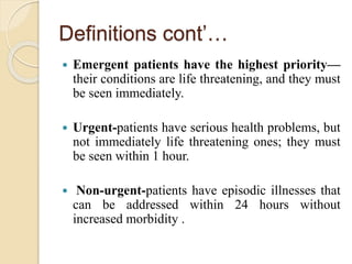
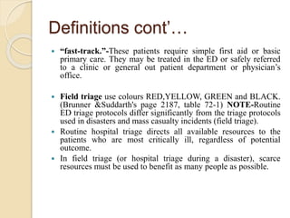
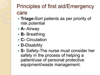
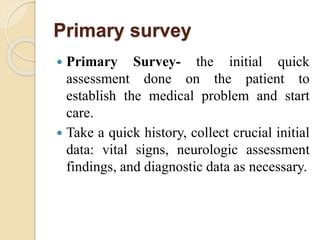
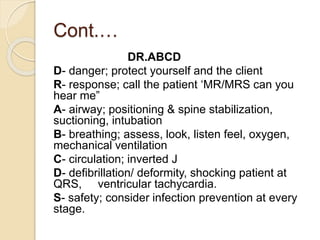
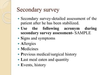
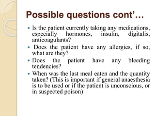
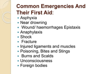
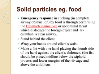
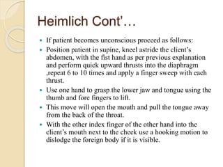
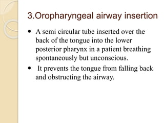
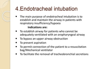
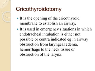
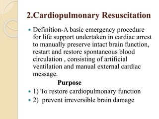
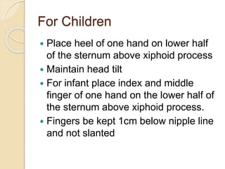
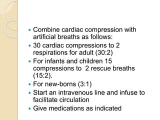
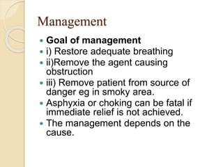
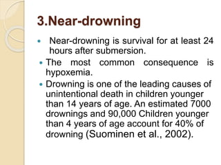
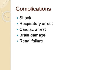
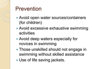
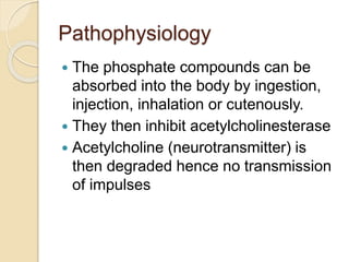
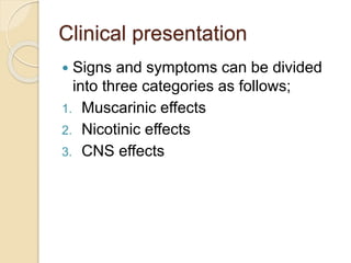
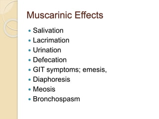
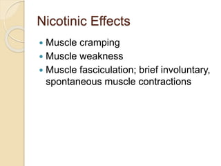
TRAUMA AND
EMERGENCY
Principles and practices of first aid
Phelix Ogadah. BScN. UEAB
By; Wangao
Topics
 Overview of trauma and emergency
 Principles of first Aid/ emergency care
 Common emergencies and their First
Aid
 (Asphyxia, Near drowning, Wound/
haemorrhages Epistaxis , Anaphylaxis,
Shock, Fracture ,Injured ligaments and
muscles, Poisoning, Bites and Stings,
Burns and Scalds, Unconsciousness,
Foreign bodies)
INTRODUCTION
 Trauma and emergency management
traditionally refers to care given to patients
with urgent and critical needs.
 However lack of access to health care facilities
may lead to many more people with non-life
threatening conditions visiting the emergency
department
 Therefore, the philosophy of emergency
management has broadened to include the
concept that an emergency is whatever the
patient or the family considers it to be.
INTRODUCTION’…
 These include people seeking emergency
care for serious life threatening illnesses
such as heart conditions
 This level of care requires training and
practice to be able to manage effectively
and efficiently
Introduction’….
 The emergency nurse establishes priorities,
monitors and continuously assesses acutely ill
and injured patients, supports and attends to
families, supervises other health personnel, and
educates patients and families on their
conditions.
 Nursing interventions are skilfully performed
interdependently with other professionals in an
emergency situation,( eg doctors,
physiotherapists etc).
Definitions:
 Trauma-(Pathology)-Wound or shock produced by
sudden physical injury as from violence or accident.
 Unintentional or intentional wound or injury
inflicted on the body from a mechanism against which
the body cannot protect itself
 Psychiatric trauma-An experience that produces
psychological injury or pain
 Emergency care-(is care that must be rendered without
delay)- Is the initial treatment given to acutely ill
patients coming to the health facility without prior plan;
(emergency department), usually presenting with life
threatening illnesses and injury
Definitions con’…
 Triage-The word triage comes from the
French word trier, meaning “to sort.”
Routinely, triage is used to sort patients in the
Emergency Department into groups based on
the severity of their health problems and the
immediacy with which these problems must be
treated or managed.
 Triage systems- Assessment is
hierarchical based on the potential for loss
of life; and has four basic categories:
emergent, urgent, non-urgent and fast tract
Definitions cont’…
 Emergent patients have the highest priority—
their conditions are life threatening, and they must
be seen immediately.
 Urgent-patients have serious health problems, but
not immediately life threatening ones; they must
be seen within 1 hour.
 Non-urgent-patients have episodic illnesses that
can be addressed within 24 hours without
increased morbidity .
Definitions cont’…
 “fast-track.”-These patients require simple first aid or basic
primary care. They may be treated in the ED or safely referred
to a clinic or general out patient department or physician’s
office.
 Field triage use colours RED,YELLOW, GREEN and BLACK.
(Brunner &Suddarth's page 2187, table 72-1) NOTE-Routine
ED triage protocols differ significantly from the triage protocols
used in disasters and mass casualty incidents (field triage).
 Routine hospital triage directs all available resources to the
patients who are most critically ill, regardless of potential
outcome.
 In field triage (or hospital triage during a disaster), scarce
resources must be used to benefit as many people as possible.
 Resuscitation-An act of restoring one to a
stable health status from unconsciousness
or from the brink of death
 Disaster- Sudden serious event or situation
with a negative impact on health and life of
the people, overwhelming the ability of the
local system to cope, requiring immediate
action.
Principles of first aid/Emergency
care
 Triage-Sort patients as per priority of
risk potential
 A- Airway
 B- Breathing
 C- Circulation
 D-Disability
 S- Safety-The nurse must consider her
safety in the process of helping a
patient/use of personal protective
equipment/waste management.
Primary survey
 Primary Survey- the initial quick
assessment done on the patient to
establish the medical problem and start
care.
 Take a quick history, collect crucial initial
data: vital signs, neurologic assessment
findings, and diagnostic data as necessary.
Cont.…
DR.ABCD
D- danger; protect yourself and the client
R- response; call the patient ‘MR/MRS can you
hear me”
A- airway; positioning & spine stabilization,
suctioning, intubation
B- breathing; assess, look, listen feel, oxygen,
mechanical ventilation
C- circulation; inverted J
D- defibrillation/ deformity, shocking patient at
QRS, ventricular tachycardia.
S- safety; consider infection prevention at every
stage.
Cont.…
A- alert, is the patient conscious,
V- voice, the patient response to voice
when called.
P- pain, the patient responds when pain
is inflicted
U- unresponsive, the patient is in coma
and doesn’t respond to stimuli.
Secondary survey
 Secondary survey-detailed assessment of the
patient after he has been stabilized.
 Use the following acronym during
secondary survey assessment- SAMPLE
 Signs and symptoms
 Allergies
 Medicines
 Previous medical/surgical history
 Last meal eaten and quantity
 Events, history
Inspection
Deformities
Contusions
Ablations- change in color
Penetrations/ punctures
Burns
Tenderness
Lacerations
Swellings
Secondary survey cont’…
 Obtain a complete health history
 P/E; head-to-toe assessment-examination,
 Diagnostic and laboratory testing and other
advanced medical procedures included in the
secondary survey.
 The following questions reflect the minimum
information that should be obtained from the
patient or from the person who accompanied
the patient to the ED and document all the
responses
for reference.
Secondary survey cont’…
 What were the circumstances, precipitating
events, location, and time of the injury or
illness?
 When did the symptoms appear?
 Was the patient unconscious after the
injury or onset of illness?
 How did the patient get to the hospital?
 What was the health status of the patient
before the injury or illness?
 Is there a medical or surgical history, a
history of admissions to the hospital?
Possible questions cont’…
 Is the patient currently taking any medications,
especially hormones, insulin, digitalis,
anticoagulants?
 Does the patient have any allergies, if so,
what are they?
 Does the patient have any bleeding
tendencies?
 When was the last meal eaten and the quantity
taken? (This is important if general anaesthesia
is to be used or if the patient is unconscious, or
in suspected poison)
Common Emergencies And
Their First Aid:
 Asphyxia
 Near drowning
 Wound/ haemorrhages Epistaxis
 Anaphylaxis
 Shock
 Fracture
 Injured ligaments and muscles
 Poisoning, Bites and Stings
 Burns and Scalds
 Unconsciousness
 Foreign bodies
Burns
Shock
Unconsciousness
Wounds/ Hemorrhages
Assignment
1.Asphyxia (suffocation or
choking)
 Definition-a situation or state of reduced
oxygen supply to the body tissues due to
interrupted breathing as occurs when the
airway is partially or completely blocked by
(food particles, secretions or other foreign
objects) strangulation
 Types-mild, moderate and severe
 Causes of asphyxia
 Food particles
 Secretions
 Foreign objects
Causes of asphyxia Cont’…
 Drowning
 Gas or smoke inhalation during fire
accidents
 Accidental coverage of the nose and
mouth by a piece of plastic
 Accidental or intentional strangulation
 being trapped in a confined spaces
with no ventilation
Pathophysiology
• Airway obstruction is caused by aspiration of
foreign bodies, anaphylaxis, viral, bacterial
infections, inhalation or chemical burns.
• In adults, aspiration of a bolus meat is the
most common cause while in children it is
caused by small toys, buttons and other
objects in addition to food, conditions like
peritonsillar abscesses, epiglottitis and other
acute infectious processes of the posterior
pharynx can result in airway obstruction
Solid particles eg. food
 Emergency response in choking (in complete
airway obstruction) by food is through performing
the Heimlich manoeuvre or abdominal thrust
which dislodges the foreign object and re-
establish a clear airway.
 Stand behind the client
 Wrap your hands around client’s waist
 Make a fist with one hand placing the thumb side
of the hand against the client’s abdomen. (the fist
should be placed midline below the xiphoid
process and lower margins of the rib cage and
above the umbilicus
Heimlich Cont’…
 Perform quick upward distinct thrusts to the
client’s abdomen.
 Each thrust should be separate and discrete
(a conscious patient can sit during the
procedure)
 Repeat the process six to ten times until the
client expels the foreign body
 If procedure fails-patient develops
respiratory distress or complete blockage
call for help
Heimlich Cont’…
 If patient becomes unconscious proceed as follows:
 Position patient in supine, kneel astride the client’s
abdomen, with the fist hand as per previous explanation
and perform quick upward thrusts into the diaphragm
,repeat 6 to 10 times and apply a finger sweep with each
thrust.
 Use one hand to grasp the lower jaw and tongue using the
thumb and fore fingers to lift.
 This move will open the mouth and pull the tongue away
from the back of the throat.
 With the other index finger of the other hand into the
client’s mouth next to the cheek use a hooking motion to
dislodge the foreign body if it is visible.
Airway Obstruction
 Definition: this is the partial or complete
occlusion of the airway which may be
acute or chronic.
 Acute upper airway obstruction is a life-
threatening medical emergency.
 If the airway is completely obstructed,
permanent brain damage or death will
occur within 3 to 5 minutes due to
secondary hypoxia.
Pathophysiology
• Airway obstruction is caused by aspiration of
foreign bodies, anaphylaxis, viral, bacterial
infections, inhalation or chemical burns.
• In adults, aspiration of a bolus meat is the
most common cause while in children it is
caused by small toys, buttons and other
objects in addition to food, conditions like
peritonsillar abscesses, epiglottitis and other
acute infectious processes of the posterior
pharynx can result in airway obstruction
Airway Cont’..
 Partial obstruction of the airway can lead to
progressive hypoxia, hypercapnia, and respiratory
and cardiac arrest.
 For an emergent or urgent health problem stabilize,
provide critical treatments, and promptly transfer
the patient to the appropriate setting i.e. intensive
care unit, operating room, general care unit which
are the priority areas of emergency care.
 Although initiation of treatment is at the ED,
ongoing definitive treatment of the underlying
problem is provided in other settings, and the sooner
the patient is stabilized and moved to the specific
area, the better.
Causes of airway obstruction
 Aspiration foreign bodies,
 anaphylaxis,
 viral or bacterial infection,
 Trauma
 inhalation or chemical burns.
 In adults, aspiration of a bolus of meat is
the most common cause of airway
obstruction.
Causes Cont’…
 In children, small toys, buttons, coins, and
other objects are commonly aspirated in
addition to food.
 Peritonsillar abscesses,-abscess between
the capsule of the tonsil and the pharynx
 Epiglottitis', and other acute infectious
processes of the posterior pharynx can
result in airway obstruction.
Clinical Presentation of airway
obstruction
Common signs and symptoms:
 Choking
 Apprehensive appearance
 Inspiratory and expiratory stridor,
 Laboured breathing,
 Use of accessory muscles (supra-sternal and
intercostal retraction),
 Flaring nostrils,
 Increasing anxiety,
 Restlessness, and confusion.
 Cyanosis and loss of consciousness develop as
hypoxia worsens.
Assessment and Diagnostic
Findings
 Look, Listen, Feel
 Assessment of the patient who has a foreign
object occluding the airway may involve
simply asking the person whether he or she is
choking and requires help.
 If patient is unconscious, inspection of the
oropharynx may reveal the obstructing
object.
 X-rays, laryngoscopy, or Bronchoscopy also
may be done.
Management
 The ED staff work collaboratively and follow the
ABCD (airway, breathing, circulation, disability)
method:
 Establish a patent airway.
 Provide adequate ventilation, employing
resuscitation measures when necessary to ensure
patient breathing (rising and falling of chest-if
not test breathing by placing the back of hand
close to patient’s mouth, if breathing, a stream of
warm air will be felt on it.
 (Trauma patients must have the cervical spine
protected and chest injuries assessed first.)
1.Head- tilt-chin-lift-maneuver
 Patient is placed supine on a firm flat
surface
 Airway is opened by either head-tilt-chin-
lift or the jaw thrust maneuver
 Head tilt chin lift which helps to tilt the
head back should be used only if it is
determined that the patients cervical spine
is not injured
2.Jaw thrust maneuver
• The angle of the patients lower jaw are
grasped and lifted displacing the mandible
forward.
• It is a safe approach to opening the airway
of a victim with suspected neck injury
because it can be accomplished without
extending the neck.
Abdominal thrust maneuver
 Referred to as the Heimlich maneuver according to the American
Heart Association. This is done on a conscious patient. The following
steps:
 Stand behind the person who is choking.
 Place both arms around the person’s waist.
 Make a fist with one hand with the thumb outside the fist.
 Place thumb side of fist against the person’s abdomen above the navel
and below the xiphoid process.
 Grasp fist with other hand.
 Quickly and forcefully exert pressure against the person’s diaphragm,
pressing upward with quick, firm thrusts.
 Apply thrusts 6 to 10 times until the obstruction is cleared.
 The pressure from the thrusts should lift the diaphragm, force air into
the lungs, and create an artificial cough powerful enough to expel the
aspirated object
Artificial Airways
1. Endotracheal tube
2. Tracheostomy
3. Pharyngeal airways
3.Oropharyngeal airway insertion
 A semi circular tube inserted over the
back of the tongue into the lower
posterior pharynx in a patient breathing
spontaneously but unconscious.
 It prevents the tongue from falling back
and obstructing the airway.
4.Endotracheal intubation
 The main purpose of endotracheal intubation is to
establish and maintain the airway in patients with
respiratory insufficiency/hypoxia
Indications are:
 To establish airway for patients who cannot be
adequately ventilated with an oropharyngeal airway
 To bypass an upper airway obstruction
 To prevent aspiration
 To permit connection of the patient to a resuscitation
bag/Mechanical ventilator
 To facilitate the removal of tracheobronchial secretions
Cricothyroidotomy
 It is the opening of the cricothyroid
membrane to establish an airway.
 It is used in emergency situations in which
endotracheal intubation is either not
possible or contra indicated eg in airway
obstruction from laryngeal edema,
hemorrhage to the neck tissue or
obstruction of the larynx.
2.Cardiopulmonary Resuscitation
 Definition-A basic emergency procedure
for life support undertaken in cardiac arrest
to manually preserve intact brain function,
restart and restore spontaneous blood
circulation , consisting of artificial
ventilation and manual external cardiac
message.
Purpose
 1) To restore cardiopulmonary function
 2) prevent irreversible brain damage
Indications
 Respiratory failure (Pulse present but
patient not breathing)
 Drowning
 Electric/ hypovolemic shock
 Anaphylactic reaction
 Drug overdose
 Cardiac arrest
 Asphyxia
Assessment
1) Client’s state of consciousness to confirm
need for resuscitation
2) Breathing-look, listen, feel
3) Pallor-to determine state of oxygenation
 Planning and preparation for patient
and self
 Review knowledge on CPR and organize
for resuscitation team and trolley
Requirement
 Emergency trolley with:
 Syringes with needles –assorted sizes
 Functional laryngoscope with assorted
blades (adult and child)
 Ambo bags and face masks (adult and
child/infant)
 Endotracheal tubes assorted sizes
 Torch
 Suction catheters-assorted sizes
 Naso-gastric tube
Requirement cont.,..
 Oxygen source
 Gloves
 Infusion equipment
 Splints/hard fracture boards/Firm surface
 Airways
 Cannulae assorted sizes
 Scissors
 Adhesive tapes or strapping
 Defibrillator/mechanical
ventilators/cardiac monitors
Implementation
 Three Ss in emergency response
 Safety
 Stimulation
 Shout for help (for teamwork)
 Put on gloves
 Lay the casualty on firm surface in supine
position without a pillow for ease of external
chest compression during cardiac message
 Assess for breathing and ensure airway is clear-
Open airway by using head tilt or jaw thrust
Manoeuvre to establish and maintain airway to
ensure ventilation
 Insert the oropharyngeal airway to prevent
obstruction by the tongue (prevents tongue
from falling back)
 Perform oropharyngeal suction if secretion
present to clear airway and prevent
aspiration.
 If patient not breathing connect ambo bag
and give two rescue breaths as you check for
chest expansion.
 Then connect oxygen as you place face mask
over nose and mouth appropriately
 Palpate or feel for carotid pulse in adults and
children and or brachial for infants for 5-10
seconds to confirm blood circulation.
 If pulse is absent start chest compression to
stimulate the heart and restore circulation:
 Place heel of one hand over lower third of
sternum, other hand on top, straighten
elbows over shoulders perpendicular to
patient’s chest.
For Children
 Place heel of one hand on lower half
of the sternum above xiphoid process
 Maintain head tilt
 For infant place index and middle
finger of one hand on the lower half of
the sternum above xiphoid process.
 Fingers be kept 1cm below nipple line
and not slanted
 Combine cardiac compression with
artificial breaths as follows:
 30 cardiac compressions to 2
respirations for adult (30:2)
 For infants and children 15
compressions to 2 rescue breaths
(15:2).
 For new-borns (3:1)
 Start an intravenous line and infuse to
facilitate circulation
 Give medications as indicated
Signs and Symptoms
 i) if the casualty/client is conscious he will
usually grasp the anterior neck and being
unable to speak or cough.
 ii) Anxiety and apprehension
 iii) Cyanosis
 iv) Stridor-a harsh crowing sound made on
inhalation caused by constriction of the
airway, may also occur in severe allergic
reactions
 Grunting-sound like air moving through fluid
Management
 Goal of management
 i) Restore adequate breathing
 ii)Remove the agent causing
obstruction
 iii) Remove patient from source of
danger eg in smoky area.
 Asphyxia or choking can be fatal if
immediate relief is not achieved.
 The management depends on the
cause.
3.Near-drowning
 Near-drowning is survival for at least 24
hours after submersion.
 The most common consequence is
hypoxemia.
 Drowning is one of the leading causes of
unintentional death in children younger
than 14 years of age. An estimated 7000
drownings and 90,000 Children younger
than 4 years of age account for 40% of
drowning (Suominen et al., 2002).
 Factors associated with drowning and
near-drowning include:
 Alcohol ingestion, inability to swim,
diving injuries, hypothermia, and
exhaustion
 Efforts to save the victim should not
be abandoned prematurely.
Near drowning cont’….
 After resuscitation, hypoxia and acidosis, are
the primary problems of a victim who has
nearly drowned, this require immediate
intervention.
 Resultant pathophysiologic changes and
pulmonary injury depend on the type of fluid
(fresh or salt water) and the volume
aspirated.
 Fresh water aspiration results in a loss of
surfactant, hence an inability to expand the
lungs.
Near drowning cont’….
 Salt water aspiration leads to pulmonary
oedema from the osmotic effects of the
salt within the lung.
 After a person survives submersion, acute
respiratory distress syndrome resulting in
hypoxia, hypercapnia, and respiratory or
metabolic acidosis can occur.
Management
 Therapeutic goals include maintaining
cerebral perfusion and adequate
oxygenation to prevent further damage to
vital organs.
 Immediate cardiopulmonary resuscitation is
the factor with the greatest influence on
survival.
 The treatment goal- prevention of hypoxia,
is accomplished by ensuring an adequate
airway and respiration, thus improving
ventilation (which helps to correct
respiratory acidosis) and oxygenation.
Management
 Arterial blood gas analyses are
performed to evaluate oxygen, carbon
dioxide, and bicarbonate levels and pH.
 These parameters determine the type
of ventilatory support needed.
 Use of endotracheal intubation with
positive pressure ventilation improves
oxygenation, prevents aspiration, and
corrects intrapulmonary shunting and
ventilation.
Complications
 Shock
 Respiratory arrest
 Cardiac arrest
 Brain damage
 Renal failure
Prevention
 Avoid open water sources/containers
(for children)
 Avoid excessive exhaustive swimming
activities
 Avoid deep waters especially for
novices in swimming
 Those unskilled should not engage in
swimming without skilled assistance
 Use of life saving jackets.
4.Anaphylaxis
 It is also known as type 1 or immediate
hypersensitivity
 Occurs in the first encounter or
exposure to immunogen
 Mediated by Ig E, basophils and mast
cells
 Has three phases
i. Sensitization
ii. Activation
iii. Effector
Sensitization
 Occurs in first encounter with an antigen
 B cells internalizes the allergen, process
and present it to the CD4 cells (Helper T
cell)
 T helper cell secrets interleukin iv (IL4)
which activates humoral response
 IL4 makes B cells to go clonal expansion
and differentiation making it to switch from
Ig M to Ig E
 Ig E then binds to Mast cells and basophils
 Sensitization then occurs when Ig E is
bound to basophils and mast cells
 It affects susceptible individuals
Activation
 Second exposure to the same antigen/
allergen
 The allergen does not bind to the B
cell but binds to FCE receptor on mast
cells and basophils
 It cross links two Ig E
 The cell is activated and begins to
synthesize leukotrienes C4 and D4
Effector phase
 Future exposure to the same antigen
 Mast cells and basophils undergo
degranulation
 Preformed and newly formed substances are
produced, eg histamine, leukotrienes,
serotonin and heparin
 This causes, vasodilatation, reduced BP,
constriction of smooth muscles (bronchus),
vascular permeability- edema, stimulation of
goblet cells increased secretion of mucous
 Death can occur in 10 minutes
 Allergens include, proteins, drugs, foods,
insect products, plants pollen, fur/ hair, dust
mold spores etc
Prevention
 Identify the allergen
 Avoid the allergen
 Use of drugs
 Antihistamine- block histamine receptors
 Cromoglycate based drugs- destabilizes the
mast cells
 Catecholamine- adrenaline (stimulates
autonomic nerve action) for penicillin allergy.
 Desensitization- giving a small dose of the
antigen to an individual to switch Ig E to Ig G.
5.Poisoning
 Organophosphate poisoning (OPP)
 They are compounds used in both
domestic and industrial use eg
insecticides, pesticides, herbicides,
anthelminthic, nerge gases
 Suicidal attempts occurs via exposure
intentionally or unintentionally
Pathophysiology
 The phosphate compounds can be
absorbed into the body by ingestion,
injection, inhalation or cutenously.
 They then inhibit acetylcholinesterase
 Acetylcholine (neurotransmitter) is
then degraded hence no transmission
of impulses
Clinical presentation
 Signs and symptoms can be divided
into three categories as follows;
1. Muscarinic effects
2. Nicotinic effects
3. CNS effects
Muscarinic Effects
 Salivation
 Lacrimation
 Urination
 Defecation
 GIT symptoms; emesis,
 Diaphoresis
 Meosis
 Bronchospasm
Nicotinic Effects
 Muscle cramping
 Muscle weakness
 Muscle fasciculation; brief involuntary,
spontaneous muscle contractions
CNS Effects
 Confusion
 Impaired memory
 Psychosis
 Restlessness
 Tremors
 paralysis
 coma
Vital signs
 Depressed
respiration
 Bradycardia
 Hypotension
 Tachycardia
 Tarchypnoea
 Hypoxia
Management of OPP
AIMS
 Identify the poison
Proper airway and oxygenation
Administer antidote
Prevent PUD, coma and aspiration
pneumonia
Prevent RDS due to bronchospasms,
bronchorrhea and laryngeal spasms
DR.ABCD
Atropinazation
 Atropine is anticholinergic preparation
 Give 1mg IV ¼ hourly until dilatation of
pupils is achieved
 IV fluids infusion full-balst, monitor
input output
 End point is reached when;
a) Pupils are fully dilated
b) Secretions are dry
c) Symptoms are reversed
Management cont.
 Use activated charcoal
 Gastric lavage should be done within 30
minutes of poisoning
 Consider corrosiveness of poison
 Benzodiazepines for convulsions and
seizers
 Mgso4 (magnesium iv sulphate),
administered for acetylcholine
functioning
 Monitor urine output, respiration and
other vital signs
 Keep the patient warm
6.Shock
7.Fracture
 A fracture may be a complete break in
the continuity of a bone or,
occasionally, it may be incomplete.
Pathophysiology
 When the bone is broken, adjacent
structures are also affected, resulting
in soft tissue edema, hemorrhage into
the muscles and joints, joint
dislocations, ruptured tendons,
severed nerves, and damaged blood
vessels.
 Body organs may be injured by the
force that caused the fracture or by
the fracture fragments.
Clinical features
 Pain at site of injury
 Swelling due to hematoma formation
 Loss of function due to pain and
deformity
 Deformity depending on force and
muscle tissue surrounding muscles
e.g. angulation, shortening of
extremity
 Bleeding due to ruptured blood
vessels
Classification
 Fracture may be subdivided, according
to their etiology, into four basic groups
1. Fracture caused solely by sudden injury
2. Fragility fractures
3. Fatigue or stress fracture
4. Pathological fractures
They are also classified according to:
 Location
 Type
 Direction or pattern of fracture line
Bone Healing Process
 After a fracture, bone healing follows a number of
stages:
 A hematoma forms between surrounding soft
tissues.
 Inflammatory process sets in with accumulation
of macrophages. This takes about five days. The
macrophages, phagocytose the hematoma.
Growth of granulation tissue begins.
 Callus formation, the osteoblasts secrete non-
lamellar osteoid. Calcium is also absorbed which
aids in hardening of bone to form callus.
 Remodeling, osteoclasts become active
removing excess callus and opening up a
medullary canal in callus. This may take up to
one month.
Bone Healing Process
 Factors enhancing bone healing:
 Adequate nutrition
 Adequate blood supply
 Absence of infection
Bone Healing Process
 Factors hindering bone healing:
 Presence of infective organisms e.g.
streptococci
 Fat embolism in medullary canal
 Excessive bone tissue fragments
 Deficient blood supply
 Continued mobility (lack of proper reduction
and immobilization)
 Age - old age due to slowing
 Nature of injury
 Type of bone lost
 Degree of immobilization
Principle Management of
Fractures
(First Aid)
 The emergency management of a fracture
involves: (DR.ABCD.RICE)
 Assessing the airway, breathing and
circulation
 Assessing any bleeding sites and controlling
bleeding
 Treatment of any life threatening injury
 Immobilization by use of splints
 Applying cold compresses
 Elevating the extremity
 Minimizing mobility
 Monitoring the patient closely
 Refer casualty to hospital for further
management
Injured ligaments and muscles
 Sprain is an injury to joints (ligaments)
 Strain is an injury to muscles
(tendons)
 Review anatomy of joints
• Identify type of injury
• Reassure the casualty
• Rest the injured part
• Apply ice to the area
• Compress the area, apply crepe bandage
• Elevate the affected limb
• Avoid weight bearing to the affected limb
• Refer to hospital for, x-ray, cast application if
indicated and also for anti-inflammatory therapy
First Aid
8.Bites and Stings
 Bites caused by animals
 Stings caused by insects
S&S
Swelling
Redness
Pain
Bleeding
Infection
8.1.Dog Bite
 Clean the bitten area using warm water and clean
thoroughly
 Puncture wounds should be irrigated with a sterile
catheter using methylated spirit and povidone.
 Iodine is also virucidal and may be used to clean the
wound
 Calm the casualty
 Find out if the dog was vaccinated
 Go to hospital for antirabies
 Follow the dog for 10 days
 Dress the wound
 Bite wounds should not be sutured immediately to
prevent more traumas from the suturing needle, which
will increase the areas for viral entry into the body
tissue.
 Suturing may be done 24 to 48 hours after the bite
using very few sutures under the cover of anti-rabies
Pathology of Rabies
 Rabies is a serious viral disease of
canines which is incidentally transmitted
to humans by the bite of a rabid animal.
It is caused by a virus known as lassa
virus type I.
 This happens when humans get bitten by
a rabid animal (dog/cat/bat)or when its
saliva comes into contact with the
mucous membranes or open wound of a
person
 All warm blooded animals are
susceptible to rabies.
Anti- Rabies Vaccine
 This is a very safe and effective treatment
following a rabid animal bite.
 The vaccine HDCV (Human Diploid cells tissue
Culture Vaccine) is administered in six doses
sub-cutaneous as follows:
 One ml immediately after exposure (day 0),
 One ml on day 3,
 One ml on day 7,
 One ml on day 14,
 One ml on day 30,
 One ml on day 90
 In order to prevent wound infection and tetanus
you should give the patient broad spectrum
antibiotics
8.2.Human Bite
 Clean the area using warm water
 Dress using sterile dressing
 Seek medical help
 Do not stich the wound immediately
8.3.Snake bite
 Identify the type of the snake and describe
 Green mamba- the most dangerous, gives neurotoxin
impairing the CNS and the brain
 Cobra-
 Bloom slang- hemotoxic, impairs blood clot features
appear within 24 hour
 Python-
 Puff ada-
 Calm the patient, put on gloves, head raised (to slow
Venus return),irrigate the area, clean area using soap and
warm water(to minimize infection), dress the area, apply
pressure on the either side,
 do not tie coz of ischemia,
 Do not suck, cut the wound
 Transfer patient to hospital.
9.Burns and Scalds
 Destruction of the skin sometimes with
underlying tissues by extremities of
temperature, exposure to electrical, chemical
and radiation sources.
 Classification of burns is usually according
to size(wallace rule of 9) and
depth(superficial partial, deep partial
thickness and full thickness
Etiology
 Fire
 Boiling water
 Hot objects or oil
 Electrical shocks
 Chemicals and acids
 Radiations
 Geothermal/ steam
Pathophysiology
 Every system is affected by thermal injury.
 The extent of dysfunction is proportional to
the total body surface area(TBSA)involved.
 The systemic response to burn injury is
biphasic, with early hypofunction followed
rapidly by hyperfunction. However, the organ
function gradually returns to normal as the
wound heals or is surgically grafted.
 Skeletal System
 Red bone marrow replaces red blood cells which is
destroyed by the burnt skin. If the burns area is too large
for the bone marrow to compensate for the loss in red
blood cells, the patient will require blood transfusions to
survive.
 Cardiovascular System
 Burning of the skin will lead to an increase in capillary
permeability, which causes an increase in blood
vasculature – this then results in a decrease of blood
pressure as well as live blood volume. This further
decreases the blood flow and oxygenation to tissues, which
then also results in edema, shock and eventually death.
 Respiratory System
 Airway obstruction caused by gross edema of the throat.
Also, these patients may have an increased respiratory rate
as a result of pulmonary edema (secondary to smoke
inhalation) or increased respiratory rate as an attempt to
compensate the increased metabolic rate.
 Endocrine System
 Increased secretions of adrenaline and nor-adrenaline in
response to the injury may lead to increased body
temperature and increased cell metabolism.
 Lymphatic System
 Inflammation increases as a result of damaged tissue,
which results in greater strain on the lymphatic system and
pitting edema.
 Immune System
 Decreased response as a result of excessive strain on the
lymphatic system and due to increased infection as a result
of burns area removing the first line of infection defense.
 Digestive
 Due to the potential hypovolaemic state in which a body
with severe burns is likely to be in, their is a decrease in
blood availability in the intestinal lining and liver. The
intestinal lining automatically increases nutrients required
to support metabolism and repair of damaged cells.
 Urinary
 The kidneys compensate for the increased fluid loss as a
result of the burn area by decreasing urine output. The
potential detriment of this change is the potential for
kidney damage as a result of poor perfusion.
Clinical manifestations
 A superficial burn destroys the epidermis, and
appears red and dry. Pain is due to damage to the
cutaneous nerve endings.
 A partial thickness burn destroys the epidermis and
part of the dermis and appears red and blistered. It
causes pain due to the exposure of the nerve endings
to the air.
 A full thickness burn destroys the dermis and may be
extended to the subcutaneous tissue, muscle or bone.
the skin may appear white, brown,or black and
leathery. Often there is no pain present as the nerve
endings have been destroyed.
Phases of burn care
Emergent/immediate resuscitative
 This is from onset of injury to
completion of fluid resuscitation.
 Priority activities here include, first
aid, prevention of shock, prevention of
respiratory distress, detection and
treatment of concomitant injury
wound assessment and initial care.
Phases of burn care
Acute phase: from beginning of diuresis to near
completion of wound closure. it entails wound
care and closure, prevention or treatment of
complications including infections and
nutritional support.
Rehabilitation: from major wound closure to
return to individuals optimal level of physical and
psychosocial adjustment. Prevention of scars and
contracture, physical, occupational and
vocational rehabilitation, functional and
cosmetic reconstruction and psychosocial
counseling
Emergency Burns Management
 Aims are;
 to save lives, minimize disabilities and
prepare patient for definitive care.
 First priority is to secure and protect the
airway.
 inhalation injury is suspected for patients
with history of facial burns and exposure
to closed room fires, risk of carbon
monoxide poisoning is increased thus
patient should be intubated.100% oxygen
is administered.
10.Unconsciousness
11.Foreign bodies
 in, ears
 Eyes
 Nose
 Private organs
 Throat (fish bone)
Anatomy of the Ear
The ear is divided into three areas
Outer
(external)
ear
Middle
ear
Inner
ear
Structure of the Eye
The wall is composed of three tunics
Sclera & Cornea
fibrous outside layer
Choroid –
middle
layer
Sensory
tunic –
(retina) inside
layer
Accessory Structures of the Eye
 Eyelids
 Meets at medial and lateral canthus
 Eyelashes
Female Reproductive System
Slide
Copyright © 2003 Pearson Education, Inc. publishing as Benjamin Cummings
Figure 16.8a
Mouth (Oral Cavity) Anatomy
 Foreign body
In the throat
END

TRAUMA AND EMERGENCY-1.pptx

  • 1.
    TRAUMA AND EMERGENCY Principles andpractices of first aid Phelix Ogadah. BScN. UEAB By; Wangao
  • 2.
    Topics  Overview oftrauma and emergency  Principles of first Aid/ emergency care  Common emergencies and their First Aid  (Asphyxia, Near drowning, Wound/ haemorrhages Epistaxis , Anaphylaxis, Shock, Fracture ,Injured ligaments and muscles, Poisoning, Bites and Stings, Burns and Scalds, Unconsciousness, Foreign bodies)
  • 3.
    INTRODUCTION  Trauma andemergency management traditionally refers to care given to patients with urgent and critical needs.  However lack of access to health care facilities may lead to many more people with non-life threatening conditions visiting the emergency department  Therefore, the philosophy of emergency management has broadened to include the concept that an emergency is whatever the patient or the family considers it to be.
  • 4.
    INTRODUCTION’…  These includepeople seeking emergency care for serious life threatening illnesses such as heart conditions  This level of care requires training and practice to be able to manage effectively and efficiently
  • 5.
    Introduction’….  The emergencynurse establishes priorities, monitors and continuously assesses acutely ill and injured patients, supports and attends to families, supervises other health personnel, and educates patients and families on their conditions.  Nursing interventions are skilfully performed interdependently with other professionals in an emergency situation,( eg doctors, physiotherapists etc).
  • 6.
    Definitions:  Trauma-(Pathology)-Wound orshock produced by sudden physical injury as from violence or accident.  Unintentional or intentional wound or injury inflicted on the body from a mechanism against which the body cannot protect itself  Psychiatric trauma-An experience that produces psychological injury or pain  Emergency care-(is care that must be rendered without delay)- Is the initial treatment given to acutely ill patients coming to the health facility without prior plan; (emergency department), usually presenting with life threatening illnesses and injury
  • 7.
    Definitions con’…  Triage-Theword triage comes from the French word trier, meaning “to sort.” Routinely, triage is used to sort patients in the Emergency Department into groups based on the severity of their health problems and the immediacy with which these problems must be treated or managed.  Triage systems- Assessment is hierarchical based on the potential for loss of life; and has four basic categories: emergent, urgent, non-urgent and fast tract
  • 8.
    Definitions cont’…  Emergentpatients have the highest priority— their conditions are life threatening, and they must be seen immediately.  Urgent-patients have serious health problems, but not immediately life threatening ones; they must be seen within 1 hour.  Non-urgent-patients have episodic illnesses that can be addressed within 24 hours without increased morbidity .
  • 9.
    Definitions cont’…  “fast-track.”-Thesepatients require simple first aid or basic primary care. They may be treated in the ED or safely referred to a clinic or general out patient department or physician’s office.  Field triage use colours RED,YELLOW, GREEN and BLACK. (Brunner &Suddarth's page 2187, table 72-1) NOTE-Routine ED triage protocols differ significantly from the triage protocols used in disasters and mass casualty incidents (field triage).  Routine hospital triage directs all available resources to the patients who are most critically ill, regardless of potential outcome.  In field triage (or hospital triage during a disaster), scarce resources must be used to benefit as many people as possible.
  • 10.
     Resuscitation-An actof restoring one to a stable health status from unconsciousness or from the brink of death  Disaster- Sudden serious event or situation with a negative impact on health and life of the people, overwhelming the ability of the local system to cope, requiring immediate action.
  • 11.
    Principles of firstaid/Emergency care  Triage-Sort patients as per priority of risk potential  A- Airway  B- Breathing  C- Circulation  D-Disability  S- Safety-The nurse must consider her safety in the process of helping a patient/use of personal protective equipment/waste management.
  • 12.
    Primary survey  PrimarySurvey- the initial quick assessment done on the patient to establish the medical problem and start care.  Take a quick history, collect crucial initial data: vital signs, neurologic assessment findings, and diagnostic data as necessary.
  • 13.
    Cont.… DR.ABCD D- danger; protectyourself and the client R- response; call the patient ‘MR/MRS can you hear me” A- airway; positioning & spine stabilization, suctioning, intubation B- breathing; assess, look, listen feel, oxygen, mechanical ventilation C- circulation; inverted J D- defibrillation/ deformity, shocking patient at QRS, ventricular tachycardia. S- safety; consider infection prevention at every stage.
  • 14.
    Cont.… A- alert, isthe patient conscious, V- voice, the patient response to voice when called. P- pain, the patient responds when pain is inflicted U- unresponsive, the patient is in coma and doesn’t respond to stimuli.
  • 15.
    Secondary survey  Secondarysurvey-detailed assessment of the patient after he has been stabilized.  Use the following acronym during secondary survey assessment- SAMPLE  Signs and symptoms  Allergies  Medicines  Previous medical/surgical history  Last meal eaten and quantity  Events, history
  • 16.
    Inspection Deformities Contusions Ablations- change incolor Penetrations/ punctures Burns Tenderness Lacerations Swellings
  • 17.
    Secondary survey cont’… Obtain a complete health history  P/E; head-to-toe assessment-examination,  Diagnostic and laboratory testing and other advanced medical procedures included in the secondary survey.  The following questions reflect the minimum information that should be obtained from the patient or from the person who accompanied the patient to the ED and document all the responses for reference.
  • 18.
    Secondary survey cont’… What were the circumstances, precipitating events, location, and time of the injury or illness?  When did the symptoms appear?  Was the patient unconscious after the injury or onset of illness?  How did the patient get to the hospital?  What was the health status of the patient before the injury or illness?  Is there a medical or surgical history, a history of admissions to the hospital?
  • 19.
    Possible questions cont’… Is the patient currently taking any medications, especially hormones, insulin, digitalis, anticoagulants?  Does the patient have any allergies, if so, what are they?  Does the patient have any bleeding tendencies?  When was the last meal eaten and the quantity taken? (This is important if general anaesthesia is to be used or if the patient is unconscious, or in suspected poison)
  • 20.
    Common Emergencies And TheirFirst Aid:  Asphyxia  Near drowning  Wound/ haemorrhages Epistaxis  Anaphylaxis  Shock  Fracture  Injured ligaments and muscles  Poisoning, Bites and Stings  Burns and Scalds  Unconsciousness  Foreign bodies
  • 21.
  • 22.
    1.Asphyxia (suffocation or choking) Definition-a situation or state of reduced oxygen supply to the body tissues due to interrupted breathing as occurs when the airway is partially or completely blocked by (food particles, secretions or other foreign objects) strangulation  Types-mild, moderate and severe  Causes of asphyxia  Food particles  Secretions  Foreign objects
  • 23.
    Causes of asphyxiaCont’…  Drowning  Gas or smoke inhalation during fire accidents  Accidental coverage of the nose and mouth by a piece of plastic  Accidental or intentional strangulation  being trapped in a confined spaces with no ventilation
  • 24.
    Pathophysiology • Airway obstructionis caused by aspiration of foreign bodies, anaphylaxis, viral, bacterial infections, inhalation or chemical burns. • In adults, aspiration of a bolus meat is the most common cause while in children it is caused by small toys, buttons and other objects in addition to food, conditions like peritonsillar abscesses, epiglottitis and other acute infectious processes of the posterior pharynx can result in airway obstruction
  • 25.
    Solid particles eg.food  Emergency response in choking (in complete airway obstruction) by food is through performing the Heimlich manoeuvre or abdominal thrust which dislodges the foreign object and re- establish a clear airway.  Stand behind the client  Wrap your hands around client’s waist  Make a fist with one hand placing the thumb side of the hand against the client’s abdomen. (the fist should be placed midline below the xiphoid process and lower margins of the rib cage and above the umbilicus
  • 26.
    Heimlich Cont’…  Performquick upward distinct thrusts to the client’s abdomen.  Each thrust should be separate and discrete (a conscious patient can sit during the procedure)  Repeat the process six to ten times until the client expels the foreign body  If procedure fails-patient develops respiratory distress or complete blockage call for help
  • 27.
    Heimlich Cont’…  Ifpatient becomes unconscious proceed as follows:  Position patient in supine, kneel astride the client’s abdomen, with the fist hand as per previous explanation and perform quick upward thrusts into the diaphragm ,repeat 6 to 10 times and apply a finger sweep with each thrust.  Use one hand to grasp the lower jaw and tongue using the thumb and fore fingers to lift.  This move will open the mouth and pull the tongue away from the back of the throat.  With the other index finger of the other hand into the client’s mouth next to the cheek use a hooking motion to dislodge the foreign body if it is visible.
  • 28.
    Airway Obstruction  Definition:this is the partial or complete occlusion of the airway which may be acute or chronic.  Acute upper airway obstruction is a life- threatening medical emergency.  If the airway is completely obstructed, permanent brain damage or death will occur within 3 to 5 minutes due to secondary hypoxia.
  • 29.
    Pathophysiology • Airway obstructionis caused by aspiration of foreign bodies, anaphylaxis, viral, bacterial infections, inhalation or chemical burns. • In adults, aspiration of a bolus meat is the most common cause while in children it is caused by small toys, buttons and other objects in addition to food, conditions like peritonsillar abscesses, epiglottitis and other acute infectious processes of the posterior pharynx can result in airway obstruction
  • 30.
    Airway Cont’..  Partialobstruction of the airway can lead to progressive hypoxia, hypercapnia, and respiratory and cardiac arrest.  For an emergent or urgent health problem stabilize, provide critical treatments, and promptly transfer the patient to the appropriate setting i.e. intensive care unit, operating room, general care unit which are the priority areas of emergency care.  Although initiation of treatment is at the ED, ongoing definitive treatment of the underlying problem is provided in other settings, and the sooner the patient is stabilized and moved to the specific area, the better.
  • 31.
    Causes of airwayobstruction  Aspiration foreign bodies,  anaphylaxis,  viral or bacterial infection,  Trauma  inhalation or chemical burns.  In adults, aspiration of a bolus of meat is the most common cause of airway obstruction.
  • 32.
    Causes Cont’…  Inchildren, small toys, buttons, coins, and other objects are commonly aspirated in addition to food.  Peritonsillar abscesses,-abscess between the capsule of the tonsil and the pharynx  Epiglottitis', and other acute infectious processes of the posterior pharynx can result in airway obstruction.
  • 33.
    Clinical Presentation ofairway obstruction Common signs and symptoms:  Choking  Apprehensive appearance  Inspiratory and expiratory stridor,  Laboured breathing,  Use of accessory muscles (supra-sternal and intercostal retraction),  Flaring nostrils,  Increasing anxiety,  Restlessness, and confusion.  Cyanosis and loss of consciousness develop as hypoxia worsens.
  • 34.
    Assessment and Diagnostic Findings Look, Listen, Feel  Assessment of the patient who has a foreign object occluding the airway may involve simply asking the person whether he or she is choking and requires help.  If patient is unconscious, inspection of the oropharynx may reveal the obstructing object.  X-rays, laryngoscopy, or Bronchoscopy also may be done.
  • 35.
    Management  The EDstaff work collaboratively and follow the ABCD (airway, breathing, circulation, disability) method:  Establish a patent airway.  Provide adequate ventilation, employing resuscitation measures when necessary to ensure patient breathing (rising and falling of chest-if not test breathing by placing the back of hand close to patient’s mouth, if breathing, a stream of warm air will be felt on it.  (Trauma patients must have the cervical spine protected and chest injuries assessed first.)
  • 36.
    1.Head- tilt-chin-lift-maneuver  Patientis placed supine on a firm flat surface  Airway is opened by either head-tilt-chin- lift or the jaw thrust maneuver  Head tilt chin lift which helps to tilt the head back should be used only if it is determined that the patients cervical spine is not injured
  • 37.
    2.Jaw thrust maneuver •The angle of the patients lower jaw are grasped and lifted displacing the mandible forward. • It is a safe approach to opening the airway of a victim with suspected neck injury because it can be accomplished without extending the neck.
  • 38.
    Abdominal thrust maneuver Referred to as the Heimlich maneuver according to the American Heart Association. This is done on a conscious patient. The following steps:  Stand behind the person who is choking.  Place both arms around the person’s waist.  Make a fist with one hand with the thumb outside the fist.  Place thumb side of fist against the person’s abdomen above the navel and below the xiphoid process.  Grasp fist with other hand.  Quickly and forcefully exert pressure against the person’s diaphragm, pressing upward with quick, firm thrusts.  Apply thrusts 6 to 10 times until the obstruction is cleared.  The pressure from the thrusts should lift the diaphragm, force air into the lungs, and create an artificial cough powerful enough to expel the aspirated object
  • 39.
    Artificial Airways 1. Endotrachealtube 2. Tracheostomy 3. Pharyngeal airways
  • 40.
    3.Oropharyngeal airway insertion A semi circular tube inserted over the back of the tongue into the lower posterior pharynx in a patient breathing spontaneously but unconscious.  It prevents the tongue from falling back and obstructing the airway.
  • 41.
    4.Endotracheal intubation  Themain purpose of endotracheal intubation is to establish and maintain the airway in patients with respiratory insufficiency/hypoxia Indications are:  To establish airway for patients who cannot be adequately ventilated with an oropharyngeal airway  To bypass an upper airway obstruction  To prevent aspiration  To permit connection of the patient to a resuscitation bag/Mechanical ventilator  To facilitate the removal of tracheobronchial secretions
  • 42.
    Cricothyroidotomy  It isthe opening of the cricothyroid membrane to establish an airway.  It is used in emergency situations in which endotracheal intubation is either not possible or contra indicated eg in airway obstruction from laryngeal edema, hemorrhage to the neck tissue or obstruction of the larynx.
  • 43.
    2.Cardiopulmonary Resuscitation  Definition-Abasic emergency procedure for life support undertaken in cardiac arrest to manually preserve intact brain function, restart and restore spontaneous blood circulation , consisting of artificial ventilation and manual external cardiac message. Purpose  1) To restore cardiopulmonary function  2) prevent irreversible brain damage
  • 44.
    Indications  Respiratory failure(Pulse present but patient not breathing)  Drowning  Electric/ hypovolemic shock  Anaphylactic reaction  Drug overdose  Cardiac arrest  Asphyxia
  • 45.
    Assessment 1) Client’s stateof consciousness to confirm need for resuscitation 2) Breathing-look, listen, feel 3) Pallor-to determine state of oxygenation  Planning and preparation for patient and self  Review knowledge on CPR and organize for resuscitation team and trolley
  • 46.
    Requirement  Emergency trolleywith:  Syringes with needles –assorted sizes  Functional laryngoscope with assorted blades (adult and child)  Ambo bags and face masks (adult and child/infant)  Endotracheal tubes assorted sizes  Torch  Suction catheters-assorted sizes  Naso-gastric tube
  • 47.
    Requirement cont.,..  Oxygensource  Gloves  Infusion equipment  Splints/hard fracture boards/Firm surface  Airways  Cannulae assorted sizes  Scissors  Adhesive tapes or strapping  Defibrillator/mechanical ventilators/cardiac monitors
  • 48.
    Implementation  Three Ssin emergency response  Safety  Stimulation  Shout for help (for teamwork)  Put on gloves  Lay the casualty on firm surface in supine position without a pillow for ease of external chest compression during cardiac message  Assess for breathing and ensure airway is clear- Open airway by using head tilt or jaw thrust Manoeuvre to establish and maintain airway to ensure ventilation
  • 49.
     Insert theoropharyngeal airway to prevent obstruction by the tongue (prevents tongue from falling back)  Perform oropharyngeal suction if secretion present to clear airway and prevent aspiration.  If patient not breathing connect ambo bag and give two rescue breaths as you check for chest expansion.  Then connect oxygen as you place face mask over nose and mouth appropriately
  • 50.
     Palpate orfeel for carotid pulse in adults and children and or brachial for infants for 5-10 seconds to confirm blood circulation.  If pulse is absent start chest compression to stimulate the heart and restore circulation:  Place heel of one hand over lower third of sternum, other hand on top, straighten elbows over shoulders perpendicular to patient’s chest.
  • 51.
    For Children  Placeheel of one hand on lower half of the sternum above xiphoid process  Maintain head tilt  For infant place index and middle finger of one hand on the lower half of the sternum above xiphoid process.  Fingers be kept 1cm below nipple line and not slanted
  • 52.
     Combine cardiaccompression with artificial breaths as follows:  30 cardiac compressions to 2 respirations for adult (30:2)  For infants and children 15 compressions to 2 rescue breaths (15:2).  For new-borns (3:1)  Start an intravenous line and infuse to facilitate circulation  Give medications as indicated
  • 53.
    Signs and Symptoms i) if the casualty/client is conscious he will usually grasp the anterior neck and being unable to speak or cough.  ii) Anxiety and apprehension  iii) Cyanosis  iv) Stridor-a harsh crowing sound made on inhalation caused by constriction of the airway, may also occur in severe allergic reactions  Grunting-sound like air moving through fluid
  • 54.
    Management  Goal ofmanagement  i) Restore adequate breathing  ii)Remove the agent causing obstruction  iii) Remove patient from source of danger eg in smoky area.  Asphyxia or choking can be fatal if immediate relief is not achieved.  The management depends on the cause.
  • 55.
    3.Near-drowning  Near-drowning issurvival for at least 24 hours after submersion.  The most common consequence is hypoxemia.  Drowning is one of the leading causes of unintentional death in children younger than 14 years of age. An estimated 7000 drownings and 90,000 Children younger than 4 years of age account for 40% of drowning (Suominen et al., 2002).
  • 56.
     Factors associatedwith drowning and near-drowning include:  Alcohol ingestion, inability to swim, diving injuries, hypothermia, and exhaustion  Efforts to save the victim should not be abandoned prematurely.
  • 57.
    Near drowning cont’…. After resuscitation, hypoxia and acidosis, are the primary problems of a victim who has nearly drowned, this require immediate intervention.  Resultant pathophysiologic changes and pulmonary injury depend on the type of fluid (fresh or salt water) and the volume aspirated.  Fresh water aspiration results in a loss of surfactant, hence an inability to expand the lungs.
  • 58.
    Near drowning cont’…. Salt water aspiration leads to pulmonary oedema from the osmotic effects of the salt within the lung.  After a person survives submersion, acute respiratory distress syndrome resulting in hypoxia, hypercapnia, and respiratory or metabolic acidosis can occur.
  • 59.
    Management  Therapeutic goalsinclude maintaining cerebral perfusion and adequate oxygenation to prevent further damage to vital organs.  Immediate cardiopulmonary resuscitation is the factor with the greatest influence on survival.  The treatment goal- prevention of hypoxia, is accomplished by ensuring an adequate airway and respiration, thus improving ventilation (which helps to correct respiratory acidosis) and oxygenation.
  • 60.
    Management  Arterial bloodgas analyses are performed to evaluate oxygen, carbon dioxide, and bicarbonate levels and pH.  These parameters determine the type of ventilatory support needed.  Use of endotracheal intubation with positive pressure ventilation improves oxygenation, prevents aspiration, and corrects intrapulmonary shunting and ventilation.
  • 61.
    Complications  Shock  Respiratoryarrest  Cardiac arrest  Brain damage  Renal failure
  • 62.
    Prevention  Avoid openwater sources/containers (for children)  Avoid excessive exhaustive swimming activities  Avoid deep waters especially for novices in swimming  Those unskilled should not engage in swimming without skilled assistance  Use of life saving jackets.
  • 63.
    4.Anaphylaxis  It isalso known as type 1 or immediate hypersensitivity  Occurs in the first encounter or exposure to immunogen  Mediated by Ig E, basophils and mast cells  Has three phases i. Sensitization ii. Activation iii. Effector
  • 64.
    Sensitization  Occurs infirst encounter with an antigen  B cells internalizes the allergen, process and present it to the CD4 cells (Helper T cell)  T helper cell secrets interleukin iv (IL4) which activates humoral response  IL4 makes B cells to go clonal expansion and differentiation making it to switch from Ig M to Ig E  Ig E then binds to Mast cells and basophils  Sensitization then occurs when Ig E is bound to basophils and mast cells  It affects susceptible individuals
  • 65.
    Activation  Second exposureto the same antigen/ allergen  The allergen does not bind to the B cell but binds to FCE receptor on mast cells and basophils  It cross links two Ig E  The cell is activated and begins to synthesize leukotrienes C4 and D4
  • 66.
    Effector phase  Futureexposure to the same antigen  Mast cells and basophils undergo degranulation  Preformed and newly formed substances are produced, eg histamine, leukotrienes, serotonin and heparin  This causes, vasodilatation, reduced BP, constriction of smooth muscles (bronchus), vascular permeability- edema, stimulation of goblet cells increased secretion of mucous  Death can occur in 10 minutes  Allergens include, proteins, drugs, foods, insect products, plants pollen, fur/ hair, dust mold spores etc
  • 67.
    Prevention  Identify theallergen  Avoid the allergen  Use of drugs  Antihistamine- block histamine receptors  Cromoglycate based drugs- destabilizes the mast cells  Catecholamine- adrenaline (stimulates autonomic nerve action) for penicillin allergy.  Desensitization- giving a small dose of the antigen to an individual to switch Ig E to Ig G.
  • 68.
    5.Poisoning  Organophosphate poisoning(OPP)  They are compounds used in both domestic and industrial use eg insecticides, pesticides, herbicides, anthelminthic, nerge gases  Suicidal attempts occurs via exposure intentionally or unintentionally
  • 69.
    Pathophysiology  The phosphatecompounds can be absorbed into the body by ingestion, injection, inhalation or cutenously.  They then inhibit acetylcholinesterase  Acetylcholine (neurotransmitter) is then degraded hence no transmission of impulses
  • 70.
    Clinical presentation  Signsand symptoms can be divided into three categories as follows; 1. Muscarinic effects 2. Nicotinic effects 3. CNS effects
  • 71.
    Muscarinic Effects  Salivation Lacrimation  Urination  Defecation  GIT symptoms; emesis,  Diaphoresis  Meosis  Bronchospasm
  • 72.
    Nicotinic Effects  Musclecramping  Muscle weakness  Muscle fasciculation; brief involuntary, spontaneous muscle contractions
  • 73.
    CNS Effects  Confusion Impaired memory  Psychosis  Restlessness  Tremors  paralysis  coma Vital signs  Depressed respiration  Bradycardia  Hypotension  Tachycardia  Tarchypnoea  Hypoxia
  • 74.
    Management of OPP AIMS Identify the poison Proper airway and oxygenation Administer antidote Prevent PUD, coma and aspiration pneumonia Prevent RDS due to bronchospasms, bronchorrhea and laryngeal spasms DR.ABCD
  • 75.
    Atropinazation  Atropine isanticholinergic preparation  Give 1mg IV ¼ hourly until dilatation of pupils is achieved  IV fluids infusion full-balst, monitor input output  End point is reached when; a) Pupils are fully dilated b) Secretions are dry c) Symptoms are reversed
  • 76.
    Management cont.  Useactivated charcoal  Gastric lavage should be done within 30 minutes of poisoning  Consider corrosiveness of poison  Benzodiazepines for convulsions and seizers  Mgso4 (magnesium iv sulphate), administered for acetylcholine functioning  Monitor urine output, respiration and other vital signs  Keep the patient warm
  • 77.
  • 78.
    7.Fracture  A fracturemay be a complete break in the continuity of a bone or, occasionally, it may be incomplete.
  • 79.
    Pathophysiology  When thebone is broken, adjacent structures are also affected, resulting in soft tissue edema, hemorrhage into the muscles and joints, joint dislocations, ruptured tendons, severed nerves, and damaged blood vessels.  Body organs may be injured by the force that caused the fracture or by the fracture fragments.
  • 80.
    Clinical features  Painat site of injury  Swelling due to hematoma formation  Loss of function due to pain and deformity  Deformity depending on force and muscle tissue surrounding muscles e.g. angulation, shortening of extremity  Bleeding due to ruptured blood vessels
  • 81.
    Classification  Fracture maybe subdivided, according to their etiology, into four basic groups 1. Fracture caused solely by sudden injury 2. Fragility fractures 3. Fatigue or stress fracture 4. Pathological fractures They are also classified according to:  Location  Type  Direction or pattern of fracture line
  • 82.
    Bone Healing Process After a fracture, bone healing follows a number of stages:  A hematoma forms between surrounding soft tissues.  Inflammatory process sets in with accumulation of macrophages. This takes about five days. The macrophages, phagocytose the hematoma. Growth of granulation tissue begins.  Callus formation, the osteoblasts secrete non- lamellar osteoid. Calcium is also absorbed which aids in hardening of bone to form callus.  Remodeling, osteoclasts become active removing excess callus and opening up a medullary canal in callus. This may take up to one month.
  • 83.
    Bone Healing Process Factors enhancing bone healing:  Adequate nutrition  Adequate blood supply  Absence of infection
  • 84.
    Bone Healing Process Factors hindering bone healing:  Presence of infective organisms e.g. streptococci  Fat embolism in medullary canal  Excessive bone tissue fragments  Deficient blood supply  Continued mobility (lack of proper reduction and immobilization)  Age - old age due to slowing  Nature of injury  Type of bone lost  Degree of immobilization
  • 85.
    Principle Management of Fractures (FirstAid)  The emergency management of a fracture involves: (DR.ABCD.RICE)  Assessing the airway, breathing and circulation  Assessing any bleeding sites and controlling bleeding  Treatment of any life threatening injury  Immobilization by use of splints  Applying cold compresses  Elevating the extremity  Minimizing mobility  Monitoring the patient closely  Refer casualty to hospital for further management
  • 86.
    Injured ligaments andmuscles  Sprain is an injury to joints (ligaments)  Strain is an injury to muscles (tendons)  Review anatomy of joints
  • 87.
    • Identify typeof injury • Reassure the casualty • Rest the injured part • Apply ice to the area • Compress the area, apply crepe bandage • Elevate the affected limb • Avoid weight bearing to the affected limb • Refer to hospital for, x-ray, cast application if indicated and also for anti-inflammatory therapy First Aid
  • 88.
    8.Bites and Stings Bites caused by animals  Stings caused by insects S&S Swelling Redness Pain Bleeding Infection
  • 89.
    8.1.Dog Bite  Cleanthe bitten area using warm water and clean thoroughly  Puncture wounds should be irrigated with a sterile catheter using methylated spirit and povidone.  Iodine is also virucidal and may be used to clean the wound  Calm the casualty  Find out if the dog was vaccinated  Go to hospital for antirabies  Follow the dog for 10 days  Dress the wound  Bite wounds should not be sutured immediately to prevent more traumas from the suturing needle, which will increase the areas for viral entry into the body tissue.  Suturing may be done 24 to 48 hours after the bite using very few sutures under the cover of anti-rabies
  • 90.
    Pathology of Rabies Rabies is a serious viral disease of canines which is incidentally transmitted to humans by the bite of a rabid animal. It is caused by a virus known as lassa virus type I.  This happens when humans get bitten by a rabid animal (dog/cat/bat)or when its saliva comes into contact with the mucous membranes or open wound of a person  All warm blooded animals are susceptible to rabies.
  • 91.
    Anti- Rabies Vaccine This is a very safe and effective treatment following a rabid animal bite.  The vaccine HDCV (Human Diploid cells tissue Culture Vaccine) is administered in six doses sub-cutaneous as follows:  One ml immediately after exposure (day 0),  One ml on day 3,  One ml on day 7,  One ml on day 14,  One ml on day 30,  One ml on day 90  In order to prevent wound infection and tetanus you should give the patient broad spectrum antibiotics
  • 92.
    8.2.Human Bite  Cleanthe area using warm water  Dress using sterile dressing  Seek medical help  Do not stich the wound immediately
  • 93.
    8.3.Snake bite  Identifythe type of the snake and describe  Green mamba- the most dangerous, gives neurotoxin impairing the CNS and the brain  Cobra-  Bloom slang- hemotoxic, impairs blood clot features appear within 24 hour  Python-  Puff ada-  Calm the patient, put on gloves, head raised (to slow Venus return),irrigate the area, clean area using soap and warm water(to minimize infection), dress the area, apply pressure on the either side,  do not tie coz of ischemia,  Do not suck, cut the wound  Transfer patient to hospital.
  • 94.
    9.Burns and Scalds Destruction of the skin sometimes with underlying tissues by extremities of temperature, exposure to electrical, chemical and radiation sources.  Classification of burns is usually according to size(wallace rule of 9) and depth(superficial partial, deep partial thickness and full thickness
  • 95.
    Etiology  Fire  Boilingwater  Hot objects or oil  Electrical shocks  Chemicals and acids  Radiations  Geothermal/ steam
  • 96.
    Pathophysiology  Every systemis affected by thermal injury.  The extent of dysfunction is proportional to the total body surface area(TBSA)involved.  The systemic response to burn injury is biphasic, with early hypofunction followed rapidly by hyperfunction. However, the organ function gradually returns to normal as the wound heals or is surgically grafted.
  • 97.
     Skeletal System Red bone marrow replaces red blood cells which is destroyed by the burnt skin. If the burns area is too large for the bone marrow to compensate for the loss in red blood cells, the patient will require blood transfusions to survive.  Cardiovascular System  Burning of the skin will lead to an increase in capillary permeability, which causes an increase in blood vasculature – this then results in a decrease of blood pressure as well as live blood volume. This further decreases the blood flow and oxygenation to tissues, which then also results in edema, shock and eventually death.
  • 98.
     Respiratory System Airway obstruction caused by gross edema of the throat. Also, these patients may have an increased respiratory rate as a result of pulmonary edema (secondary to smoke inhalation) or increased respiratory rate as an attempt to compensate the increased metabolic rate.  Endocrine System  Increased secretions of adrenaline and nor-adrenaline in response to the injury may lead to increased body temperature and increased cell metabolism.  Lymphatic System  Inflammation increases as a result of damaged tissue, which results in greater strain on the lymphatic system and pitting edema.
  • 99.
     Immune System Decreased response as a result of excessive strain on the lymphatic system and due to increased infection as a result of burns area removing the first line of infection defense.  Digestive  Due to the potential hypovolaemic state in which a body with severe burns is likely to be in, their is a decrease in blood availability in the intestinal lining and liver. The intestinal lining automatically increases nutrients required to support metabolism and repair of damaged cells.  Urinary  The kidneys compensate for the increased fluid loss as a result of the burn area by decreasing urine output. The potential detriment of this change is the potential for kidney damage as a result of poor perfusion.
  • 100.
    Clinical manifestations  Asuperficial burn destroys the epidermis, and appears red and dry. Pain is due to damage to the cutaneous nerve endings.  A partial thickness burn destroys the epidermis and part of the dermis and appears red and blistered. It causes pain due to the exposure of the nerve endings to the air.  A full thickness burn destroys the dermis and may be extended to the subcutaneous tissue, muscle or bone. the skin may appear white, brown,or black and leathery. Often there is no pain present as the nerve endings have been destroyed.
  • 101.
    Phases of burncare Emergent/immediate resuscitative  This is from onset of injury to completion of fluid resuscitation.  Priority activities here include, first aid, prevention of shock, prevention of respiratory distress, detection and treatment of concomitant injury wound assessment and initial care.
  • 102.
    Phases of burncare Acute phase: from beginning of diuresis to near completion of wound closure. it entails wound care and closure, prevention or treatment of complications including infections and nutritional support. Rehabilitation: from major wound closure to return to individuals optimal level of physical and psychosocial adjustment. Prevention of scars and contracture, physical, occupational and vocational rehabilitation, functional and cosmetic reconstruction and psychosocial counseling
  • 103.
    Emergency Burns Management Aims are;  to save lives, minimize disabilities and prepare patient for definitive care.  First priority is to secure and protect the airway.  inhalation injury is suspected for patients with history of facial burns and exposure to closed room fires, risk of carbon monoxide poisoning is increased thus patient should be intubated.100% oxygen is administered.
  • 104.
  • 105.
    11.Foreign bodies  in,ears  Eyes  Nose  Private organs  Throat (fish bone)
  • 106.
    Anatomy of theEar The ear is divided into three areas Outer (external) ear Middle ear Inner ear
  • 107.
    Structure of theEye The wall is composed of three tunics Sclera & Cornea fibrous outside layer Choroid – middle layer Sensory tunic – (retina) inside layer
  • 108.
    Accessory Structures ofthe Eye  Eyelids  Meets at medial and lateral canthus  Eyelashes
  • 109.
    Female Reproductive System Slide Copyright© 2003 Pearson Education, Inc. publishing as Benjamin Cummings Figure 16.8a
  • 110.
    Mouth (Oral Cavity)Anatomy  Foreign body In the throat
  • 111.

Editor's Notes

  • #8 Triage is an advanced skill that requires training to be able to classify different illnesses and injuries to ensure that patients most in need of care promptly receive it.
  • #32 Upper airway obstruction has a number of causes
  • #57  submersion in cold water. This is possible because of a decrease in metabolic demands or the diving reflex.