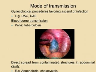
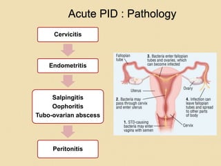
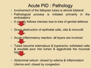
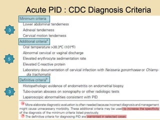
SEXUALLY TRANSMITTED
DISEASES
1
GYNAECOLOGIC
INFECTIONS
Dr. A.M. Otara
Senior Lecturer Reproductive Health
Department
Specialist Obstetrician& Gynaecologist
SCOPE
 Lower genital tract infections
 Pelvic inflammatory disease
 Pelvic Abscess
LOWER GENITAL TRACT
INFECTIONS
Lower Genital Tract Infections
Learning objectives
At the end of this chapter you should be able to:
 Define lower genital tract infections.
 Outline the characteristics of an abnormal vaginal
discharge.
 Discuss the causes and management of abnormal
vaginal discharge.
 Discuss the causes and management of ulcerative
genital infections.
 Outline the approaches to the control of Sexually
transmitted infections.
 Abnormal per vaginal discharge
 Genital Ulcers
Vaginal Discharge
 Physiological
 Abnormal
Characteristics
◦ Amount
◦ Odour
◦ Associated features :
 dysuria, dysperunia
 itching, burning sensation
vulval soreness
Physiological discharge
Discharge from each organ and its
characteristics
◦ Uterine cavity-clear, more copious
towards end of cycle
◦ Cervix – has leucocytes and quantity is
influenced by function of ovary and
vascularity of the cervix.
◦ Vaginal wall -cells join up in the
secretions therefore semi-solid, opaque &
white in colour. It may contain some
bacteria (Doderlins bacilli) and amount
increases at sexual stimulation.
◦ Vulval-from Bartholins, sebaceous and
sweat glands. The amount varies with
ovarian function
Physiological Discharge
 Secretory phase of menstrual cycle
 Sexual excitement –pre-coital
Leucorrhea
 Excess vaginal discharge.
 Heavier than usual, but clear.
Quantity is 0.67g-1g at ovulation
Leucorrhea
It is noticed at various stages: –
 At birth; may be mucoid – occurring 1st-10th/7
after birth of the neonate. Treatment –reassure
mother about baby
 Puberty-abnormal discharge due to increased
activity of hypothalamus and ovarian stimulation by
LH to produce estrogens.
 Hormones –family planning hormones: COCs.
 IUCD
 Douching, FP creams, gels –stimulates
secretion.
 Cervical Ectopion-Displacement of squamo-
columnar junction due to cervical erosion .
 Cervical cautery .
Leucorrhea
 When wet it is white or creamy. When
dry it has a brownish yellow stain on
under clothing .
 Some leucocytes and pus cells may
be seen.
 It is a nuisance as it stains clothes,
causes soreness but there is no
pruritus and has no smell.
Abnormal Vaginal Discharge
Non-infections causes
 Chemical irritation; allergy to condom
material, douching, contraceptives
 Atrophic vaginitis in post-menopausal
women
 Foreign bodies-tampons,
 Neoplasia-Benign e.g. Fibroids,
Malignant e.g. CA Cervix
Abnormal Vaginal Discharge
Infections
◦ T.Vaginalis
◦ Candida albicans
◦ N.gonorrhea
◦ Viral –HSV -rare
Inflammatory Vaginal
discharge
The discharge can be mucopurulent or
purulent, smelly, associated with
vulvitis, dysperunia.
There is a large numbers of
polymorphs in the pus
Sexualy Transmitted Infections
Definition
Sexually transmitted diseases are
diseases which are predominantly
transmitted through sexual contact .
STDs affect any of the genital organs in
females and males.
17
Magnitude
Worldwide at least 250 million new
cases of STDs occur each year.
Trichomoniasis : 120m cases / year.
Chlamydia 50 m cases /year
18
Gender issues
Women are at higher risk compared
to men
 Limited ability to negotiate safe sex.
 Limited access to information and
health care.
 Larger vaginal surface area more
vulnerable to infections.
 STIs often asymptomatic in women.
 Blood transfusion during delivery
predispose to HIV and Hepatitis B. 19
Trichomoniasis
 Caused by T. vaginalis a flagellates that reproduce
by binary fusion
 Incidence -5 % of women in FP clinic ,13-25% of
women in gynaecological clinic, 50-75% in
prostitute ,amongst male-also common, 5% of
babies born of infected mothers
 Features-Greenish yellow discharge, frothy (air
bubbles), profuse –on Cusco exam – big pool of
fluid in the posterior fornix
 Itching can be much and may cause vulval
ulcers/cracks
 Wet prep under microscope
 Treatment:-
◦ Metronidazole -200mg tds 7/7 or 400mg bid 7/7
--No alcohol.
◦ Tinidazole (fasigyn)4 tabs stat, can be taken as
tablets or pessaries-commonly fasigyn + nystatin
 Give dose for spouse as well
Candidiasis/moniliasis
 Very common
 Discharge is white thick and in in clamps.
There is vulval itchiness.
 Covers both vulva & vagina –vulvo-vaginitis
,can also be found in ano-rectal area.
 Vaginal swab for culture
 Speculum exam –vaginal wall lined with
thick discharge
 Can cause ballanitis (inflammation of
prepuce and glans penis)
Candidiasis/moniliasis
It is favoured by some conditions:
 Pregnancy glycosuria
 Diabetes Mellitus
 Oral contraceptive use
 Menstruation
 Antibiotic therapy-broad spectrum-Tetracycline,
cephasporins.
 Immune deffiency -HIV, cytotoxics
Prevalence in pregnancy increases in course of
gestation and factors in pregnancy making ideal
environment for candida include;
 Increased estrogen
 glycosuria –decreased glucose tolerance
 slight increase in vaginal fluid PH
Candidiasis/moniliasis
 Treatment is generally topical unless the infection
does not respond or recurs frequently.
 Systemic treatment and suppression with
imidazoles are effective, although long-term use
can lead to colonization with resistant candida
species
 Examples
◦ Nystatin tabs, vaginally or oral – has poor absorption
◦ Clotrimazole,Econazole,Miconazole,Bifonazole–Pessaries
or cream
◦ Per oral ; Itraconazole, Fluconazole, Ketoconazole
 Other reasons for apparent treatment failure or
breakthrough include a second etiologic agent and
noninfectious conditions.
Gonorrhoea
 Caused by N. gonorrhea, a gram negative
intracellular diplococcus.
 Sexual spread to the uterine cervix.
Incubation period is 2-14days.
 Majority of men are symptomatic and 2/3 of
women are asymptomatic.
 Features include; per urethral discharge –
purulent, dysuria, dysperunia. Pelvic
inflammation with backache & lower
abdominal pain
 Manifests in 1st 10/7 of menstrual period
Chlamydia
 Caused by C. trachomatis, Gram negative, intra-
cellular obligate organism, infects epithelial cells of
the urethra, conjunctiva, cervix and fallopian tubes
(pathogen of mucus membranes).
 Serovariants that produce different lesions:L1-3:
LGV;
A, B, Ba, C :Trachoma; B-K:cervicitis, salpingitis,
non-gonococcal urethritis, neonatal conjunctivitis
and pneumonia
 Isolated in upto 70% of men with non-specific
urethritis.
 Thought to be 2° infection to patients with
gonorrhea
 Causes cervicitis and vaginitis, but has been
isolated in cervix of asymptomatic women or
fallopian tubes.
 Associated with occasional pain when erect in men
Bacterial vaginosis
Gardnerella
KOH -fishy smell
Ulcerative lnfections
 Syphylis
 Chancroid
◦ Haemophilus ducreyi infection is
especially common in women from
developing countries.
 Herpes Simplex Virus II
Herpes Simplex Virus II
 Vaginal discharge –clear, watery
associated with pain
 Vesicles at cervix & vulva –burst after
a few days and may ulcerate
 Viral inclusion bodies in cytological
smears
 Treatment –Acyclovir 200mg,
Famciclovir
 Supportive therapy: treat 2° infections
, analgesics – painful
HIV
 Women represent one of the fastest
growing populations infected with the
human immunodeficiency virus (HIV)
 There remain significant gender-based
differences in the disease. These include:
◦ Differences in viral load early in infection
◦ Differences in selected opportunistic infections
◦ A number of female-specific complications
◦ Issues related to HIV and pregnancy
◦ The psychosocial impact and the environment in
which HIV/AIDS occurs in women
HIV
 For many women, gynecologic complaints
are the initial manifestation of HIV/AIDS.
 These conditions, which also exist in
uninfected women, can occur with higher
frequency and severity in women with HIV.
◦ Candidia vaginitis
◦ Abnormal cervical cytology
◦ Pelvic inflammatory disease
◦ Genital ulcer disease (eg, HSV, chancroid,
syphilis)
◦ Menstrual disorders
Approaches to the control of
Sexually Transmitted Infections
 STIs constitute a huge and economic
burden for developing countries: 75-85% of
the estimated 340 million annual new cases
of curable STIs occur in these countries.
 The control of STIs can reduce HIV
transmission.
 STI control programmes have 3 objectives;
◦ To interrupt transmission of STIs
◦ To prevent the development of disease
complications and sequelae
◦ To reduce the transmission of HIV infection
Approaches to the control of
Sexually Transmitted Infections
Interventions to reduce incidence and prevalence of
STIs include;
 Primary prevention by;
◦ Information, education and communication campaigns
◦ Condom promotion
◦ Use of safe microbicides
◦ Vaccines
 Screening and case finding amongst vulnerable
groups e.g. pregnant women
 STI case management using syndromic approach
 Targeted interventions for populations at risk e.g
sex workers
(4 Cs: counseling,condoms, compliance with
treatment, contact tracing)
 Targeted periodic mass treatments.
PELVIC INFLAMMATORY
DISEASE
Pelvic Inflammatory Disease
Learning objectives
At the end of this chapter you should be able to:
 Define pelvic inflammatory disease(PID).
 Discuss the epidemiology of PID.
 Outline the aetiological agents in PID.
 Discuss the pathogenesis of PID.
 Discuss the patho-physiology of PID
 Describe the diagnosis of PID
 Outline the differential diagnosis of PID
 Discuss the treatment and follow-up of a patient
with PID and its’ complications.
Definition
 An infection of the upper genital tract:-
cervix, uterus, tubes, ovaries &
surrounding peritoneum.
 Usually bilateral
 Sexually active women
 Age; common in <25yrs, in
postmenopausal associated with
malignancy
Epidemiology
 No accurate data on prevalence
 1 million women have an episode of
acute PID in a year
 100,000 become infertile annually as a
result of PID
 150 deaths annually from PID
complications
 PID is uncommon in pregnancy
Aetiology
 Causative Organisms; C. trachomatis,
N. gonnorhea, H. Influenza, Group B
& D streptococcus, E. coli,
Mycoplasma hominis, anaerobic gram
+ve cocci, Bacteroides sp, Clostridium
sp.
 Sexual spread through the uterine
cervix to the tubal mucosa where it
attaches to the epithelium and invades
the cells.
Spread Mechanisms
 There is free passage from external
into the peritoneal cavity.
 Potential infectious sites adjacent to
vagina ;
◦ urethra anteriorly
◦ the anus posteriorly .
Defense Mechanisms
There are natural defenses that prevent
easy passage of organisms:-
 Vulva
◦ inherent resistance to infection, unless
traumatized
◦ secretion by apocrine glands –rich in
undecylenic acid a strong antifungal
secretion
◦ Natural apposition of the two vulva lips
(lateral)
Defense Mechanisms
 Vagina
◦ Natural apposition – (AP)
◦ Stratified squamous epithelium
◦ Acidity-enhanced by vaginal flora eg
Doderlin’s bacilli(lactobacilli)-act on
Lactogen producing lactic acid which
decreases pH.
 Cervix –mucus plug.
 Uterine cavity –cyclic shedding of
endometrium
Defense Mechanisms
The efficiency of these mechanisms are
reduced with:
 Age –very young –lack apocrine
glands and Lactobacilli. Menopause-
reduced defense
 Menses –Mucus plug shed off,
alkalinity increases, Gonococcus is
more virulent if contracted at this time
Defense Mechanisms
Puerperium –Period upto 6/52 post delivery
decreased defense due to :
 raw area left by placenta acting as an
hideout
 breakdown in cervical, vaginal epithelium
 bruising and devitalization of tissues at birth
 Gapping of vagina and cervix post delivery
 Lochia –decreases vaginal acidity therefore
pathogens multiply
 Blood clots & decidua – good culture media
Risk Factors
 Multiple sexual partners > 2 in 30days
 Previous PID
 Non-use of barrier contraception
 IUD increases risk 6fold in 3weeks of
insertion
 Immuno-suppression
 Recent instrumentation of the genital
tract
Pathophysiology
 Bacterial colonization of the cervix ascends
to the uterus, fallopian tubes and ovaries
 Gonnorhea and chlamydia are typically
isolated from the cervix in PID, but rare in
the ovarian tissue; they facilitate infection of
the adnexia by other bacteria( polymicrobial
process)
 Vaginal flora associated with PID include;
anaerobes, G. vaginalis, H. influenza, G-ve
rods, M. hominis,
Mode of transmission
Ascending infection (Canalicular spread)
 Ascent of gonococcal & chlamydial organisms by
surface extension from the lower genital tract through
the cervical canal by way of the endometrium to the
fallopian tubes
 Facilitated by the sexually transmitted vectors such as
sperms & trichomonads
 Reflux of menstrual blood along with gonococci into the
Mode of transmission
Through uterine lymphatic & blood vessels across
parametrium
 Mycoplasma hominis
 Secondary organisms
Mode of transmission
Gynecological procedures favoring ascend of infection
 E.g. D&C, D&E
Blood-borne transmission
 Pelvic tuberculosis
Direct spread from contaminated structures in abdominal
cavity
 E.g. Appendicitis, cholecystitis
Acute PID : Pathology
Cervicitis
Endometritis
Salpingitis
Oophoritis
Tubo-ovarian abscess
Peritonitis
Acute PID : Pathology
 Involvement of the fallopian tubes is almost bilateral
 Pathological process is initiated primarily in the
endosalpinx
 It usually follows menses due to loss of genital defence
 Gross destruction of epithelial cells, cilia & microvilli
 Acute inflammatory reaction: all layers are involved
 Tubes become edematous & hyperemic; exfoliated cells
& exudate pour into lumen & agglutinate the mucosal
folds
 Abdominal ostium: closed by edema & inflammation
Uterine end: closed by congestion
Acute PID : Pathology
 Depending on the virulence: watery or purulent exudate
 Hydrosalpinx or Pyosalpinx
 Deeper penetration & more destruction
 Possibilities
Oophoritis
Tubo-ovarian abscess
Peritonitis
Pelvic abscess
or
Resolution in 2-3 weeks with/without chronic
sequelae
Acute PID : CDC Diagnosis Criteria
3
5
3
Acute PID : Presentation & Diagnosis
Investigations
 Complete blood count: Wbc count> 10,000/ml,
 C – reactive protein; elevated
 Erythrocyte sedimentation rate: elevated
 Urine Pregnancy Test (UPT), urinalysis
 Urine culture
 Urine NAATs
 Vaginal/endocervical wet mount
1. WBCs suggest PID
2. Cervical chlamydia and gonorrhea testing (gram negative
diplococci.)
3. Nucleic acid amplification tests (NAATs) for organisms
 Faecal occult blood test
 Tests for tuberculosis
 Tests for syphilis
 Tests for HIV
Acute PID : Presentation & Diagnosis
Imaging
 Transvaginal ultrasonography is the imaging modality of
choice
 Trans abdominal ultrasonography for DD
 Abdominal CT or MRI : thickened tubes, fluid
Diagnostic procedures
 Culdocentesis
 Endometrial biopsy
 Diagnostic laparoscopy:the definitive test; hyperaemia of
the external tubal surface, tubal edema, sticky exudate
 Transvaginal U/S, MRI-
Acute PID : Staging
(I-IDSOG-USA recommends following stages)
Stage I
 Women who fulfil the CDC major diagnostic criteria and
>1 of its minor criteria but who do not have overt
peritonitis (as demonstrated by the absence of rebound
tenderness) and who have not had any prior
documented STD upper tract infections
Stage II
 The above criteria, with peritonitis
Stage III
 Women with demonstrable tubo-ovarian complex or
tubo-ovarian abscess evident on either physical or
ultrasonographic examination
Stage IV
 Women with ruptured tubo-ovarian abscesses
Differential Diagnosis
 Infections;appendicitis,diverticulitis,cys
titis, septic abortion
 Tumors/malignancy
 Ectopic pregnancy
 Adnexal torsion
 Degenerating fibroid
 Endometriosis
 UTI
 Regional enteritis or ulcerative cotitis
Complications
 Peritonitis –local pelvic peritonitis or spreads to
spleen, liver as Fitz-Hugh-Curtis (perihepatitis-
’Violin strings ‘ on laparoscopy), Generalized
peritonitis.
 Paralytic ilieus leading to chronic distension then
electrolyte imbalance
 Abscess formation
◦ Pyosalpinx –tube
◦ Tubovarian abscess
◦ POD abscess
 Long-term; Infertility (commonest cause),
adhesions in the pelvis , chronic pelvic pain,
intestinal obstruction due to adhesions.
 Gonococcal arthritis –rare
 Septic shock
Treatment
Out-patient
 If mild or moderate- oral or parentral
therapy has similar efficacy
 Analgesics incase of fever with
minimal abdominal findings give–
Iboprufen, mefnamic acid,ASA
 Bed rest for 1 week
Treatment
Outpatient treatment
 High dose antibiotics
◦ Penicillin -4.8 mU with probencid lg oral dose followed with
10 – 14 days of,Doxycycline 100mg –
◦ -Cefoxitim 2g 1m start followed by Doxycycline or
tetracycline
◦ Amoxicillin or Ampicillin stat, followed with Doxycycline or
tetracycline.
◦ Quinolones- Ofloxacin400mg bid or Levofloxacin 500mg od
+/- Metronidazole 500mg bd for 14 days
◦ 3rd/4th gen Cephalosporins e.g. i.m. cefoxitin stat +
Doxcycline 100mg bd +/- Metronidazole 500mg bd for 14
days
 Treat the partner
 Review patient in 3-4 days – If not better admit.
Treatment
In-patient
 CDC criteria for hospitalization-surgical
emergency, pregnancy, no response to oral
antibiotics, unable to tolerate or follow Rx,
vomiting, tubo-ovarian abscess
 3rd Generation cephalosporin-
cefotetan,cefoxitin 2g i.v bd+ Doxcyline
100mg po or iv bd
 Clindamycin 900mg iv tds+Gentamicin
1.5mg/kg tds
 No
improvement;laparascopy/laparotomy
Prevention
 Health education.
 Risks of multiple partners .
 Youth separately, ie.isolated clinics.
 Contact tracing & early detection in
male with asymptomatic.
Follow-up
 Improvement within 72 hours, re-
evaluate
 Contact tracing, treatment and
education
 Referral
 25% risk of long-term sequelae;
infertility ectopic pregnancy and
chronic pelvic pain
 Fitz-Hugh-Curtis syndrome- fibrous
peri-hepatic adhesions
PELVIC ABSCESS
Pelvic Abscess
Learning objectives
At the end of this chapter you should be able to:
 Define pelvic abscess.
 Discuss the pathogenesis of Pelvic abscess.
 Describe the diagnosis of Pelvic abscess
 Discuss the treatment and follow-up of a patient
with Pelvic Abscess and its’ complications.
Introduction
 A collection of pus within pelvis created by
adherence of adjacent organs.
 Most abscesses occur in the pouch of
Douglas, on the adnexia as tubo-ovarian
abscess and rarely broad ligament
abscess.
 Most abscesses are polymicrobial.
 Follows recurrent PID,abortion, puerperal
sepsis, gynaecologic cancer, gynaecologic
surgery and procedures.
 Common in HIV/AIDS.
Clinical presentation
 Lower abdominal pain and swelling
 Cervical discharge.
 Painful defecation and lower back pain.
 Fever, nausea, tachycardia, malaise
 Fluctuant mass filling the cul-de-sac.
 Laboratory tests are of little value in
diagnosis; leucocytosis, ESR, C-reactive
proteins.
 Diagnosis: paracentesis, culdocentesis.
Pelvic ultrasound scan.
Treatment
 Supportive care with intravenous fluids, parenteral
antibiotics, naso-gastric intubation, transfusion.
 Surgery; emergency surgery in suspicion of rupture
of abscess to avoid fulminant peritonitis.
 U/S or CT guided percutenous drainage of the pus
and irrigation may be successful in 75-84% of
cases.
 Laparotomy- break the pus locules, releases
adhesions, drainage of pus. Salpingectomy if there
is pyosalpinx.
 Posterior colpotomy with dissection of the sacules
then drainage.
Complications
 Recurrence
 Bowel obstruction
 Ectopic pregnancy
 Infertility
 Chronic pelvic pain
 Visceral injury at laparotomy
Management : Surgery in PID
(Main complications in Stage IV PID : Ruptured abscess)
During operation
1. Septic shock
2. Injury to small bowel
3. Injury to rectum
Post-operative
1. Pus collected again
2. Chest empyema
3. Septicemia
4. Septic shock
5.. Recto-vaginal fistula
6. Wound abscess or infection
7. Pneumonia
8. Renal failure
9. Liver failure
Prevention
 Non-pharmacologic; health education,
prevention of STI, barrier methods,
contact tracing, avoid IUCD in high
risk.
 Prompt diagnosis and treatment of
PID
 Vaccines; chlamydia

GYNAECOLOGIC INFECTIONS 2016 (2).ppt

  • 1.
  • 2.
    GYNAECOLOGIC INFECTIONS Dr. A.M. Otara SeniorLecturer Reproductive Health Department Specialist Obstetrician& Gynaecologist
  • 3.
    SCOPE  Lower genitaltract infections  Pelvic inflammatory disease  Pelvic Abscess
  • 4.
  • 5.
    Lower Genital TractInfections Learning objectives At the end of this chapter you should be able to:  Define lower genital tract infections.  Outline the characteristics of an abnormal vaginal discharge.  Discuss the causes and management of abnormal vaginal discharge.  Discuss the causes and management of ulcerative genital infections.  Outline the approaches to the control of Sexually transmitted infections.
  • 6.
     Abnormal pervaginal discharge  Genital Ulcers
  • 7.
    Vaginal Discharge  Physiological Abnormal Characteristics ◦ Amount ◦ Odour ◦ Associated features :  dysuria, dysperunia  itching, burning sensation vulval soreness
  • 8.
    Physiological discharge Discharge fromeach organ and its characteristics ◦ Uterine cavity-clear, more copious towards end of cycle ◦ Cervix – has leucocytes and quantity is influenced by function of ovary and vascularity of the cervix. ◦ Vaginal wall -cells join up in the secretions therefore semi-solid, opaque & white in colour. It may contain some bacteria (Doderlins bacilli) and amount increases at sexual stimulation. ◦ Vulval-from Bartholins, sebaceous and sweat glands. The amount varies with ovarian function
  • 9.
    Physiological Discharge  Secretoryphase of menstrual cycle  Sexual excitement –pre-coital
  • 10.
    Leucorrhea  Excess vaginaldischarge.  Heavier than usual, but clear. Quantity is 0.67g-1g at ovulation
  • 11.
    Leucorrhea It is noticedat various stages: –  At birth; may be mucoid – occurring 1st-10th/7 after birth of the neonate. Treatment –reassure mother about baby  Puberty-abnormal discharge due to increased activity of hypothalamus and ovarian stimulation by LH to produce estrogens.  Hormones –family planning hormones: COCs.  IUCD  Douching, FP creams, gels –stimulates secretion.  Cervical Ectopion-Displacement of squamo- columnar junction due to cervical erosion .  Cervical cautery .
  • 12.
    Leucorrhea  When wetit is white or creamy. When dry it has a brownish yellow stain on under clothing .  Some leucocytes and pus cells may be seen.  It is a nuisance as it stains clothes, causes soreness but there is no pruritus and has no smell.
  • 13.
    Abnormal Vaginal Discharge Non-infectionscauses  Chemical irritation; allergy to condom material, douching, contraceptives  Atrophic vaginitis in post-menopausal women  Foreign bodies-tampons,  Neoplasia-Benign e.g. Fibroids, Malignant e.g. CA Cervix
  • 14.
    Abnormal Vaginal Discharge Infections ◦T.Vaginalis ◦ Candida albicans ◦ N.gonorrhea ◦ Viral –HSV -rare
  • 15.
    Inflammatory Vaginal discharge The dischargecan be mucopurulent or purulent, smelly, associated with vulvitis, dysperunia. There is a large numbers of polymorphs in the pus
  • 16.
  • 17.
    Definition Sexually transmitted diseasesare diseases which are predominantly transmitted through sexual contact . STDs affect any of the genital organs in females and males. 17
  • 18.
    Magnitude Worldwide at least250 million new cases of STDs occur each year. Trichomoniasis : 120m cases / year. Chlamydia 50 m cases /year 18
  • 19.
    Gender issues Women areat higher risk compared to men  Limited ability to negotiate safe sex.  Limited access to information and health care.  Larger vaginal surface area more vulnerable to infections.  STIs often asymptomatic in women.  Blood transfusion during delivery predispose to HIV and Hepatitis B. 19
  • 20.
    Trichomoniasis  Caused byT. vaginalis a flagellates that reproduce by binary fusion  Incidence -5 % of women in FP clinic ,13-25% of women in gynaecological clinic, 50-75% in prostitute ,amongst male-also common, 5% of babies born of infected mothers  Features-Greenish yellow discharge, frothy (air bubbles), profuse –on Cusco exam – big pool of fluid in the posterior fornix  Itching can be much and may cause vulval ulcers/cracks  Wet prep under microscope  Treatment:- ◦ Metronidazole -200mg tds 7/7 or 400mg bid 7/7 --No alcohol. ◦ Tinidazole (fasigyn)4 tabs stat, can be taken as tablets or pessaries-commonly fasigyn + nystatin  Give dose for spouse as well
  • 21.
    Candidiasis/moniliasis  Very common Discharge is white thick and in in clamps. There is vulval itchiness.  Covers both vulva & vagina –vulvo-vaginitis ,can also be found in ano-rectal area.  Vaginal swab for culture  Speculum exam –vaginal wall lined with thick discharge  Can cause ballanitis (inflammation of prepuce and glans penis)
  • 22.
    Candidiasis/moniliasis It is favouredby some conditions:  Pregnancy glycosuria  Diabetes Mellitus  Oral contraceptive use  Menstruation  Antibiotic therapy-broad spectrum-Tetracycline, cephasporins.  Immune deffiency -HIV, cytotoxics Prevalence in pregnancy increases in course of gestation and factors in pregnancy making ideal environment for candida include;  Increased estrogen  glycosuria –decreased glucose tolerance  slight increase in vaginal fluid PH
  • 23.
    Candidiasis/moniliasis  Treatment isgenerally topical unless the infection does not respond or recurs frequently.  Systemic treatment and suppression with imidazoles are effective, although long-term use can lead to colonization with resistant candida species  Examples ◦ Nystatin tabs, vaginally or oral – has poor absorption ◦ Clotrimazole,Econazole,Miconazole,Bifonazole–Pessaries or cream ◦ Per oral ; Itraconazole, Fluconazole, Ketoconazole  Other reasons for apparent treatment failure or breakthrough include a second etiologic agent and noninfectious conditions.
  • 24.
    Gonorrhoea  Caused byN. gonorrhea, a gram negative intracellular diplococcus.  Sexual spread to the uterine cervix. Incubation period is 2-14days.  Majority of men are symptomatic and 2/3 of women are asymptomatic.  Features include; per urethral discharge – purulent, dysuria, dysperunia. Pelvic inflammation with backache & lower abdominal pain  Manifests in 1st 10/7 of menstrual period
  • 25.
    Chlamydia  Caused byC. trachomatis, Gram negative, intra- cellular obligate organism, infects epithelial cells of the urethra, conjunctiva, cervix and fallopian tubes (pathogen of mucus membranes).  Serovariants that produce different lesions:L1-3: LGV; A, B, Ba, C :Trachoma; B-K:cervicitis, salpingitis, non-gonococcal urethritis, neonatal conjunctivitis and pneumonia  Isolated in upto 70% of men with non-specific urethritis.  Thought to be 2° infection to patients with gonorrhea  Causes cervicitis and vaginitis, but has been isolated in cervix of asymptomatic women or fallopian tubes.  Associated with occasional pain when erect in men
  • 26.
  • 27.
    Ulcerative lnfections  Syphylis Chancroid ◦ Haemophilus ducreyi infection is especially common in women from developing countries.  Herpes Simplex Virus II
  • 28.
    Herpes Simplex VirusII  Vaginal discharge –clear, watery associated with pain  Vesicles at cervix & vulva –burst after a few days and may ulcerate  Viral inclusion bodies in cytological smears  Treatment –Acyclovir 200mg, Famciclovir  Supportive therapy: treat 2° infections , analgesics – painful
  • 29.
    HIV  Women representone of the fastest growing populations infected with the human immunodeficiency virus (HIV)  There remain significant gender-based differences in the disease. These include: ◦ Differences in viral load early in infection ◦ Differences in selected opportunistic infections ◦ A number of female-specific complications ◦ Issues related to HIV and pregnancy ◦ The psychosocial impact and the environment in which HIV/AIDS occurs in women
  • 30.
    HIV  For manywomen, gynecologic complaints are the initial manifestation of HIV/AIDS.  These conditions, which also exist in uninfected women, can occur with higher frequency and severity in women with HIV. ◦ Candidia vaginitis ◦ Abnormal cervical cytology ◦ Pelvic inflammatory disease ◦ Genital ulcer disease (eg, HSV, chancroid, syphilis) ◦ Menstrual disorders
  • 31.
    Approaches to thecontrol of Sexually Transmitted Infections  STIs constitute a huge and economic burden for developing countries: 75-85% of the estimated 340 million annual new cases of curable STIs occur in these countries.  The control of STIs can reduce HIV transmission.  STI control programmes have 3 objectives; ◦ To interrupt transmission of STIs ◦ To prevent the development of disease complications and sequelae ◦ To reduce the transmission of HIV infection
  • 32.
    Approaches to thecontrol of Sexually Transmitted Infections Interventions to reduce incidence and prevalence of STIs include;  Primary prevention by; ◦ Information, education and communication campaigns ◦ Condom promotion ◦ Use of safe microbicides ◦ Vaccines  Screening and case finding amongst vulnerable groups e.g. pregnant women  STI case management using syndromic approach  Targeted interventions for populations at risk e.g sex workers (4 Cs: counseling,condoms, compliance with treatment, contact tracing)  Targeted periodic mass treatments.
  • 33.
  • 34.
    Pelvic Inflammatory Disease Learningobjectives At the end of this chapter you should be able to:  Define pelvic inflammatory disease(PID).  Discuss the epidemiology of PID.  Outline the aetiological agents in PID.  Discuss the pathogenesis of PID.  Discuss the patho-physiology of PID  Describe the diagnosis of PID  Outline the differential diagnosis of PID  Discuss the treatment and follow-up of a patient with PID and its’ complications.
  • 35.
    Definition  An infectionof the upper genital tract:- cervix, uterus, tubes, ovaries & surrounding peritoneum.  Usually bilateral  Sexually active women  Age; common in <25yrs, in postmenopausal associated with malignancy
  • 36.
    Epidemiology  No accuratedata on prevalence  1 million women have an episode of acute PID in a year  100,000 become infertile annually as a result of PID  150 deaths annually from PID complications  PID is uncommon in pregnancy
  • 37.
    Aetiology  Causative Organisms;C. trachomatis, N. gonnorhea, H. Influenza, Group B & D streptococcus, E. coli, Mycoplasma hominis, anaerobic gram +ve cocci, Bacteroides sp, Clostridium sp.  Sexual spread through the uterine cervix to the tubal mucosa where it attaches to the epithelium and invades the cells.
  • 38.
    Spread Mechanisms  Thereis free passage from external into the peritoneal cavity.  Potential infectious sites adjacent to vagina ; ◦ urethra anteriorly ◦ the anus posteriorly .
  • 39.
    Defense Mechanisms There arenatural defenses that prevent easy passage of organisms:-  Vulva ◦ inherent resistance to infection, unless traumatized ◦ secretion by apocrine glands –rich in undecylenic acid a strong antifungal secretion ◦ Natural apposition of the two vulva lips (lateral)
  • 40.
    Defense Mechanisms  Vagina ◦Natural apposition – (AP) ◦ Stratified squamous epithelium ◦ Acidity-enhanced by vaginal flora eg Doderlin’s bacilli(lactobacilli)-act on Lactogen producing lactic acid which decreases pH.  Cervix –mucus plug.  Uterine cavity –cyclic shedding of endometrium
  • 41.
    Defense Mechanisms The efficiencyof these mechanisms are reduced with:  Age –very young –lack apocrine glands and Lactobacilli. Menopause- reduced defense  Menses –Mucus plug shed off, alkalinity increases, Gonococcus is more virulent if contracted at this time
  • 42.
    Defense Mechanisms Puerperium –Periodupto 6/52 post delivery decreased defense due to :  raw area left by placenta acting as an hideout  breakdown in cervical, vaginal epithelium  bruising and devitalization of tissues at birth  Gapping of vagina and cervix post delivery  Lochia –decreases vaginal acidity therefore pathogens multiply  Blood clots & decidua – good culture media
  • 43.
    Risk Factors  Multiplesexual partners > 2 in 30days  Previous PID  Non-use of barrier contraception  IUD increases risk 6fold in 3weeks of insertion  Immuno-suppression  Recent instrumentation of the genital tract
  • 44.
    Pathophysiology  Bacterial colonizationof the cervix ascends to the uterus, fallopian tubes and ovaries  Gonnorhea and chlamydia are typically isolated from the cervix in PID, but rare in the ovarian tissue; they facilitate infection of the adnexia by other bacteria( polymicrobial process)  Vaginal flora associated with PID include; anaerobes, G. vaginalis, H. influenza, G-ve rods, M. hominis,
  • 45.
    Mode of transmission Ascendinginfection (Canalicular spread)  Ascent of gonococcal & chlamydial organisms by surface extension from the lower genital tract through the cervical canal by way of the endometrium to the fallopian tubes  Facilitated by the sexually transmitted vectors such as sperms & trichomonads  Reflux of menstrual blood along with gonococci into the
  • 46.
    Mode of transmission Throughuterine lymphatic & blood vessels across parametrium  Mycoplasma hominis  Secondary organisms
  • 47.
    Mode of transmission Gynecologicalprocedures favoring ascend of infection  E.g. D&C, D&E Blood-borne transmission  Pelvic tuberculosis Direct spread from contaminated structures in abdominal cavity  E.g. Appendicitis, cholecystitis
  • 48.
    Acute PID :Pathology Cervicitis Endometritis Salpingitis Oophoritis Tubo-ovarian abscess Peritonitis
  • 49.
    Acute PID :Pathology  Involvement of the fallopian tubes is almost bilateral  Pathological process is initiated primarily in the endosalpinx  It usually follows menses due to loss of genital defence  Gross destruction of epithelial cells, cilia & microvilli  Acute inflammatory reaction: all layers are involved  Tubes become edematous & hyperemic; exfoliated cells & exudate pour into lumen & agglutinate the mucosal folds  Abdominal ostium: closed by edema & inflammation Uterine end: closed by congestion
  • 50.
    Acute PID :Pathology  Depending on the virulence: watery or purulent exudate  Hydrosalpinx or Pyosalpinx  Deeper penetration & more destruction  Possibilities Oophoritis Tubo-ovarian abscess Peritonitis Pelvic abscess or Resolution in 2-3 weeks with/without chronic sequelae
  • 51.
    Acute PID :CDC Diagnosis Criteria 3 5 3
  • 52.
    Acute PID :Presentation & Diagnosis Investigations  Complete blood count: Wbc count> 10,000/ml,  C – reactive protein; elevated  Erythrocyte sedimentation rate: elevated  Urine Pregnancy Test (UPT), urinalysis  Urine culture  Urine NAATs  Vaginal/endocervical wet mount 1. WBCs suggest PID 2. Cervical chlamydia and gonorrhea testing (gram negative diplococci.) 3. Nucleic acid amplification tests (NAATs) for organisms  Faecal occult blood test  Tests for tuberculosis  Tests for syphilis  Tests for HIV
  • 53.
    Acute PID :Presentation & Diagnosis Imaging  Transvaginal ultrasonography is the imaging modality of choice  Trans abdominal ultrasonography for DD  Abdominal CT or MRI : thickened tubes, fluid Diagnostic procedures  Culdocentesis  Endometrial biopsy  Diagnostic laparoscopy:the definitive test; hyperaemia of the external tubal surface, tubal edema, sticky exudate  Transvaginal U/S, MRI-
  • 54.
    Acute PID :Staging (I-IDSOG-USA recommends following stages) Stage I  Women who fulfil the CDC major diagnostic criteria and >1 of its minor criteria but who do not have overt peritonitis (as demonstrated by the absence of rebound tenderness) and who have not had any prior documented STD upper tract infections Stage II  The above criteria, with peritonitis Stage III  Women with demonstrable tubo-ovarian complex or tubo-ovarian abscess evident on either physical or ultrasonographic examination Stage IV  Women with ruptured tubo-ovarian abscesses
  • 56.
    Differential Diagnosis  Infections;appendicitis,diverticulitis,cys titis,septic abortion  Tumors/malignancy  Ectopic pregnancy  Adnexal torsion  Degenerating fibroid  Endometriosis  UTI  Regional enteritis or ulcerative cotitis
  • 57.
    Complications  Peritonitis –localpelvic peritonitis or spreads to spleen, liver as Fitz-Hugh-Curtis (perihepatitis- ’Violin strings ‘ on laparoscopy), Generalized peritonitis.  Paralytic ilieus leading to chronic distension then electrolyte imbalance  Abscess formation ◦ Pyosalpinx –tube ◦ Tubovarian abscess ◦ POD abscess  Long-term; Infertility (commonest cause), adhesions in the pelvis , chronic pelvic pain, intestinal obstruction due to adhesions.  Gonococcal arthritis –rare  Septic shock
  • 58.
    Treatment Out-patient  If mildor moderate- oral or parentral therapy has similar efficacy  Analgesics incase of fever with minimal abdominal findings give– Iboprufen, mefnamic acid,ASA  Bed rest for 1 week
  • 59.
    Treatment Outpatient treatment  Highdose antibiotics ◦ Penicillin -4.8 mU with probencid lg oral dose followed with 10 – 14 days of,Doxycycline 100mg – ◦ -Cefoxitim 2g 1m start followed by Doxycycline or tetracycline ◦ Amoxicillin or Ampicillin stat, followed with Doxycycline or tetracycline. ◦ Quinolones- Ofloxacin400mg bid or Levofloxacin 500mg od +/- Metronidazole 500mg bd for 14 days ◦ 3rd/4th gen Cephalosporins e.g. i.m. cefoxitin stat + Doxcycline 100mg bd +/- Metronidazole 500mg bd for 14 days  Treat the partner  Review patient in 3-4 days – If not better admit.
  • 60.
    Treatment In-patient  CDC criteriafor hospitalization-surgical emergency, pregnancy, no response to oral antibiotics, unable to tolerate or follow Rx, vomiting, tubo-ovarian abscess  3rd Generation cephalosporin- cefotetan,cefoxitin 2g i.v bd+ Doxcyline 100mg po or iv bd  Clindamycin 900mg iv tds+Gentamicin 1.5mg/kg tds  No improvement;laparascopy/laparotomy
  • 61.
    Prevention  Health education. Risks of multiple partners .  Youth separately, ie.isolated clinics.  Contact tracing & early detection in male with asymptomatic.
  • 62.
    Follow-up  Improvement within72 hours, re- evaluate  Contact tracing, treatment and education  Referral  25% risk of long-term sequelae; infertility ectopic pregnancy and chronic pelvic pain  Fitz-Hugh-Curtis syndrome- fibrous peri-hepatic adhesions
  • 63.
  • 64.
    Pelvic Abscess Learning objectives Atthe end of this chapter you should be able to:  Define pelvic abscess.  Discuss the pathogenesis of Pelvic abscess.  Describe the diagnosis of Pelvic abscess  Discuss the treatment and follow-up of a patient with Pelvic Abscess and its’ complications.
  • 65.
    Introduction  A collectionof pus within pelvis created by adherence of adjacent organs.  Most abscesses occur in the pouch of Douglas, on the adnexia as tubo-ovarian abscess and rarely broad ligament abscess.  Most abscesses are polymicrobial.  Follows recurrent PID,abortion, puerperal sepsis, gynaecologic cancer, gynaecologic surgery and procedures.  Common in HIV/AIDS.
  • 66.
    Clinical presentation  Lowerabdominal pain and swelling  Cervical discharge.  Painful defecation and lower back pain.  Fever, nausea, tachycardia, malaise  Fluctuant mass filling the cul-de-sac.  Laboratory tests are of little value in diagnosis; leucocytosis, ESR, C-reactive proteins.  Diagnosis: paracentesis, culdocentesis. Pelvic ultrasound scan.
  • 67.
    Treatment  Supportive carewith intravenous fluids, parenteral antibiotics, naso-gastric intubation, transfusion.  Surgery; emergency surgery in suspicion of rupture of abscess to avoid fulminant peritonitis.  U/S or CT guided percutenous drainage of the pus and irrigation may be successful in 75-84% of cases.  Laparotomy- break the pus locules, releases adhesions, drainage of pus. Salpingectomy if there is pyosalpinx.  Posterior colpotomy with dissection of the sacules then drainage.
  • 68.
    Complications  Recurrence  Bowelobstruction  Ectopic pregnancy  Infertility  Chronic pelvic pain  Visceral injury at laparotomy
  • 69.
    Management : Surgeryin PID (Main complications in Stage IV PID : Ruptured abscess) During operation 1. Septic shock 2. Injury to small bowel 3. Injury to rectum Post-operative 1. Pus collected again 2. Chest empyema 3. Septicemia 4. Septic shock 5.. Recto-vaginal fistula 6. Wound abscess or infection 7. Pneumonia 8. Renal failure 9. Liver failure
  • 70.
    Prevention  Non-pharmacologic; healtheducation, prevention of STI, barrier methods, contact tracing, avoid IUCD in high risk.  Prompt diagnosis and treatment of PID  Vaccines; chlamydia