CONTENTS
• Introduction
• Transportmodel: Permeability-Solubility-charge state and pH
partition hypothesis
• Properties of GIT
• pH microclimate
• Intracellular pH environment
• Tight junction complex
3.
Introduction
• During thediscovery and the development of drug molecules,
the pharmacokinetic properties of the drug molecules play an
important role.
• For the easy study of the developing drug molecules, the
“Transport model” is used for the pharmacokinetic profiling.
• This model is centrally embracing solubility and
permeability, with qualification related to pH and dissolution.
• To rationalize the critical components related to oral
absorption, the BCS is used, based on in vitro transport model.
4.
Cont.
• The BCScan be rationalized by considering Fick's first law,
applied to membranes.
• When molecules are introduced on one side of a lipid membrane
barrier (e.g., epithelial cell wall) and no such molecules are on the
other side, passive diffusion will drive the molecules across the
membrane.
• When certain simplifying assumptions are made, the flux equation
in Fick's law reduces simply to a product of permeability and
solubility.
• Permeability (Pe): A kinetic parameter related to lipophilicity (as
indicated by the partition and distribution coefficients, log P and
log D). Retention (R) of lipophilic molecules by the membrane
(which is related to lipophilicity and may predict PK volumes of
distribution) influences the characterization of permeability.
• Solubility (S): A thermodynamic parameter, and is closely related
to dissolution, a kinetic parameter.
5.
Transport Model
• Thetransport of the drug molecules from the membrane is
explained by Fick’s first law, applied to a membrane shows,
passive diffusion of a solute is the product of the diffusivity
and the concentration gradient of the solute inside the
membrane.
• The amount of the uncharged molecules present at a given pH,
that directly contributes to the flux, it depends on several
important factors, such as pH, binding to indigenous carriers
(proteins and bile acids), self-binding (aggregate or micelle
formation), and solubility (a solid-state form of self-binding).
• Consider a vessel divided into two chambers, separated by a
homogeneous lipid membrane. The left side is the donor
compartment, where the sample molecules are first
introduced; the right side is the acceptor compartment, which
at the start has no sample molecules. Fick’s first law applied to
homogeneous membranes at steady state.
7.
Cont.
• Fick’s firstlaw: The amount of material flowing through a unit
cross section, h, of a barrier in unit time, t, is known as the
flux, J.
J = DmdCm/dx = Dm[ Cmᴼ- Cmᴴ] / h …..(1)
• where J is the flux, where Cmᴼ and Cmᴴ are the
concentrations of the uncharged form of the solute within the
membrane at the two water- membrane boundaries (at
positions x = 0 and x = h in where h is the thickness of the
membrane), and where Dm is the diffusivity of the solute
within the membrane.
• But because of inconvenient measurement of the concentration
of solute within different parts of membrane, the considering
the distribution coefficient between bulk water and membrane,
Kd.
J = DmKd(CD- CA) / h .....(2)
• This eq.(2) is used to predicts, how quikly the molecule pass
through simple membrane
8.
Cont.
• Now convertingthe above eq. in form of the effective
permeability, Pe, as following eq.
Pe = Dm Kd / h …..(3)
• So now the relevance of eq. 2 to solubility comes in the
concentration terms. Consider “sink” conditions, where CAis
essentially zero, eq. (2) reduces to the following flux equation
J = Pe CD …..(4)
• Now for the uncharged molecules
J = PoCo < PoSo …..(5)
• Here, Poand Coare the intrinsic permeability and
concentration of the uncharged species, respectively.
• Po is independent of pH but, Co is. Co is always less than or
equal to intrinsic solubility, So.
• Now for uncharged species the eq. (3) will be
Po= Dm Kp/ h
9.
Cont.
• Charge state:The charge state or ionization of the molecules
depended on the pH, and which affects the solubility and the
permeation of the molecules.
• pKa, an ionization constant, a pH at which a substance
exists as 50% ionized form and 50% unionized form, it is
relating the pH to the charge state of a molecule, and it can
predict the absorption, distribution and elimination of the drug
molecules.
• For acidic drugs,
pH= pKa + log [ionized form] / [unionized form]
• For basic drugs,
pH= pka + log [unionized form] / [ionized form]
• So for eg, for acidic drug, if the pH is higher than the pKa,
there will be more ionized form, and if it is lower, then there
will more unionized form present
10.
Cont.
• So theknowledge of pka is very useful. For eg.pH (which is
normally 5.7 - 5.8) can be altered with oral doses of NH4Cl or
NaHCO3 to satisfy reabsorption of uncharged species for
therapeutic reasons, or to ease excretion of ionized species in
toxicological emergencies.
• Weak acids may be excreted in alkaline urine and weak bases
may be eliminated in acidic urine, so it lifesaving with
overdoses of barbiturates, amphetamines, and narcotics.
• To understand the concept of charge state an another eg is:
Illustrating this concept using an acid (ketoprofen), a base
(verapamil) and an ampholyte (piroxicam), with assumption
that the concentrations are set to their respective doses.
And the graph of logJ Vs pH is prepared for the
understanding of the concept, which are as follows:
Properties of GIT
•The GI tract exhibits a considerable pH gradient, and the pH-
partition hypothesis predicts that the absorption of ionizable
drugs may be location specific.
• pH of the various parts of GIT is depended on the fasting state
and fed state.
• The acidified contents of the stomach are neutralized in the
duodenum by the infusion of bicarbonate ions through the
pancreatic duct.
• Fatty foods trigger the release of bile acids, phospholipids and
biliary proteins via the hepatic/bile ducts into the duodenum.
• Bile acids and lecithin combine to form mixed micelles, which
help to solubilize lipid molecules, such as cholesterol (or
highly lipophilic drugs).
15.
Cont.
• Maximal absorptionof drug products takes place in the
jejunum and ileum over a period of 3 - 5 hr, in a pH range 4.5 -
8.0. This suggests that weak acids to be better absorbed in the
jejunum, and weak bases in the ileum.
• Another reason for the better absorption in the intestine region
is the increase in surface area.
• 4- to 10-fold expansion of the surface is produced by the villi
structures,
• The layer of epithelial cells lining the villi structures separate
the lumen from the circulatory system.
• A schematic view of the surface of the epithelial cells shows
further 10- to 30-fold surface expansion structures, in the form
of microvilli on the luminal side of the cell layer
18.
Cont.
• The microvillihave glycoproteins (the glycocalyx) protruding into the
luminal fluid.
• The mucus layer play a role in regulating epithelial cell surface pH ,
and is composed of a high molecular weight (2x10⁶ Da) glycoprotein,
which is 90% oligosaccharide.
• The glycocalyx and the mucus layer make up the structure of the
unstirred water layer (UWL). The thickness of the UWL is estimated
to be 30 - 100 mm in vivo.
• The membrane surface facing the lumen is called the apical surface,
and the membrane surface on the side facing blood is called the
basolateral surface.
• The intestinal cells are joined at the tight junctions. These junctions
have pores that can allow small molecules (MW < 200 Da) to diffuse
through in the aqueous solution.
• In the jejunum, the pores are about 7 - 9 Å in size. In the ileum the
junctions are tighter, and pores are about 3 - 4 Å in size.
19.
Cont.
• Moreover, inaddition to pH considerations, the Gl fluids
contain various materials that have been shown to influence
absorption, particularly bile salts, enzymes, and mucin.
• Bile salts: Highly surface active,
- May enhance the rate &/or extent of absorption of
poorly water-soluble substances by increasing the rate of
dissolution in the GI fluids.
- May also reduce drug absorption (e.g.,neomycin
and kanamycin) through the formation of water-insoluble,
nonabsorbable complexes.
• Enzymes: certain enzymes needed for food metabolism in GI
fluid may act on certain drugs
- For eg. Pancreatic enzymes hydrolyze
chloramphenicol palmitate, hydrolysis of orally administerd
cocaine by esterase enzymes in the gut.
20.
Cont.
• Mucin: aviscous mucopolysaccharide
- It lines and protects the intestinal epithelium
- Nonspecifically binds to certain drugs and prevents or reduces
its absorption.
• Three important comparison of the GI absorption.
1. By comparing gastric absorption at pH 3 and pH 6 where surface area
and factors other than pH are constant, one sees that the general
principle is supported; acid drugs are more rapidly absorbed from
acidic solution, whereas basic drugs are more rapidly absorbed from
relatively alkaline solution.
2. At the same pH (i.e., pH 6) acidic and basic drugs are more rapidly
absorbed from the intestine compared to the stomach, by virtue of the
larger intestinal surface area.
3. Acidic drugs are more rapidly absorbed from the intestine (pH 6),
although there is substantial ionization, compared to the rate of gastric
absorption, even at a pH where the drug is in a far more acidic solution
(pH 3). Again, this is primarily a result of surface area differences.
pH – PartitionTheory
– The theory states that, the drug compounds of molecular
weight more than 100, which are primarily transported
by passive diffusion across the biomembrane, the
absorption is governed by:
1. The drug dissociation constant (pka).
2. The lipid solubility of the unionized drug (a function of
drug KO/W).
3. The pH at the site of absorption.
23.
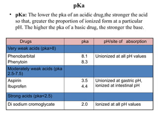
pKa
• pKa: Thelower the pka of an acidic drug,the stronger the acid
so that, greater the proportion of ionized form at a particular
pH. The higher the pka of a basic drug, the stronger the base.
Drugs pka pH/site of absorption
Very weak acids (pka>8)
Phenobarbital
Phenytoin
8.1
8.3
Unionized at all pH values
Moderately weak acids (pka
2.5-7.5)
Aspirin
Ibuprofen
3.5
4.4
Unionized at gastric pH,
ionized at intestinal pH
Strong acids (pka<2.5)
Di sodium cromoglycate 2.0 Ionized at all pH values
24.
drug pka pH/site of
absorption
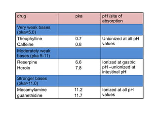
Very weak bases
(pka<5.0)
Theophylline
Caffeine
0.7
0.8
Unionized at all pH
values
Moderately weak
bases (pka 5-11)
Reserpine
Heroin
6.6
7.8
Ionized at gastric
pH –unionized at
intestinal pH
Stronger bases
(pka>11.0)
Mecamylamine
guanethidine
11.2
11.7
Ionized at all pH
values
25.
Cont.
• Comparison ofintestinal drug absorption in rat at several pH
values
pka pH=4 pH=5 pH=7 pH=8
ACIDS
Salicylic
Benzoic
3.0
4.2
64
62
35
36
30
35
10
5
BASES
Aniline
Quinine
4.6
4.2
40
09
48
11
58
41
61
54
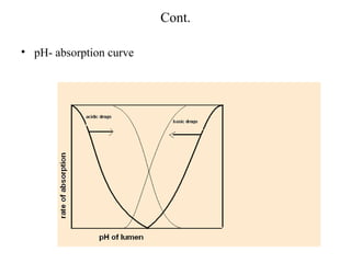
Deviation from thepH partition theory
• The deviation from the theory is observed with the deviation
of the inflection point of the pH-absorption curve.
• For a simple acid or base, the inflection point of the pH-
absorption curve should occur at a pH that is equal to the pka
of the drug.
• The three factors that may contribute to the deviations are:
1. Absorption of the ionized form of the drug.
2. Presence of an aqueous unstirred diffusion layer adjacent to
the cell membrane.
3. Difference between luminal pH and pH at the surface of the
cell membrane.
28.
Cont.
• Unstirred layer[USL]: There is usually a stagnant layer
adjacent to the GI membrane, that acts as an additional
diffusion barrier.
• It rapidly permeating substances through membrane by
diffusion, could actually be rate-limited.
• Osmotic volume flow across membranes is also affected by
USL, because the movement of the solvent will carry
dissolved solutes along with it.
• Concentration gradients of the solute induced within the USL
in both the cases: the solute permeation and osmosis
• The pH of this layer is about 5.2 to 6.2.
29.
Cont.
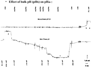
• pH- microclimate(pHm): The another factor that affects the
deviation of the pH absorption curve, which is the virtual or
microclimate pH at the surface of epical cell membrane.
• The apparent pKa values observed in the absorption-pH curve
were shifted to higher values for acids compared with the true
pKa, and to the lower values for bases.
• This deviation can be explained by the effect of an acid layer on
the apical side of cells, known as acid pH microclimate.
• It varies between 5.2- 6.2 within the different sections of intestine.
• The thickness of microclimate pH is 600-800 mm.
• For the particular segment it is very reproducible.
30.
Cont.
• The microclimate-pHhypothesis is supported by the fact that
H+
ions are secreted into intestinal lumen.
• So Na+
/H+
antiporter mechanism, dependent on cellular
metabolism, is responsible for the acid pH microclimate.
• But the maintenance of the low-pH microclimate is due to the
presence of an ampholyte, the mucus, at the surface lining of
the intestine, rather than H+
ion secretion.
• The presence and the thickness of this mucus layer alters the
pH microclimate.
• The effect of various factors on the pH microclimate is to be
studied for the prediction of the drug absorption.
Cont.
• Effect ofsodium on pHm : For the experiment the 148 meq/l
NaCl medium was taken and pHm was measured.
• Then 10 ml of that solution is replaced with isosmotic
mannitol solution and pHm was measured.
• This replacements are done seven times and NaCl
concentration was reduced from 148 to 30 meq/l and pHm was
measured every time.
• But there was no change in pHm.
• The reverse order of replacement (from 0 to 140 meq/l) also
failed to alter the pHm.
33.
Cont.
• Alteration ofpHm : Studies suggests that the maintenance of
the acidic pH is not due to H+ ion secretion but due to the
restrictive barrier, a physical structure, at the surface of the
intestine.
• A higher pHm was found, when that barrier is removed by
physical mean (vigorously washed).
• For the standardization, the knowledge of the effect of
washing and mechanical shearing is required.
• For the experiment, the rat intestine, after gentle washing,
everted on the metal rode, and then spun in phosphate buffer,
at 1000 rpm, for 30, 60 and 90 sec.
35.
Cont.
• Effect ofglucose on pHm : The effect of glucose is depended
on the initial pHm.
• When the initial pHm is low, the glucose at any concentration
does not affect the pHm.
• But when the initial pHm is high, the glucose at low
concentration can affect the pHm.
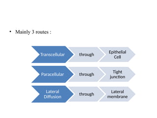
• Mainly 3routes :
Transcellular through
Epithelial
Cell
Paracellular through
Tight
junction
Lateral
Diffusion
through
Lateral
membrane
40.
Tight junction complex
•Epithelial tight junctions (TJs) are the key structures
regulating paracellular trafficking of macromolecules.
• The TJ is multiprotein complex that forms a selective
permeable seal between adjacent epithelial cells.
• It is the boundary between apical and basolateral membrane
domains.
• TJs are the multiple protein complexes, located at the apical
ends of the lateral membranes of intestinal epithelial cells.
• They have pores that can allow small molecules (MW < 200
Da) to diffuse through in the aqueous solution.
• In the jejunum, the pores are about 7 - 9 Å in size.
• In the ileum the junctions are tighter, and pores are about 3 - 4
Å in size.
42.
Cont.
• The TJcomplex consists of transmembrane and intracellular
scaffold proteins.
• The transmembrane proteins (claudins, occludin, and junctional
adhesion molecules [JAMs]) create a permselective barrier in the
paracellular pathways.
• The actual cell-cell adhesions occur in the adheren junctions,
located further away from the apical side.
• Apparently three calciums contiguously link 10-residue portions
of cadheren proteins spanning from two adjoining cell walls.
• Calcium-binding agents can open the junctions by interactions
with the cadheren complex.


![Cont.
• Fick’s first law: The amount of material flowing through a unit
cross section, h, of a barrier in unit time, t, is known as the
flux, J.
J = DmdCm/dx = Dm[ Cmᴼ- Cmᴴ] / h …..(1)
• where J is the flux, where Cmᴼ and Cmᴴ are the
concentrations of the uncharged form of the solute within the
membrane at the two water- membrane boundaries (at
positions x = 0 and x = h in where h is the thickness of the
membrane), and where Dm is the diffusivity of the solute
within the membrane.
• But because of inconvenient measurement of the concentration
of solute within different parts of membrane, the considering
the distribution coefficient between bulk water and membrane,
Kd.
J = DmKd(CD- CA) / h .....(2)
• This eq.(2) is used to predicts, how quikly the molecule pass
through simple membrane](https://image.slidesharecdn.com/transportmodel-250402044816-ccfaa121/85/Transport-Model-pptx-7-320.jpg)
![Cont.
• Charge state: The charge state or ionization of the molecules
depended on the pH, and which affects the solubility and the
permeation of the molecules.
• pKa, an ionization constant, a pH at which a substance
exists as 50% ionized form and 50% unionized form, it is
relating the pH to the charge state of a molecule, and it can
predict the absorption, distribution and elimination of the drug
molecules.
• For acidic drugs,
pH= pKa + log [ionized form] / [unionized form]
• For basic drugs,
pH= pka + log [unionized form] / [ionized form]
• So for eg, for acidic drug, if the pH is higher than the pKa,
there will be more ionized form, and if it is lower, then there
will more unionized form present](https://image.slidesharecdn.com/transportmodel-250402044816-ccfaa121/85/Transport-Model-pptx-9-320.jpg)


![Cont.
• Unstirred layer [USL]: There is usually a stagnant layer
adjacent to the GI membrane, that acts as an additional
diffusion barrier.
• It rapidly permeating substances through membrane by
diffusion, could actually be rate-limited.
• Osmotic volume flow across membranes is also affected by
USL, because the movement of the solvent will carry
dissolved solutes along with it.
• Concentration gradients of the solute induced within the USL
in both the cases: the solute permeation and osmosis
• The pH of this layer is about 5.2 to 6.2.](https://image.slidesharecdn.com/transportmodel-250402044816-ccfaa121/85/Transport-Model-pptx-28-320.jpg)


![Cont.
• The TJ complex consists of transmembrane and intracellular
scaffold proteins.
• The transmembrane proteins (claudins, occludin, and junctional
adhesion molecules [JAMs]) create a permselective barrier in the
paracellular pathways.
• The actual cell-cell adhesions occur in the adheren junctions,
located further away from the apical side.
• Apparently three calciums contiguously link 10-residue portions
of cadheren proteins spanning from two adjoining cell walls.
• Calcium-binding agents can open the junctions by interactions
with the cadheren complex.](https://image.slidesharecdn.com/transportmodel-250402044816-ccfaa121/85/Transport-Model-pptx-42-320.jpg)


