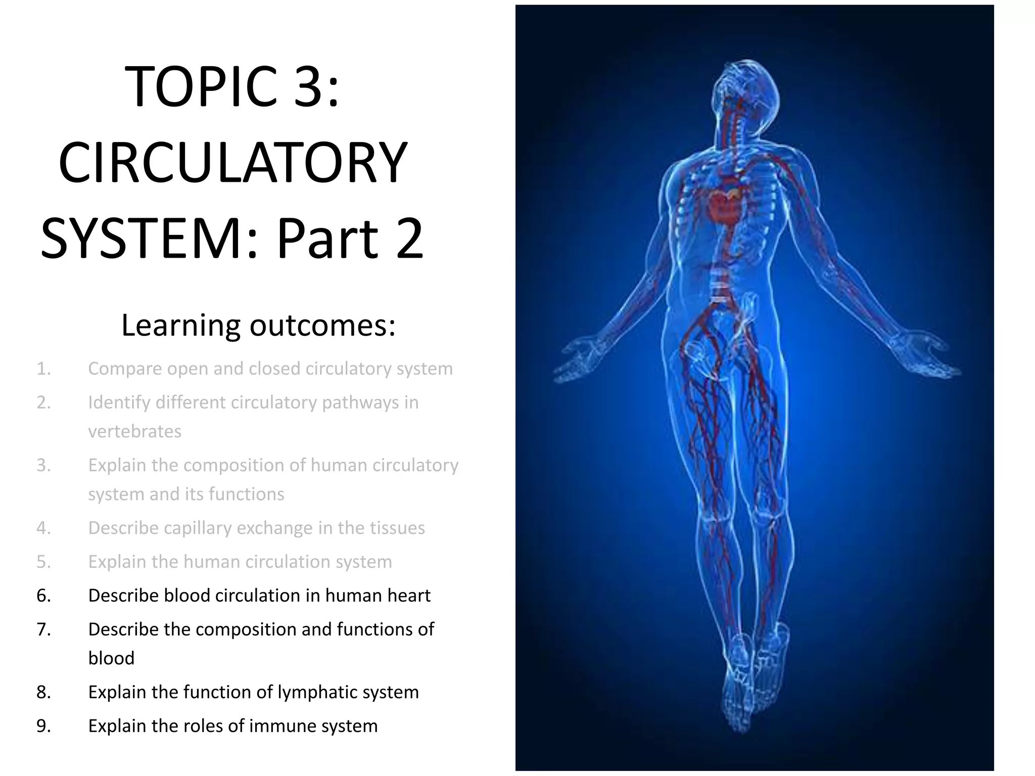
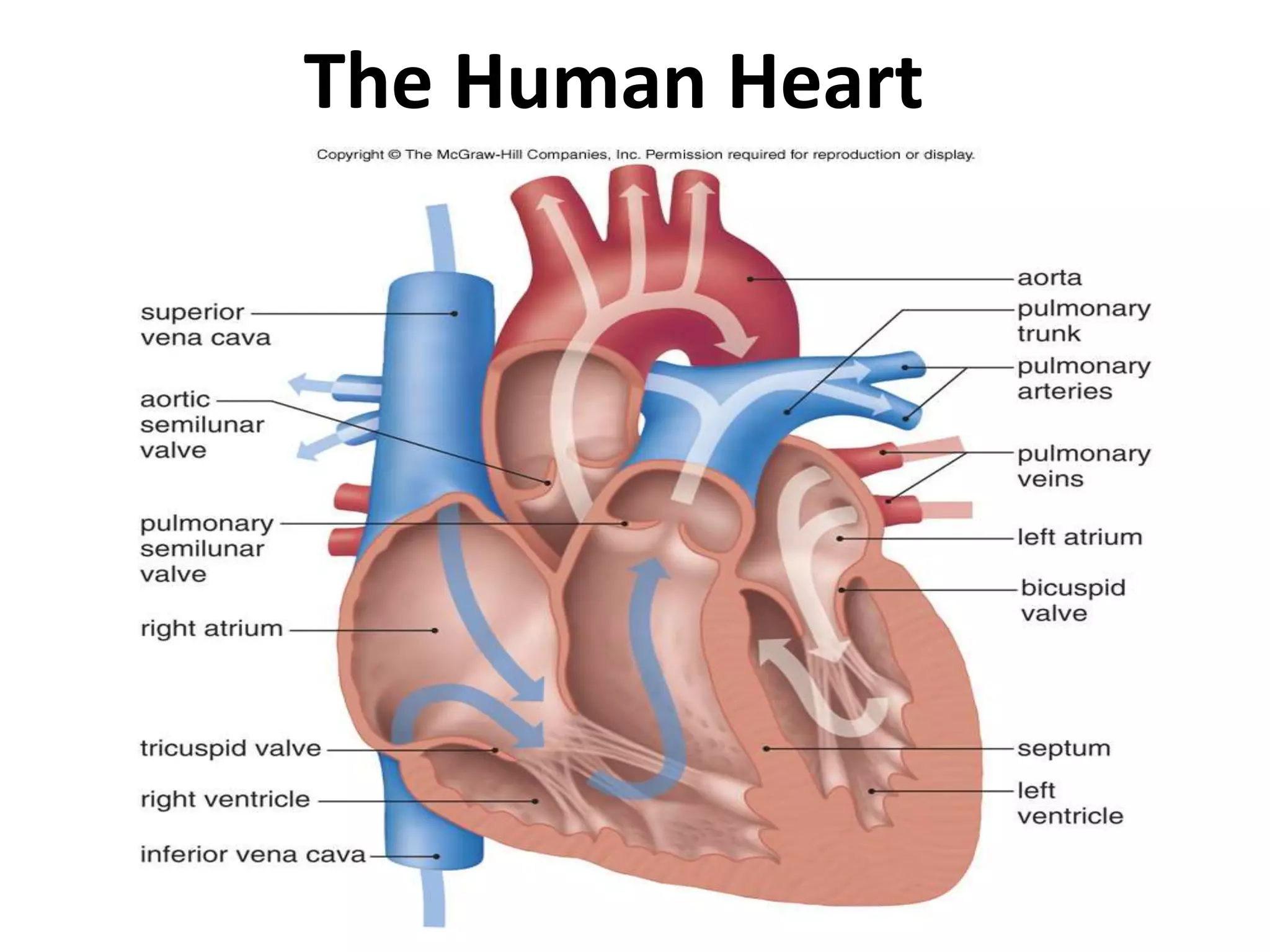
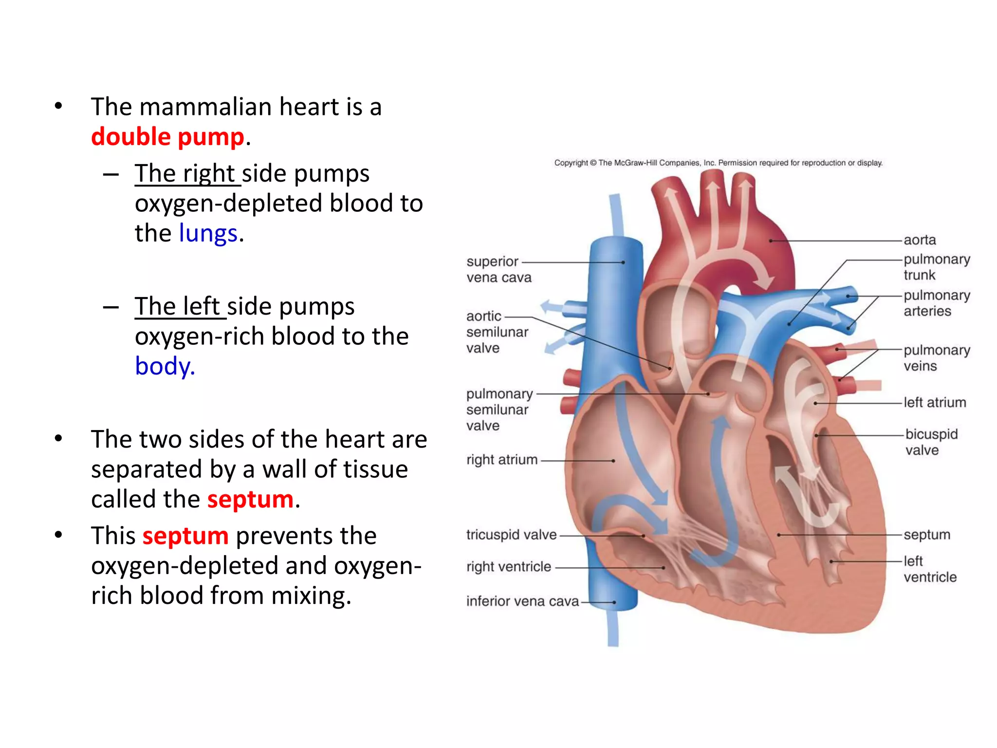
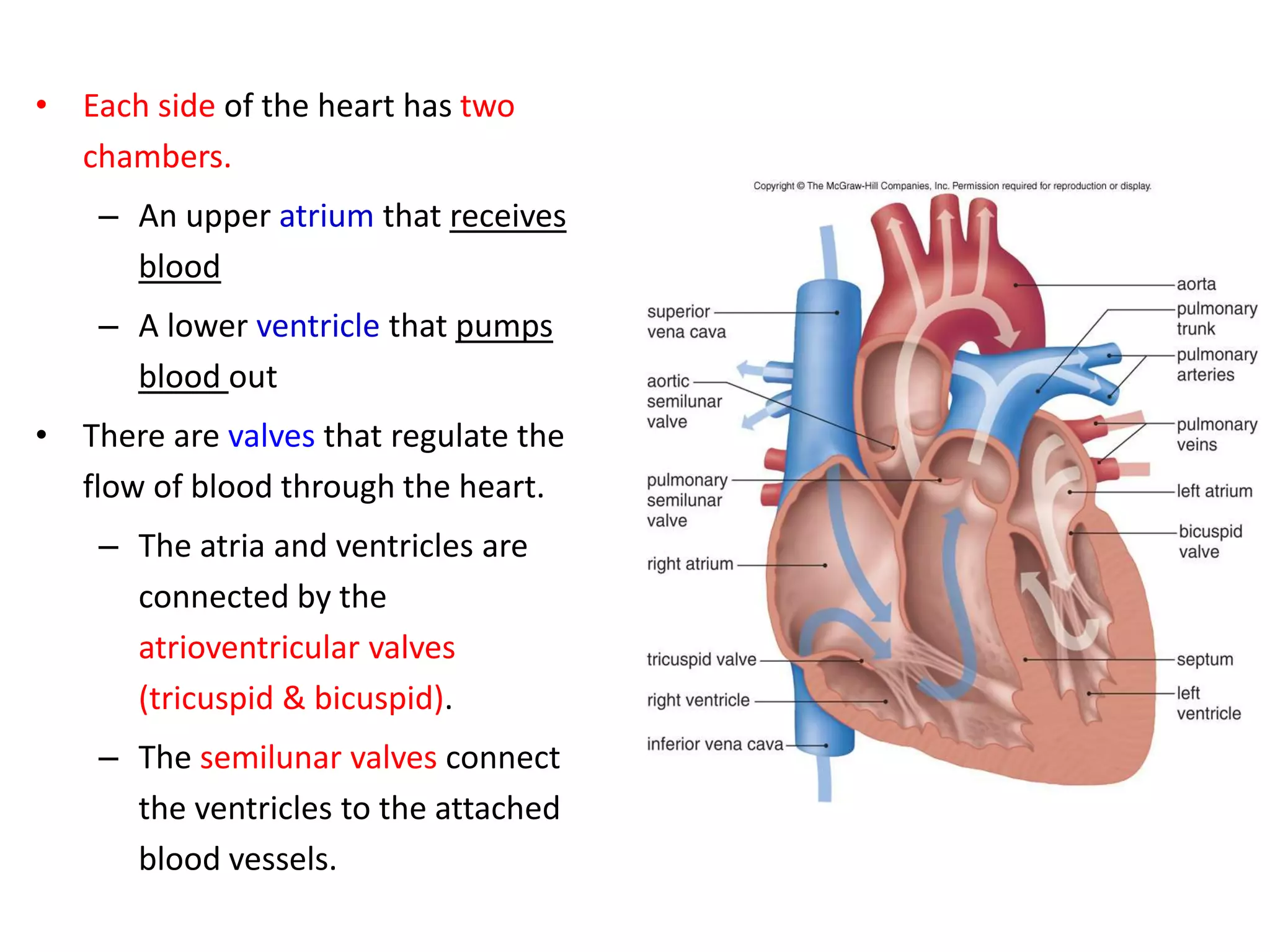
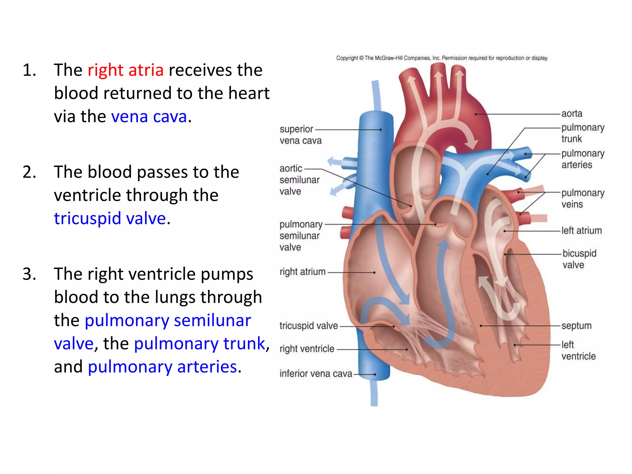
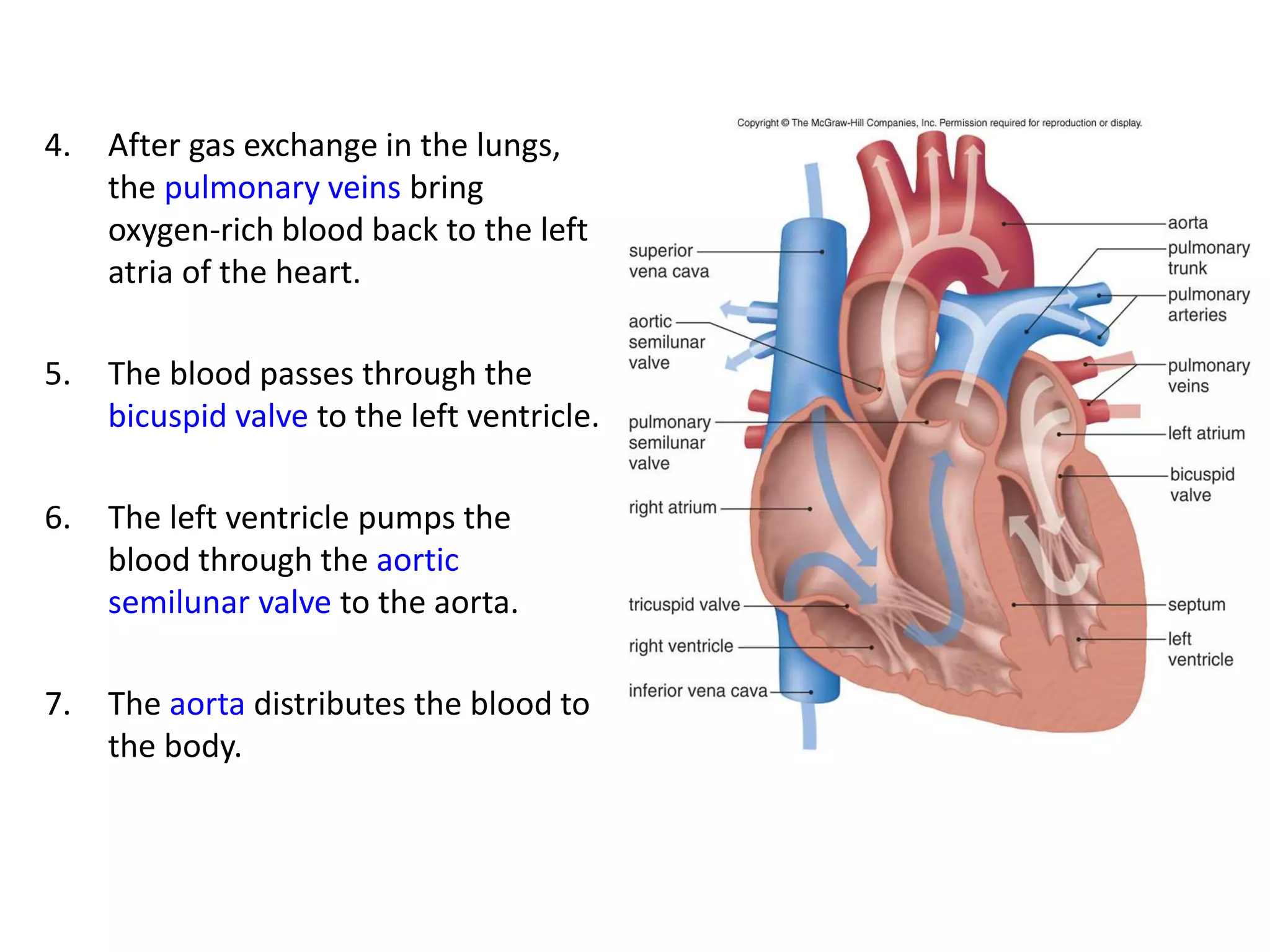
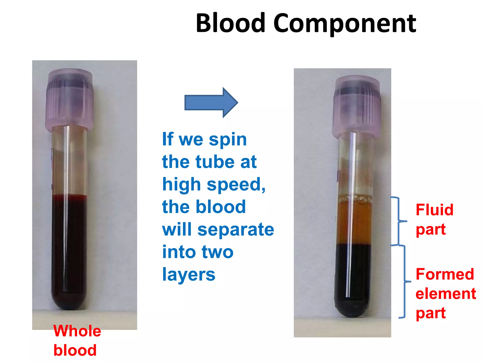
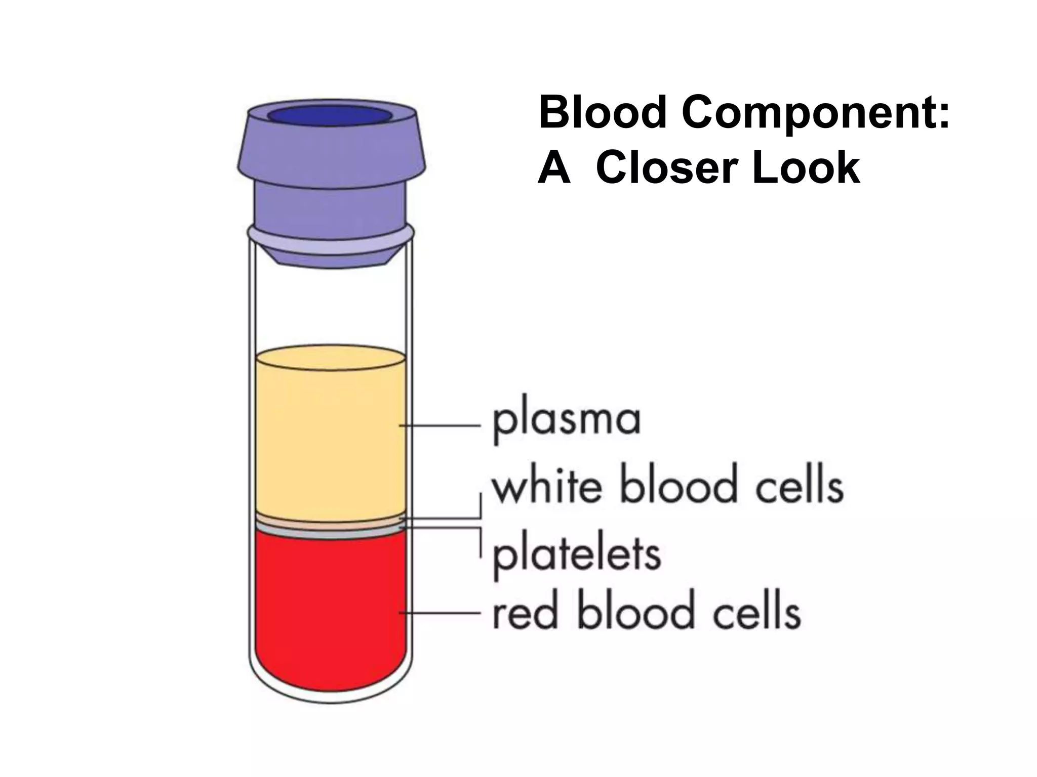
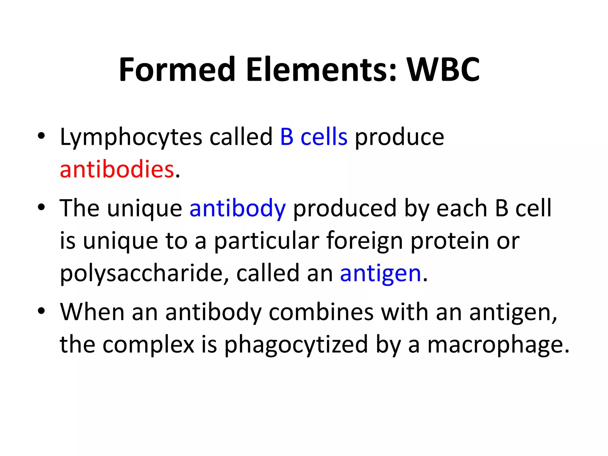
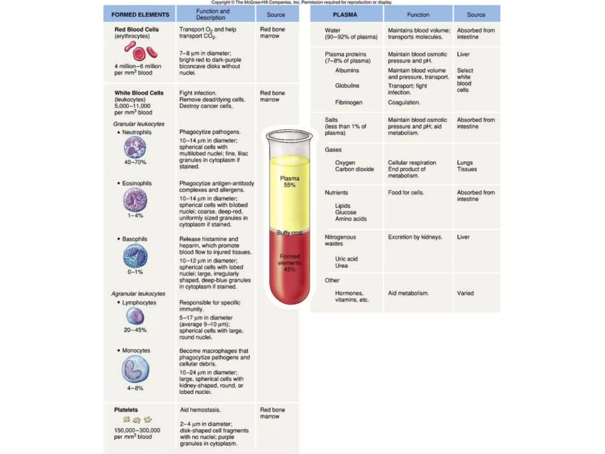
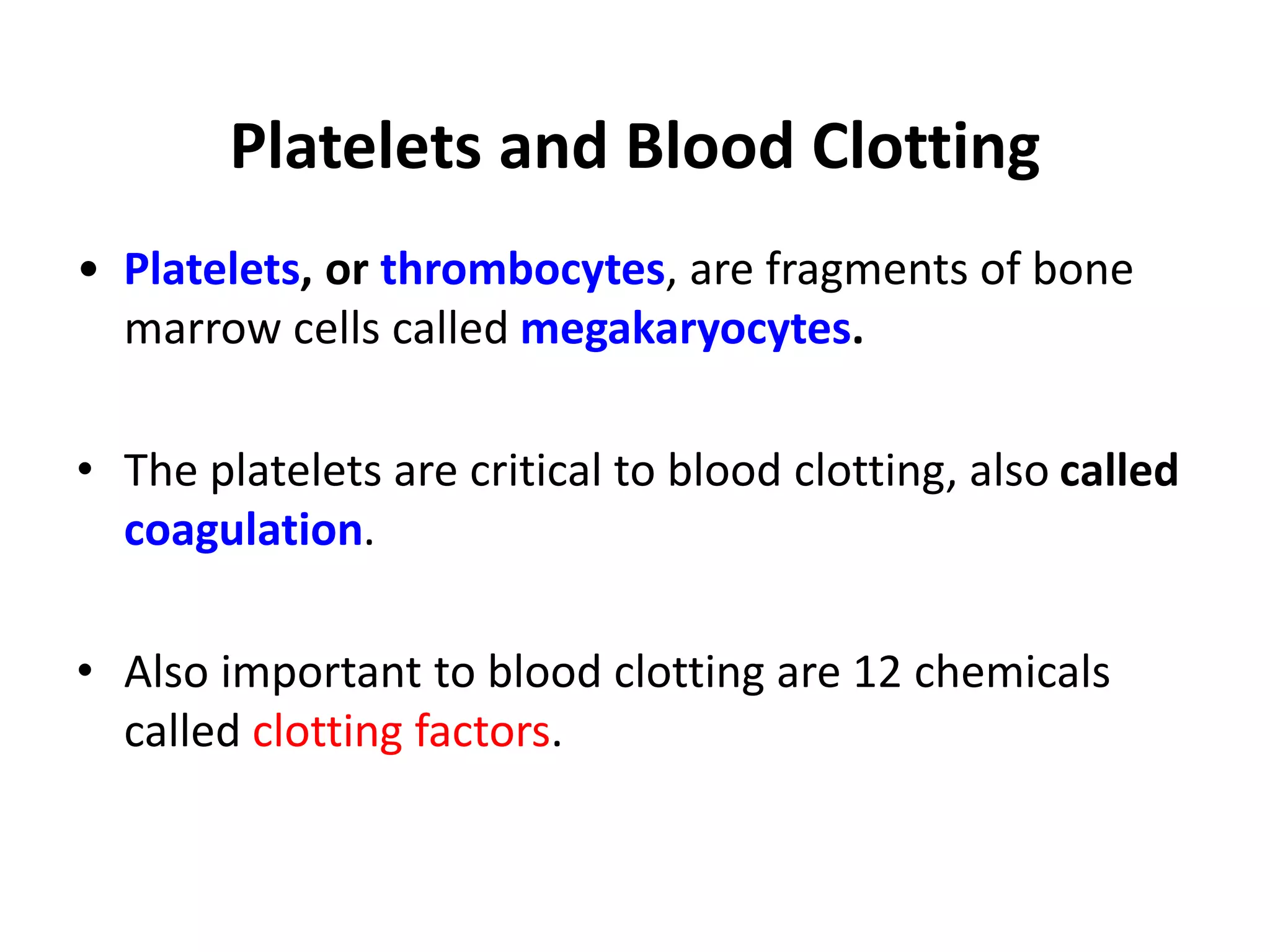
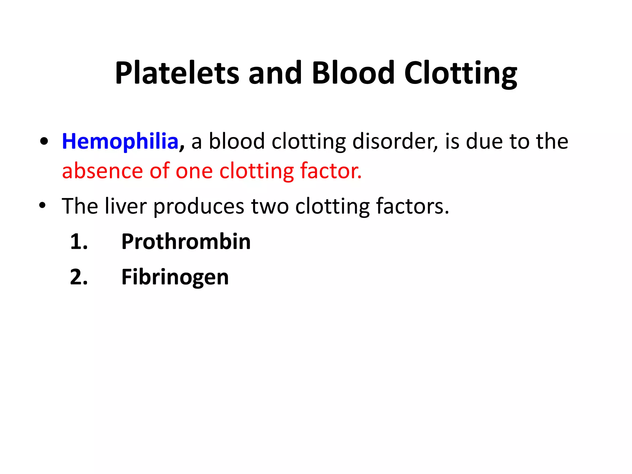
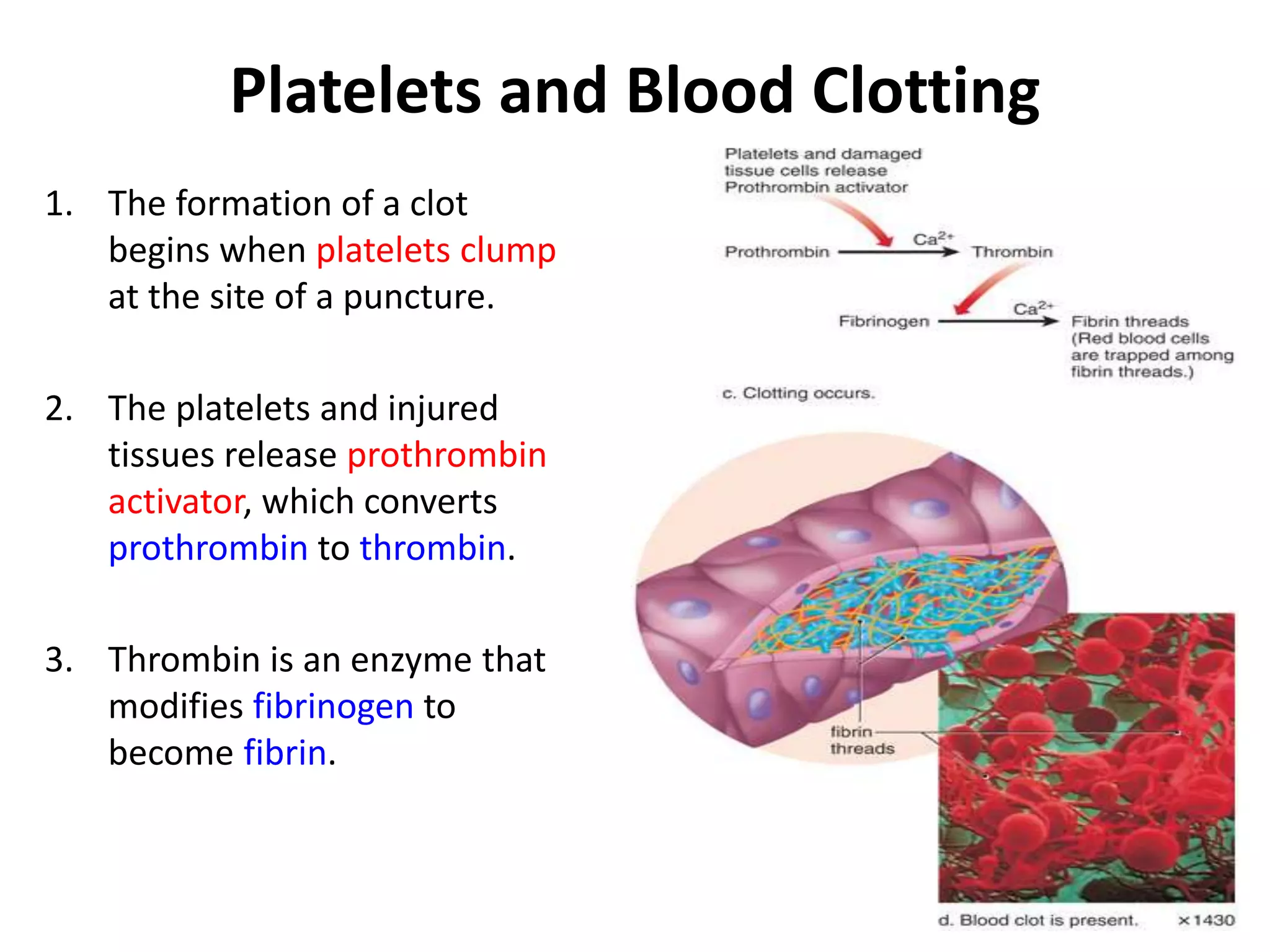
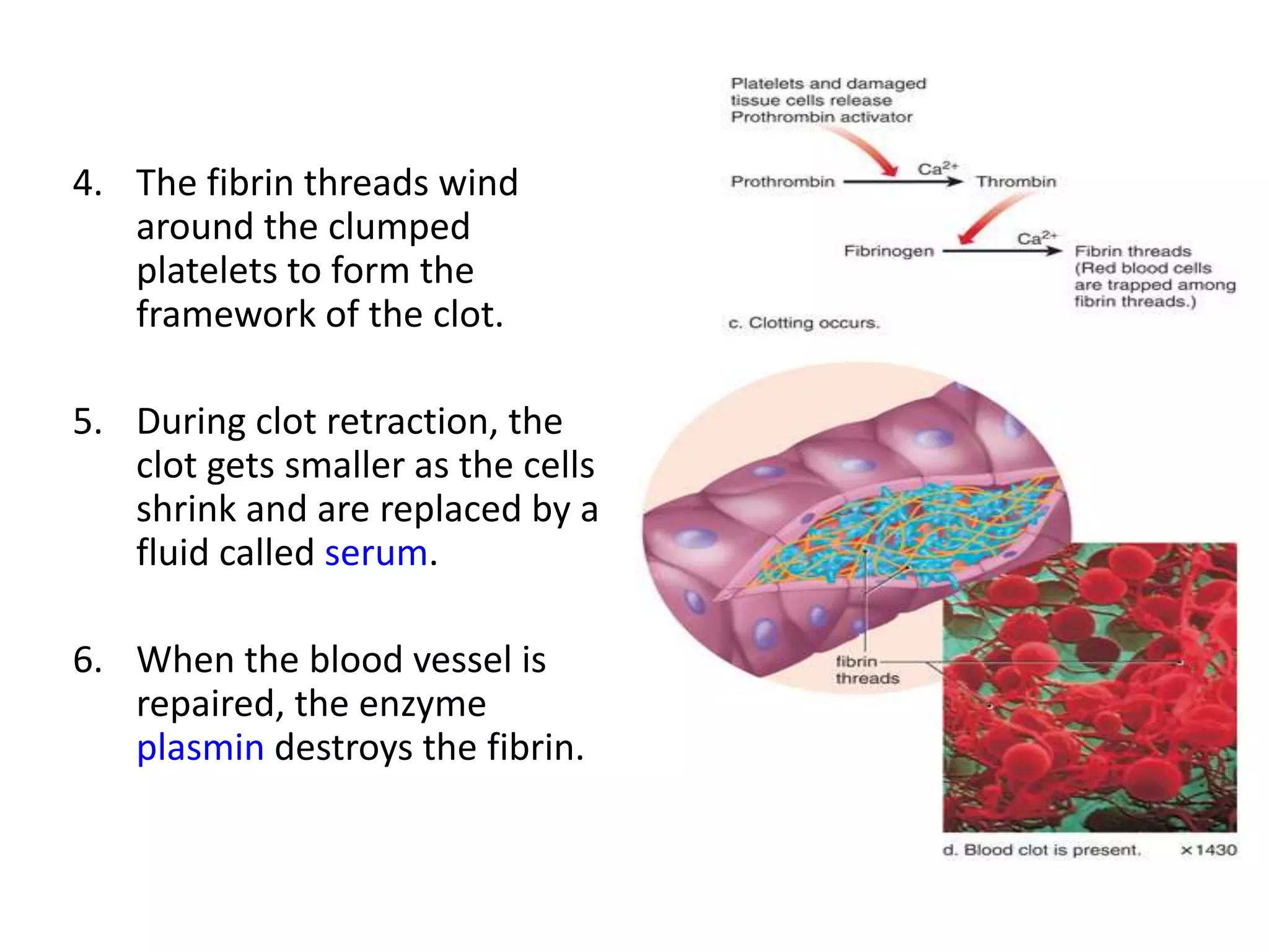
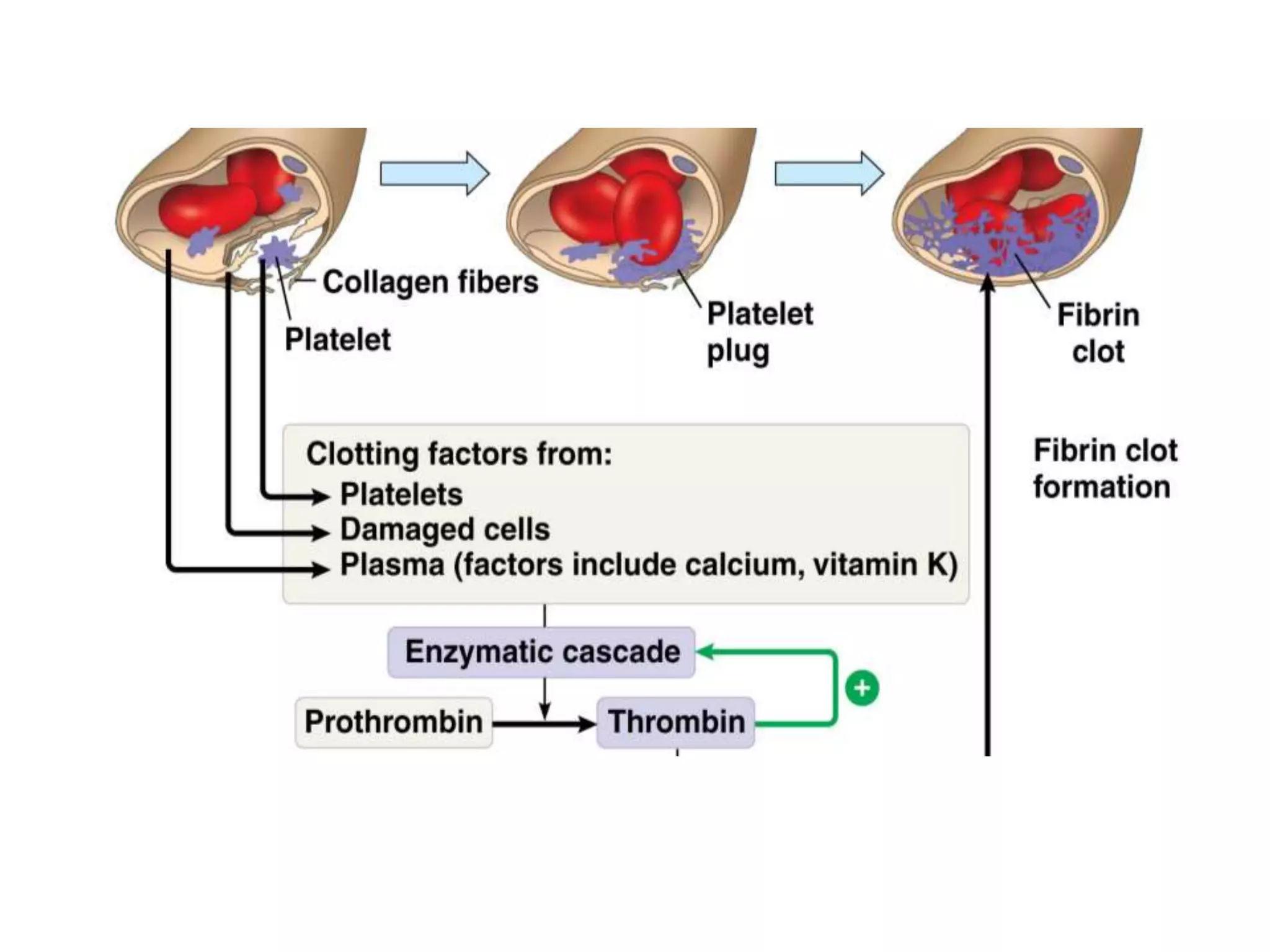
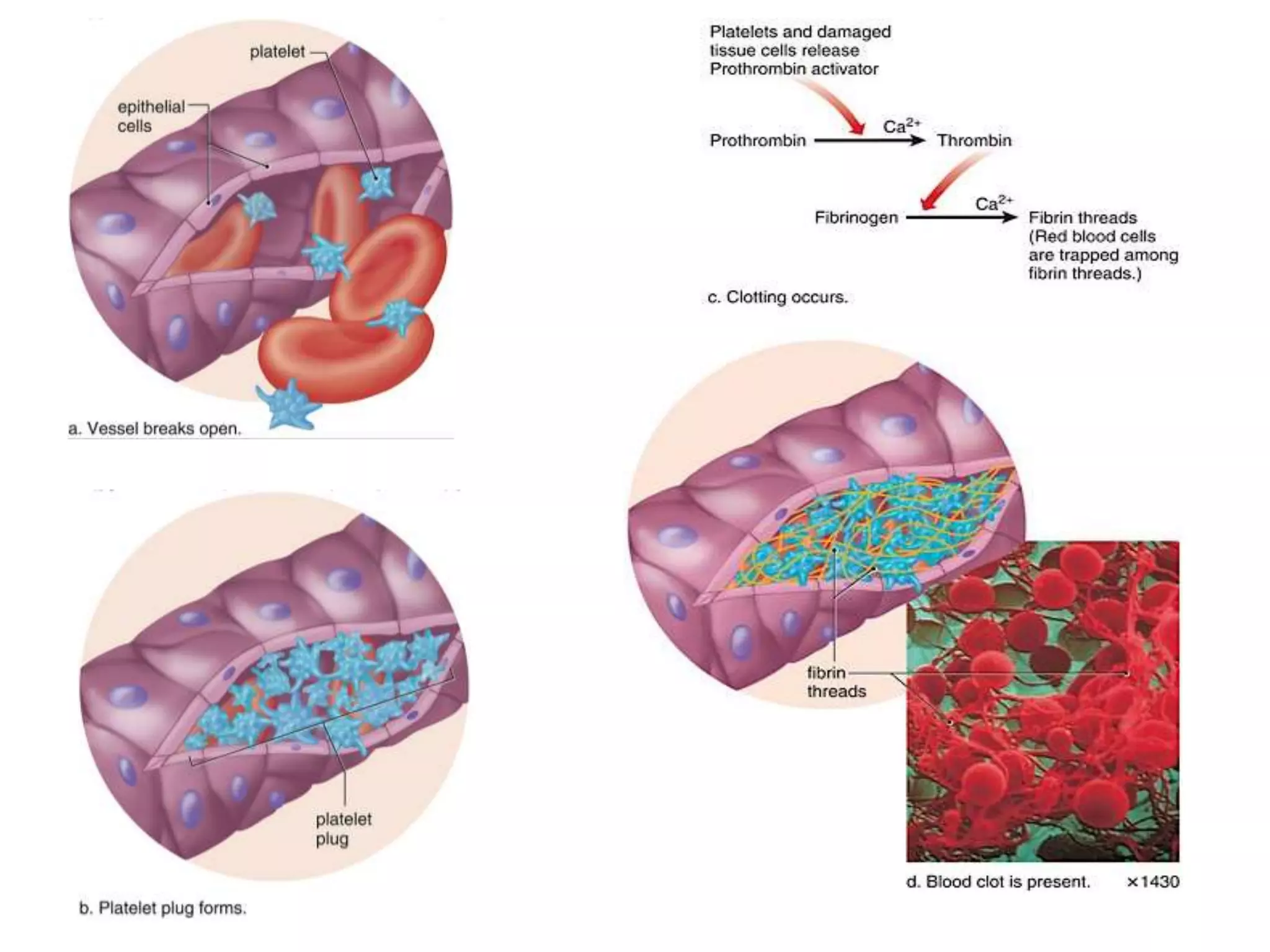
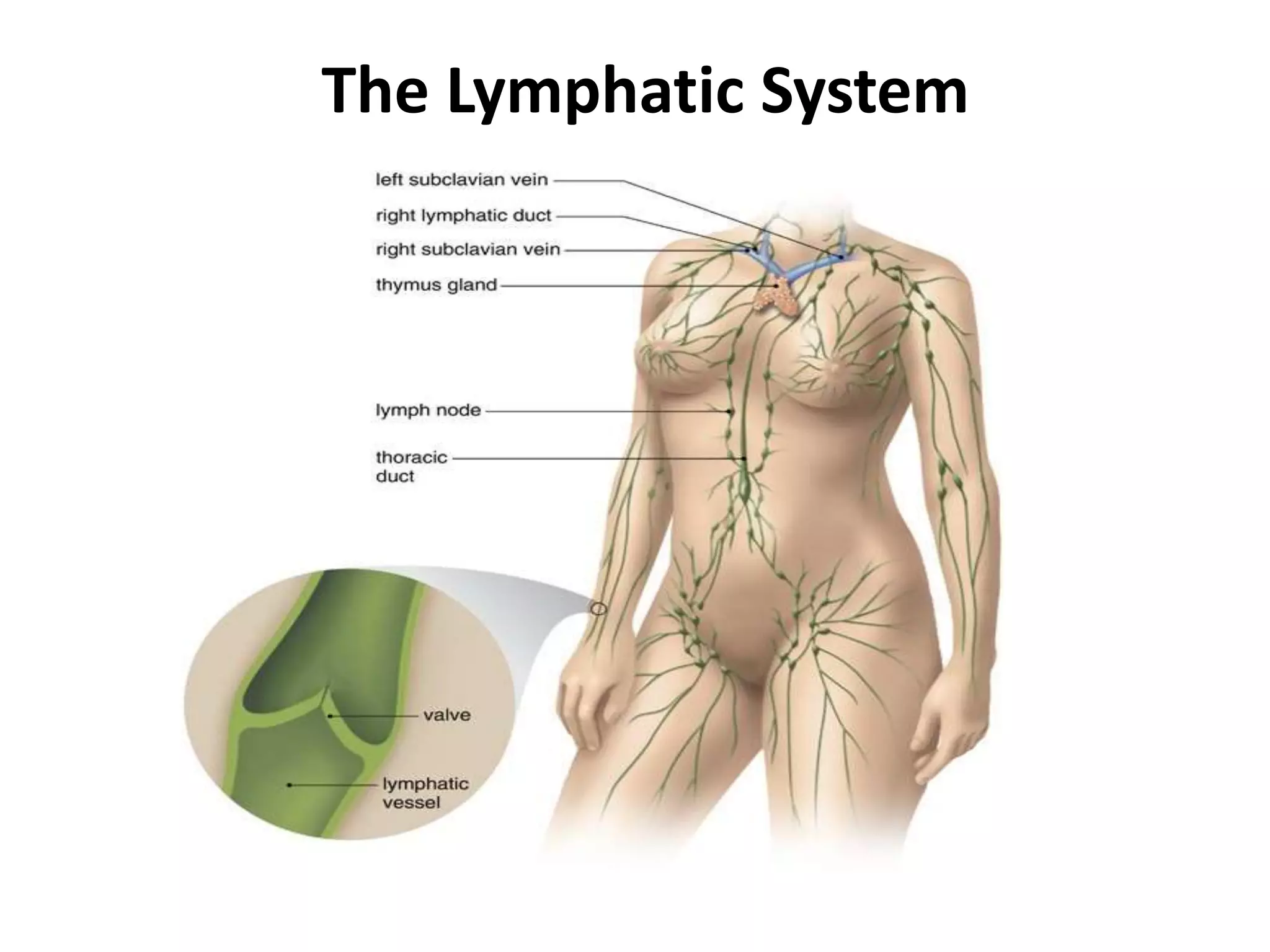
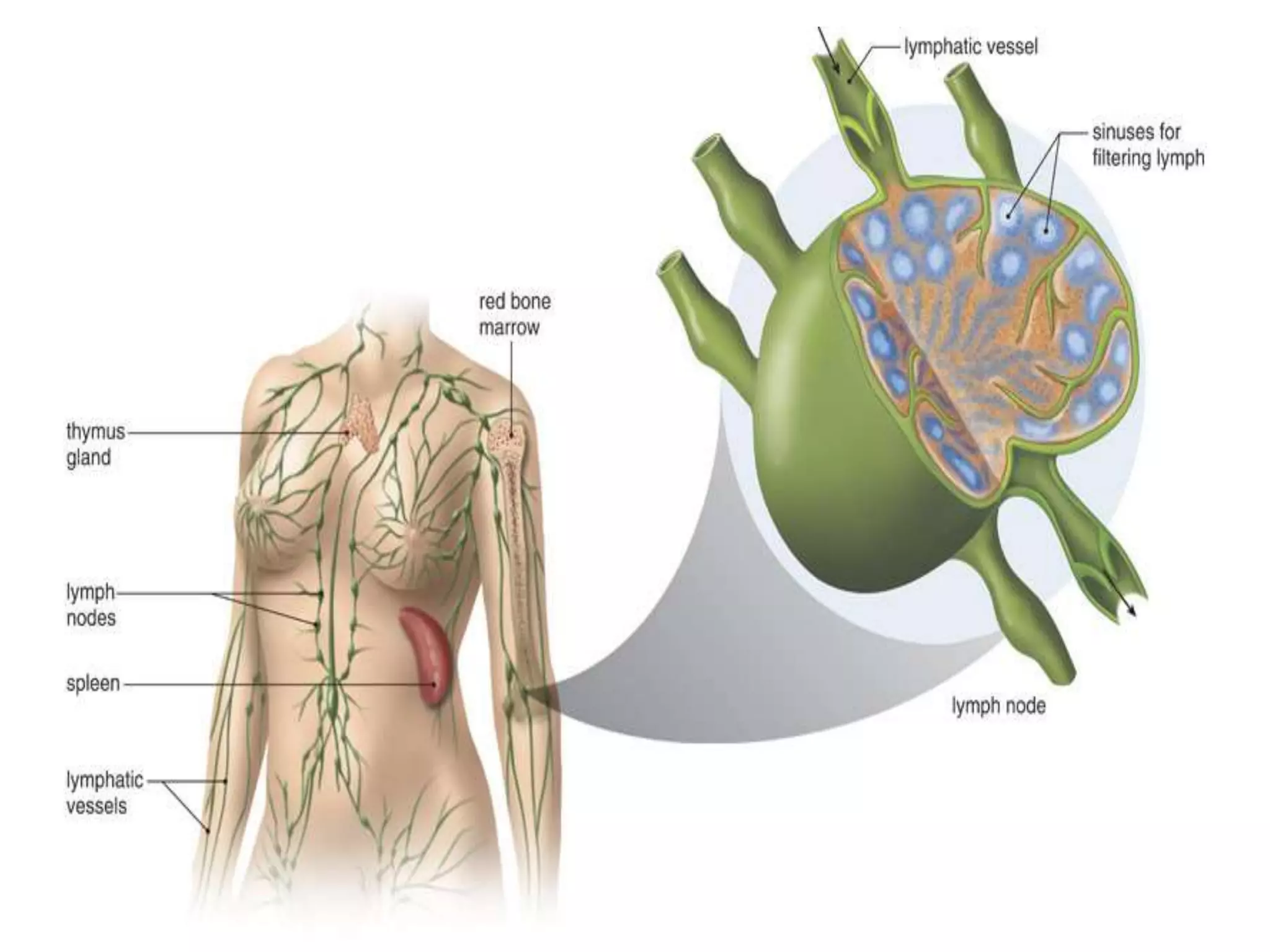
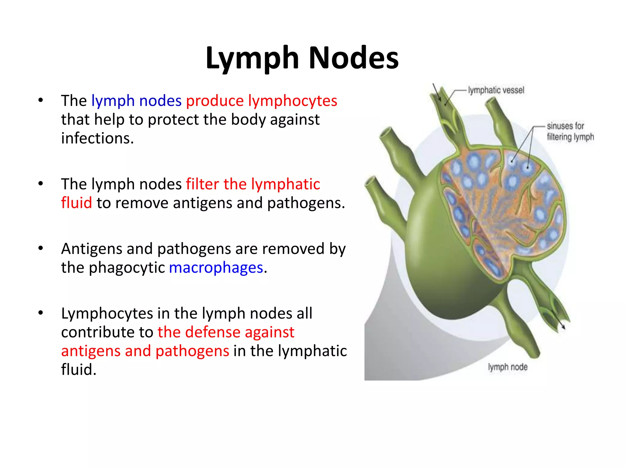
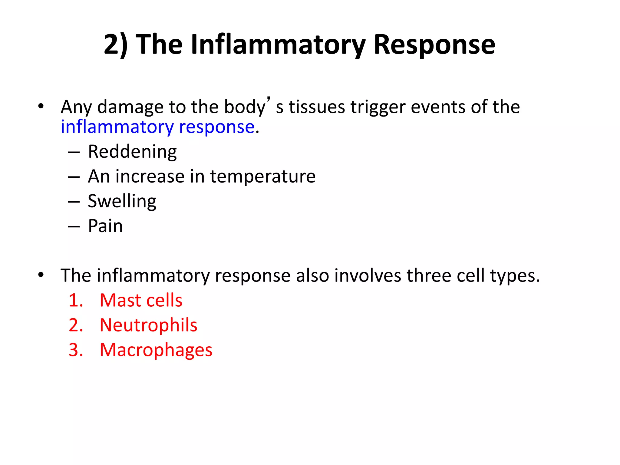
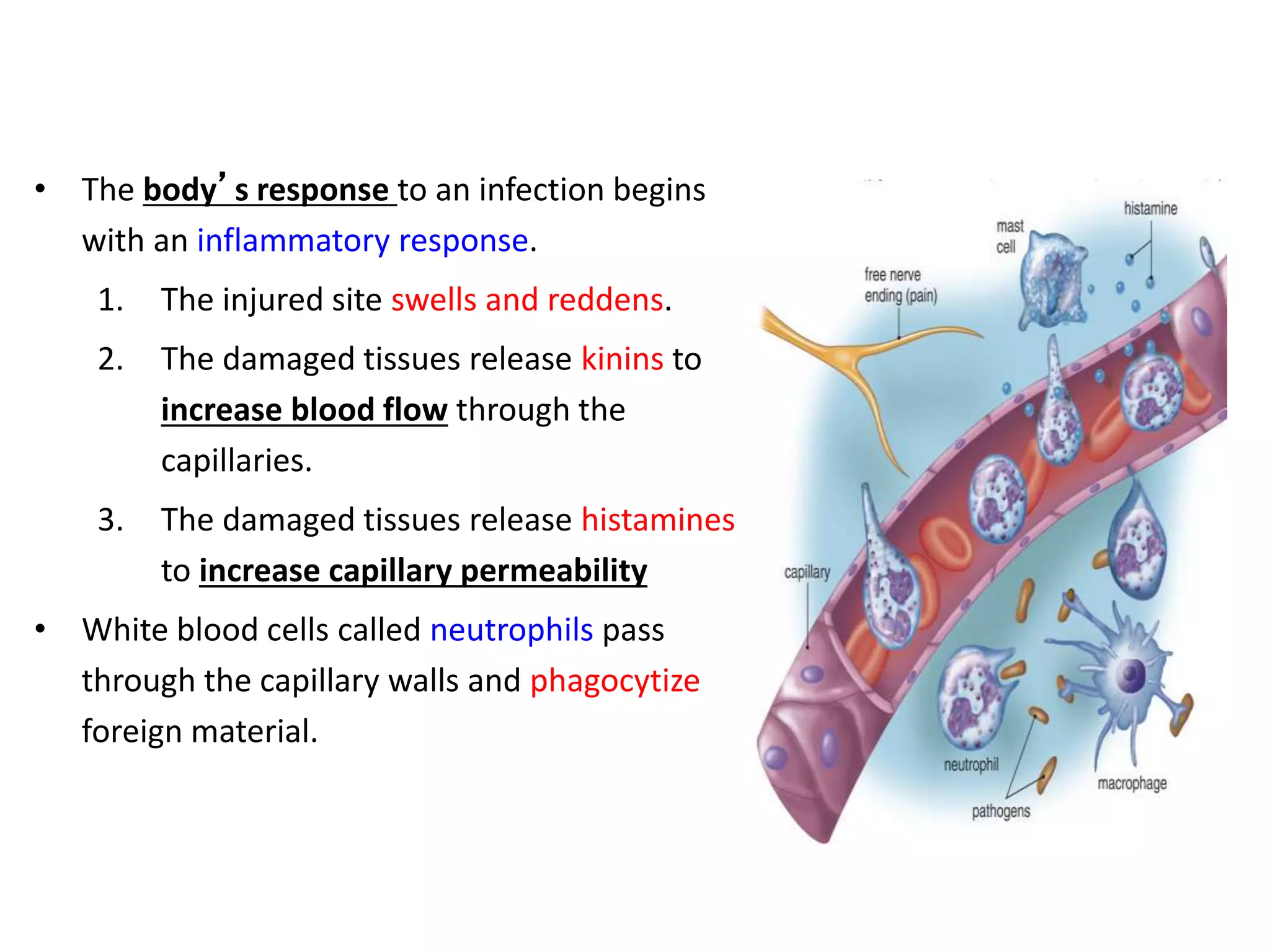
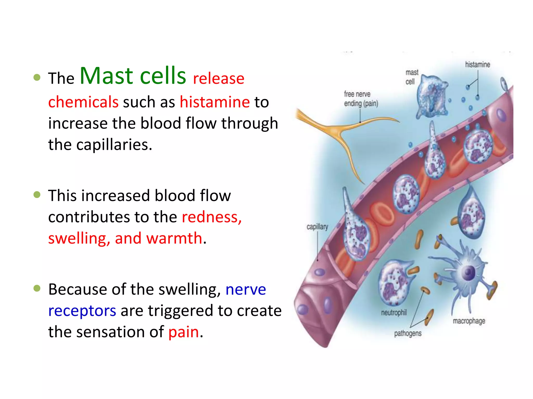
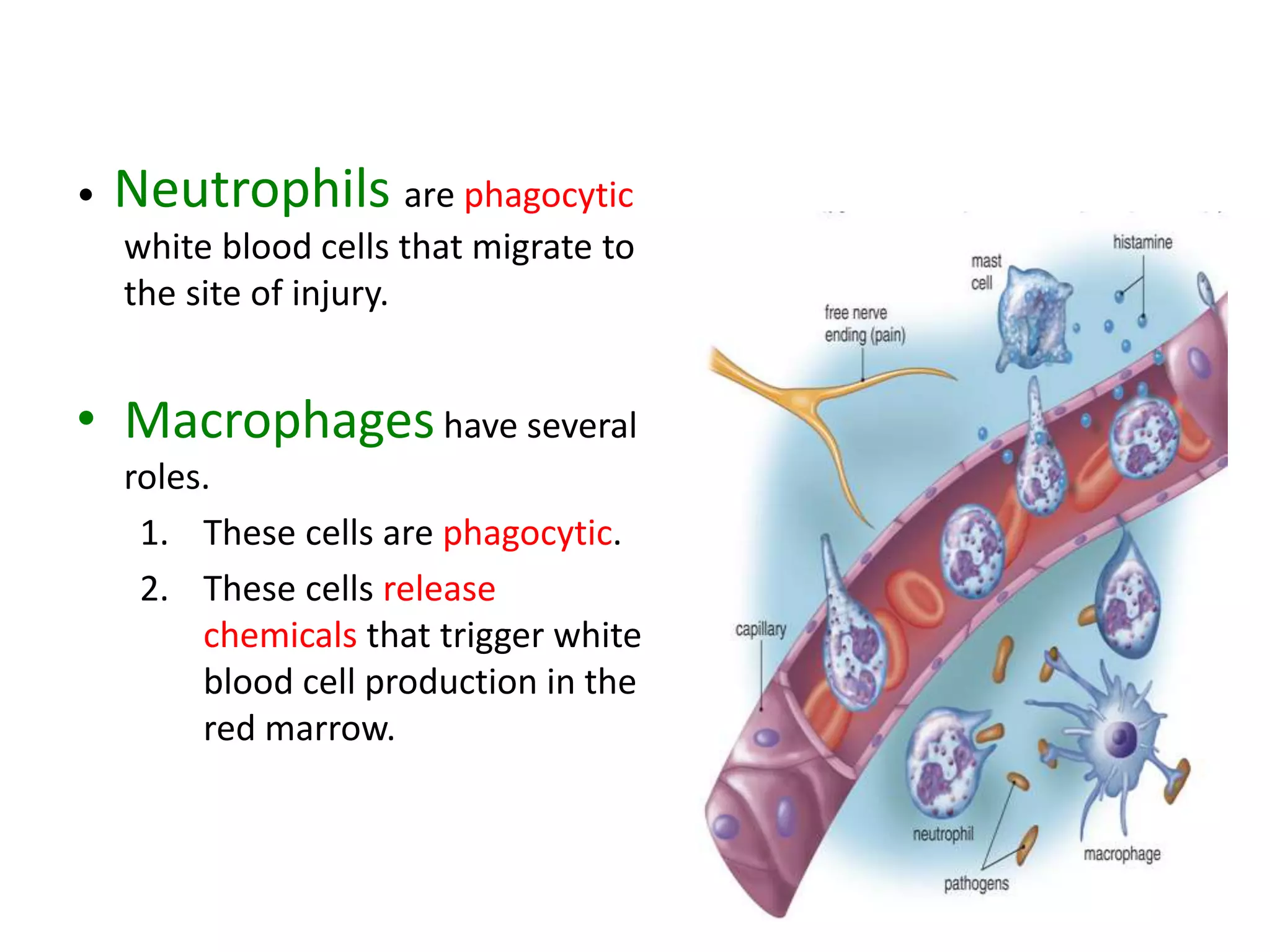
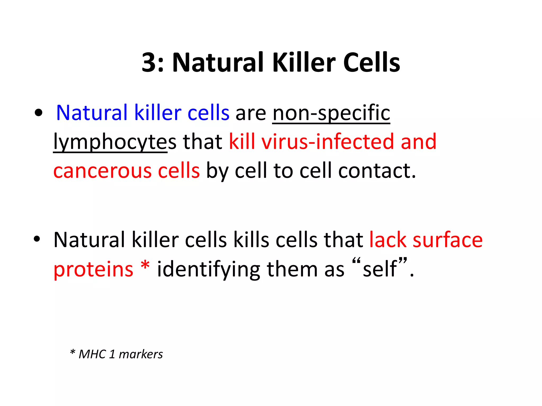
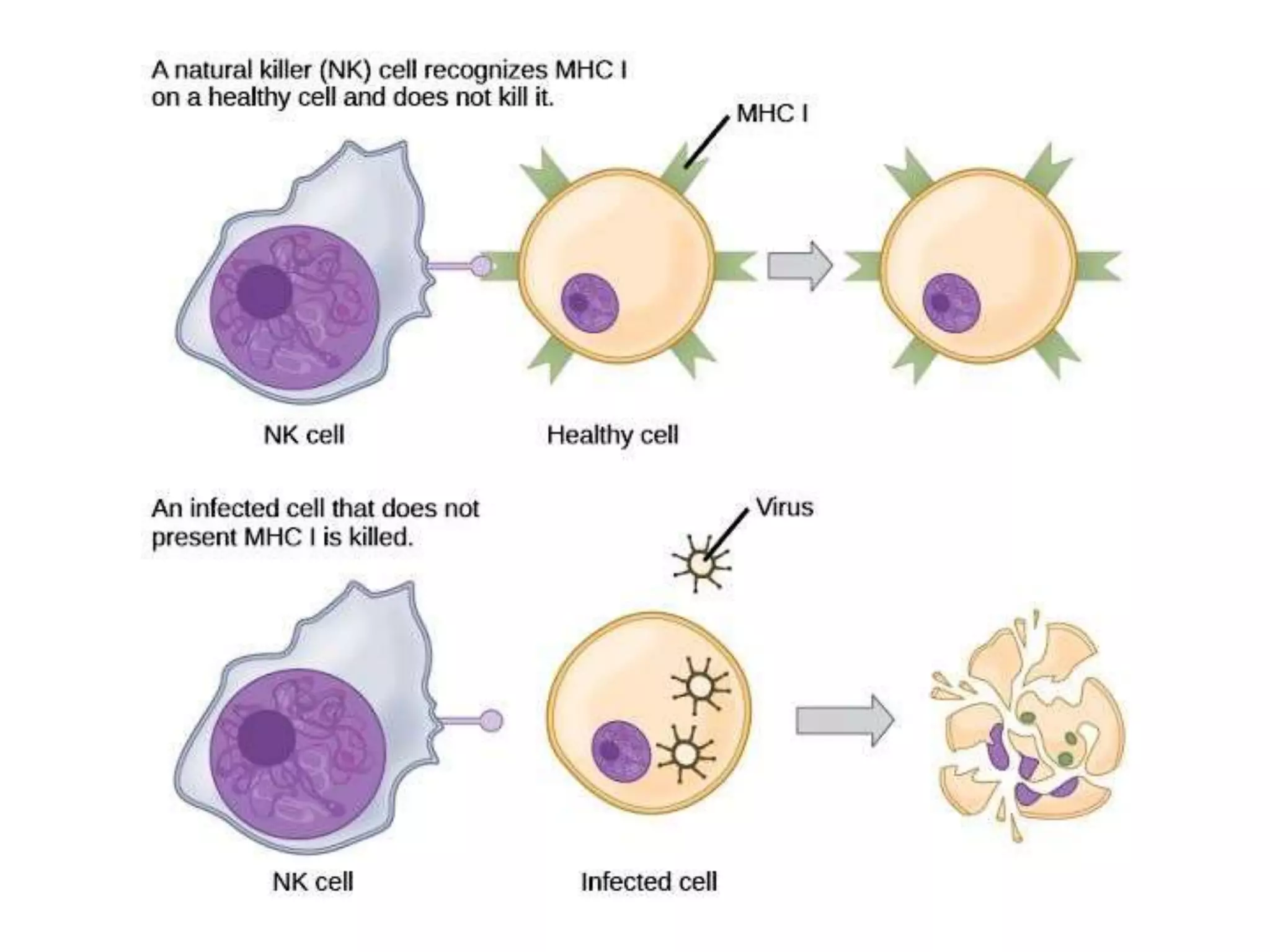
TOPIC 3:
CIRCULATORY
SYSTEM: Part 2
Learning outcomes:
1. Compare open and closed circulatory system
2. Identify different circulatory pathways in
vertebrates
3. Explain the composition of human circulatory
system and its functions
4. Describe capillary exchange in the tissues
5. Explain the human circulation system
6. Describe blood circulation in human heart
7. Describe the composition and functions of
blood
8. Explain the function of lymphatic system
9. Explain the roles of immune system
Peer to Peer
1. Heart and Blood flow
2. Blood and its elements
3. Platelet and blood clotting
4. Lymphatic systems
5. Non specific defenses
The Human Heart
• The mammalian heart is a
double pump.
– The right side pumps
oxygen-depleted blood to
the lungs.
– The left side pumps
oxygen-rich blood to the
body.
• The two sides of the heart are
separated by a wall of tissue
called the septum.
• This septum prevents the
oxygen-depleted and oxygen-
rich blood from mixing.
• Each side of the heart has two
chambers.
– An upper atrium that receives
blood
– A lower ventricle that pumps
blood out
• There are valves that regulate the
flow of blood through the heart.
– The atria and ventricles are
connected by the
atrioventricular valves
(tricuspid & bicuspid).
– The semilunar valves connect
the ventricles to the attached
blood vessels.
1. The right atria receives the
blood returned to the heart
via the vena cava.
2. The blood passes to the
ventricle through the
tricuspid valve.
3. The right ventricle pumps
blood to the lungs through
the pulmonary semilunar
valve, the pulmonary trunk,
and pulmonary arteries.
4. After gas exchange in the lungs,
the pulmonary veins bring
oxygen-rich blood back to the left
atria of the heart.
5. The blood passes through the
bicuspid valve to the left ventricle.
6. The left ventricle pumps the
blood through the aortic
semilunar valve to the aorta.
7. The aorta distributes the blood to
the body.
Blood: A Transport Medium
• The blood serves several functions.
1. Blood serves as a transport medium to
capillaries for fluid and solute exchange.
2. The blood distributes components of the
immune system.
3. The blood helps regulate body temperature.
4. The blood can form clots to prevent excessive
blood losses from wounds.
Blood Component
Whole
blood
If we spin
the tube at
high speed,
the blood
will separate
into two
layers
Fluid
part
Formed
element
part
Blood Component:
A Closer Look
Plasma
• Plasma consists of two main components.
–Water (>90%)
–Protein (<10%)
• The plasma also contains nutrients, wastes,
and salts.
• The salts help buffer the blood pH and
maintain the osmotic potential of the blood.
Formed Elements: RBC
• There are three types of formed elements.
1. Red blood cells
2. White blood cells
3. Platelets
• The red blood cells are small cells that contain
hemoglobin for oxygen transport.
• A decrease in the number of red blood cells in
the body can lead to anemia.
Formed Elements: RBC
• Red blood cells are produced by the marrow
of the skull, ribs, vertebrae, and long bones.
• The kidneys secrete erythropoietin, which
stimulates red blood cell production.
• As red blood cells are released from the
bones, they become specialized for oxygen
transport.
• Red blood cells are eventually recycled to
produce new red blood cells.
Formed Elements: RBC
Formed Elements: WBC
• The white blood cells that fight infection are
also called leukocytes.
• White blood cells are found not only in the
blood but in the lymphatic system.
• The life span of white blood cells varies from a
few days to years.
Formed Elements: WBC
• White blood cells called monocytes are
transformed into macrophages, another type
of phagocytizing cells.
• Macrophages also release growth factors that
increase the number of white blood cells
called lymphocytes.
• Lymphocytes called T cells specialize in the
destruction of virus-infected cells.
Formed Elements: WBC
• Lymphocytes called B cells produce
antibodies.
• The unique antibody produced by each B cell
is unique to a particular foreign protein or
polysaccharide, called an antigen.
• When an antibody combines with an antigen,
the complex is phagocytized by a macrophage.
Platelets and Blood Clotting
• Platelets, or thrombocytes, are fragments of bone
marrow cells called megakaryocytes.
• The platelets are critical to blood clotting, also called
coagulation.
• Also important to blood clotting are 12 chemicals
called clotting factors.
Platelets and Blood Clotting
• Hemophilia, a blood clotting disorder, is due to the
absence of one clotting factor.
• The liver produces two clotting factors.
1. Prothrombin
2. Fibrinogen
Platelets and Blood Clotting
1. The formation of a clot
begins when platelets clump
at the site of a puncture.
2. The platelets and injured
tissues release prothrombin
activator, which converts
prothrombin to thrombin.
3. Thrombin is an enzyme that
modifies fibrinogen to
become fibrin.
4. The fibrin threads wind
around the clumped
platelets to form the
framework of the clot.
5. During clot retraction, the
clot gets smaller as the cells
shrink and are replaced by a
fluid called serum.
6. When the blood vessel is
repaired, the enzyme
plasmin destroys the fibrin.
The Lymphatic System
• The lymphatic system has several functions in the
human body.
1. This system returns excess fluid (lymph) from the
tissues to the heart.
2. Fat is taken up from the digestive tract.
3. The lymphatic system works with the immune
system.
• The lymphatic vessels are an extensive network of
ducts and vessels throughout the body.
The Lymphatic System
Lymph Nodes
• The lymph nodes produce lymphocytes
that help to protect the body against
infections.
• The lymph nodes filter the lymphatic
fluid to remove antigens and pathogens.
• Antigens and pathogens are removed by
the phagocytic macrophages.
• Lymphocytes in the lymph nodes all
contribute to the defense against
antigens and pathogens in the lymphatic
fluid.
Role of Blood and Lymphatic
Components
in Immune System
Nonspecific Defenses
• The body has a variety of nonspecific defenses
that help protect the body from infection.
1. Barriers to entry
2. The inflammatory response
3. Natural killer cells
1) Barriers to Entry
• There are two primary barriers that prevent entry of
pathogens into the body.
1. The skin- outer layer of skin, harden die/dead cells
form, a protecting barrier against invasion by
pathogen.
2. The mucous membranes- Nasal cavity & trachea
secreted mucous to trap the dust article& spore
• In addition to mechanically blocking pathogen entry,
the skin has oil glands secrete chemicals to counter
pathogens.
2) The Inflammatory Response
• Any damage to the body’s tissues trigger events of the
inflammatory response.
– Reddening
– An increase in temperature
– Swelling
– Pain
• The inflammatory response also involves three cell types.
1. Mast cells
2. Neutrophils
3. Macrophages
• The body’s response to an infection begins
with an inflammatory response.
1. The injured site swells and reddens.
2. The damaged tissues release kinins to
increase blood flow through the
capillaries.
3. The damaged tissues release histamines
to increase capillary permeability
• White blood cells called neutrophils pass
through the capillary walls and phagocytize
foreign material.
 The Mast cells release
chemicals such as histamine to
increase the blood flow through
the capillaries.
 This increased blood flow
contributes to the redness,
swelling, and warmth.
 Because of the swelling, nerve
receptors are triggered to create
the sensation of pain.
• Neutrophils are phagocytic
white blood cells that migrate to
the site of injury.
• Macrophageshave several
roles.
1. These cells are phagocytic.
2. These cells release
chemicals that trigger white
blood cell production in the
red marrow.
3: Natural Killer Cells
• Natural killer cells are non-specific
lymphocytes that kill virus-infected and
cancerous cells by cell to cell contact.
• Natural killer cells kills cells that lack surface
proteins * identifying them as “self”.
* MHC 1 markers
Topic 3 Circulatory System part 2

Topic 3 Circulatory System part 2

  • 1.
    TOPIC 3: CIRCULATORY SYSTEM: Part2 Learning outcomes: 1. Compare open and closed circulatory system 2. Identify different circulatory pathways in vertebrates 3. Explain the composition of human circulatory system and its functions 4. Describe capillary exchange in the tissues 5. Explain the human circulation system 6. Describe blood circulation in human heart 7. Describe the composition and functions of blood 8. Explain the function of lymphatic system 9. Explain the roles of immune system
  • 2.
    Peer to Peer 1.Heart and Blood flow 2. Blood and its elements 3. Platelet and blood clotting 4. Lymphatic systems 5. Non specific defenses
  • 3.
  • 4.
    • The mammalianheart is a double pump. – The right side pumps oxygen-depleted blood to the lungs. – The left side pumps oxygen-rich blood to the body. • The two sides of the heart are separated by a wall of tissue called the septum. • This septum prevents the oxygen-depleted and oxygen- rich blood from mixing.
  • 5.
    • Each sideof the heart has two chambers. – An upper atrium that receives blood – A lower ventricle that pumps blood out • There are valves that regulate the flow of blood through the heart. – The atria and ventricles are connected by the atrioventricular valves (tricuspid & bicuspid). – The semilunar valves connect the ventricles to the attached blood vessels.
  • 6.
    1. The rightatria receives the blood returned to the heart via the vena cava. 2. The blood passes to the ventricle through the tricuspid valve. 3. The right ventricle pumps blood to the lungs through the pulmonary semilunar valve, the pulmonary trunk, and pulmonary arteries.
  • 7.
    4. After gasexchange in the lungs, the pulmonary veins bring oxygen-rich blood back to the left atria of the heart. 5. The blood passes through the bicuspid valve to the left ventricle. 6. The left ventricle pumps the blood through the aortic semilunar valve to the aorta. 7. The aorta distributes the blood to the body.
  • 8.
    Blood: A TransportMedium • The blood serves several functions. 1. Blood serves as a transport medium to capillaries for fluid and solute exchange. 2. The blood distributes components of the immune system. 3. The blood helps regulate body temperature. 4. The blood can form clots to prevent excessive blood losses from wounds.
  • 9.
    Blood Component Whole blood If wespin the tube at high speed, the blood will separate into two layers Fluid part Formed element part
  • 10.
  • 11.
    Plasma • Plasma consistsof two main components. –Water (>90%) –Protein (<10%) • The plasma also contains nutrients, wastes, and salts. • The salts help buffer the blood pH and maintain the osmotic potential of the blood.
  • 12.
    Formed Elements: RBC •There are three types of formed elements. 1. Red blood cells 2. White blood cells 3. Platelets • The red blood cells are small cells that contain hemoglobin for oxygen transport. • A decrease in the number of red blood cells in the body can lead to anemia.
  • 13.
    Formed Elements: RBC •Red blood cells are produced by the marrow of the skull, ribs, vertebrae, and long bones. • The kidneys secrete erythropoietin, which stimulates red blood cell production. • As red blood cells are released from the bones, they become specialized for oxygen transport. • Red blood cells are eventually recycled to produce new red blood cells.
  • 14.
  • 15.
    Formed Elements: WBC •The white blood cells that fight infection are also called leukocytes. • White blood cells are found not only in the blood but in the lymphatic system. • The life span of white blood cells varies from a few days to years.
  • 16.
    Formed Elements: WBC •White blood cells called monocytes are transformed into macrophages, another type of phagocytizing cells. • Macrophages also release growth factors that increase the number of white blood cells called lymphocytes. • Lymphocytes called T cells specialize in the destruction of virus-infected cells.
  • 17.
    Formed Elements: WBC •Lymphocytes called B cells produce antibodies. • The unique antibody produced by each B cell is unique to a particular foreign protein or polysaccharide, called an antigen. • When an antibody combines with an antigen, the complex is phagocytized by a macrophage.
  • 19.
    Platelets and BloodClotting • Platelets, or thrombocytes, are fragments of bone marrow cells called megakaryocytes. • The platelets are critical to blood clotting, also called coagulation. • Also important to blood clotting are 12 chemicals called clotting factors.
  • 20.
    Platelets and BloodClotting • Hemophilia, a blood clotting disorder, is due to the absence of one clotting factor. • The liver produces two clotting factors. 1. Prothrombin 2. Fibrinogen
  • 21.
    Platelets and BloodClotting 1. The formation of a clot begins when platelets clump at the site of a puncture. 2. The platelets and injured tissues release prothrombin activator, which converts prothrombin to thrombin. 3. Thrombin is an enzyme that modifies fibrinogen to become fibrin.
  • 22.
    4. The fibrinthreads wind around the clumped platelets to form the framework of the clot. 5. During clot retraction, the clot gets smaller as the cells shrink and are replaced by a fluid called serum. 6. When the blood vessel is repaired, the enzyme plasmin destroys the fibrin.
  • 25.
    The Lymphatic System •The lymphatic system has several functions in the human body. 1. This system returns excess fluid (lymph) from the tissues to the heart. 2. Fat is taken up from the digestive tract. 3. The lymphatic system works with the immune system. • The lymphatic vessels are an extensive network of ducts and vessels throughout the body.
  • 26.
  • 28.
    Lymph Nodes • Thelymph nodes produce lymphocytes that help to protect the body against infections. • The lymph nodes filter the lymphatic fluid to remove antigens and pathogens. • Antigens and pathogens are removed by the phagocytic macrophages. • Lymphocytes in the lymph nodes all contribute to the defense against antigens and pathogens in the lymphatic fluid.
  • 29.
    Role of Bloodand Lymphatic Components in Immune System
  • 30.
    Nonspecific Defenses • Thebody has a variety of nonspecific defenses that help protect the body from infection. 1. Barriers to entry 2. The inflammatory response 3. Natural killer cells
  • 31.
    1) Barriers toEntry • There are two primary barriers that prevent entry of pathogens into the body. 1. The skin- outer layer of skin, harden die/dead cells form, a protecting barrier against invasion by pathogen. 2. The mucous membranes- Nasal cavity & trachea secreted mucous to trap the dust article& spore • In addition to mechanically blocking pathogen entry, the skin has oil glands secrete chemicals to counter pathogens.
  • 34.
    2) The InflammatoryResponse • Any damage to the body’s tissues trigger events of the inflammatory response. – Reddening – An increase in temperature – Swelling – Pain • The inflammatory response also involves three cell types. 1. Mast cells 2. Neutrophils 3. Macrophages
  • 35.
    • The body’sresponse to an infection begins with an inflammatory response. 1. The injured site swells and reddens. 2. The damaged tissues release kinins to increase blood flow through the capillaries. 3. The damaged tissues release histamines to increase capillary permeability • White blood cells called neutrophils pass through the capillary walls and phagocytize foreign material.
  • 36.
     The Mastcells release chemicals such as histamine to increase the blood flow through the capillaries.  This increased blood flow contributes to the redness, swelling, and warmth.  Because of the swelling, nerve receptors are triggered to create the sensation of pain.
  • 37.
    • Neutrophils arephagocytic white blood cells that migrate to the site of injury. • Macrophageshave several roles. 1. These cells are phagocytic. 2. These cells release chemicals that trigger white blood cell production in the red marrow.
  • 38.
    3: Natural KillerCells • Natural killer cells are non-specific lymphocytes that kill virus-infected and cancerous cells by cell to cell contact. • Natural killer cells kills cells that lack surface proteins * identifying them as “self”. * MHC 1 markers