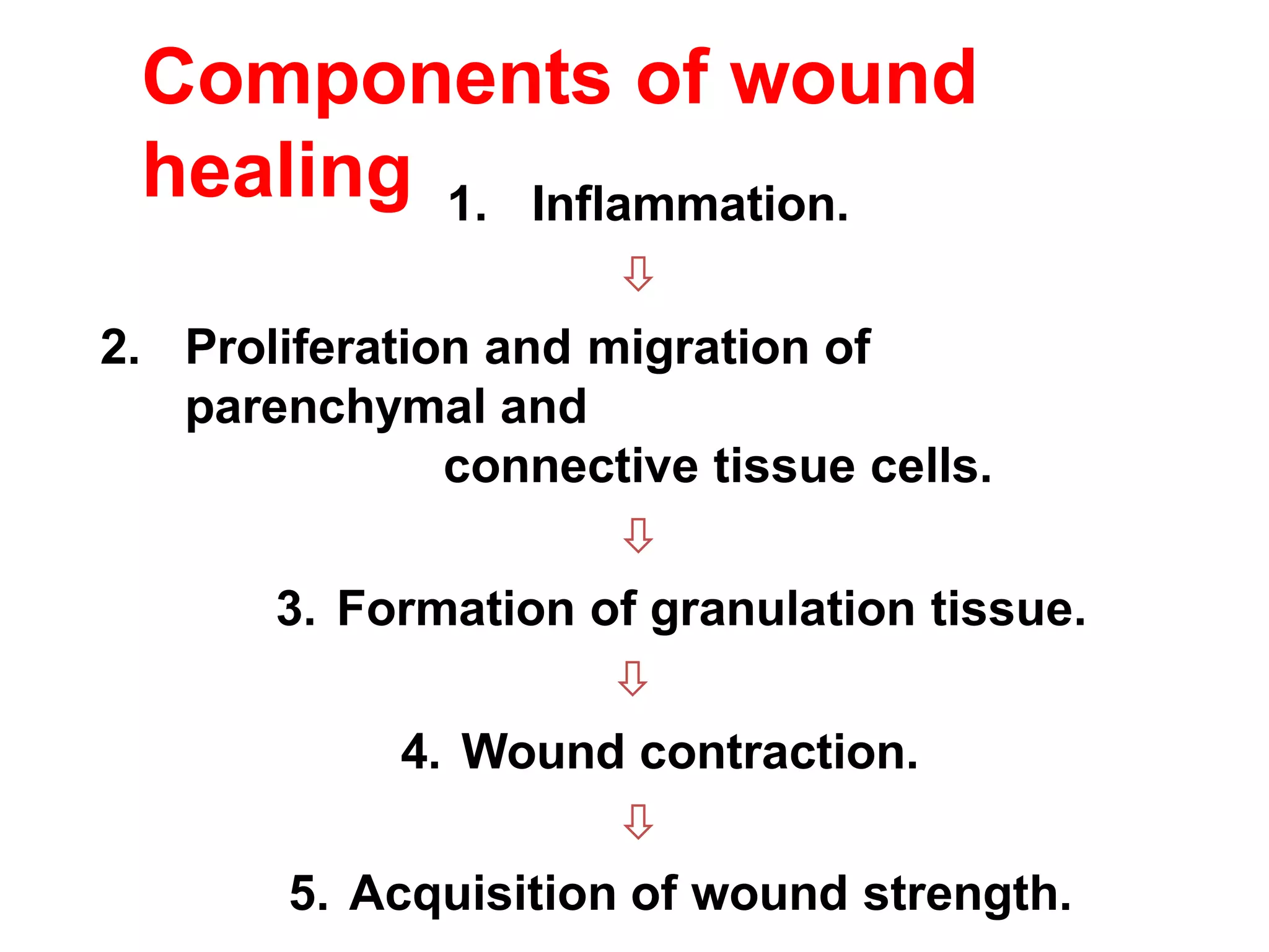
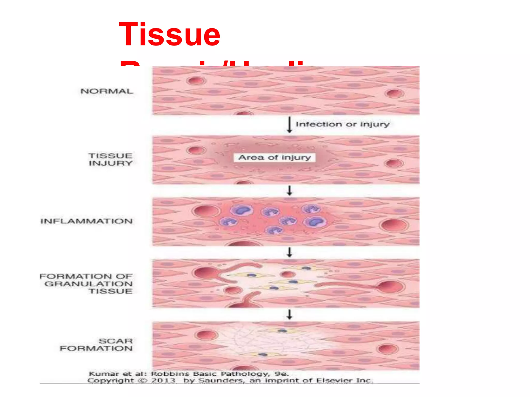
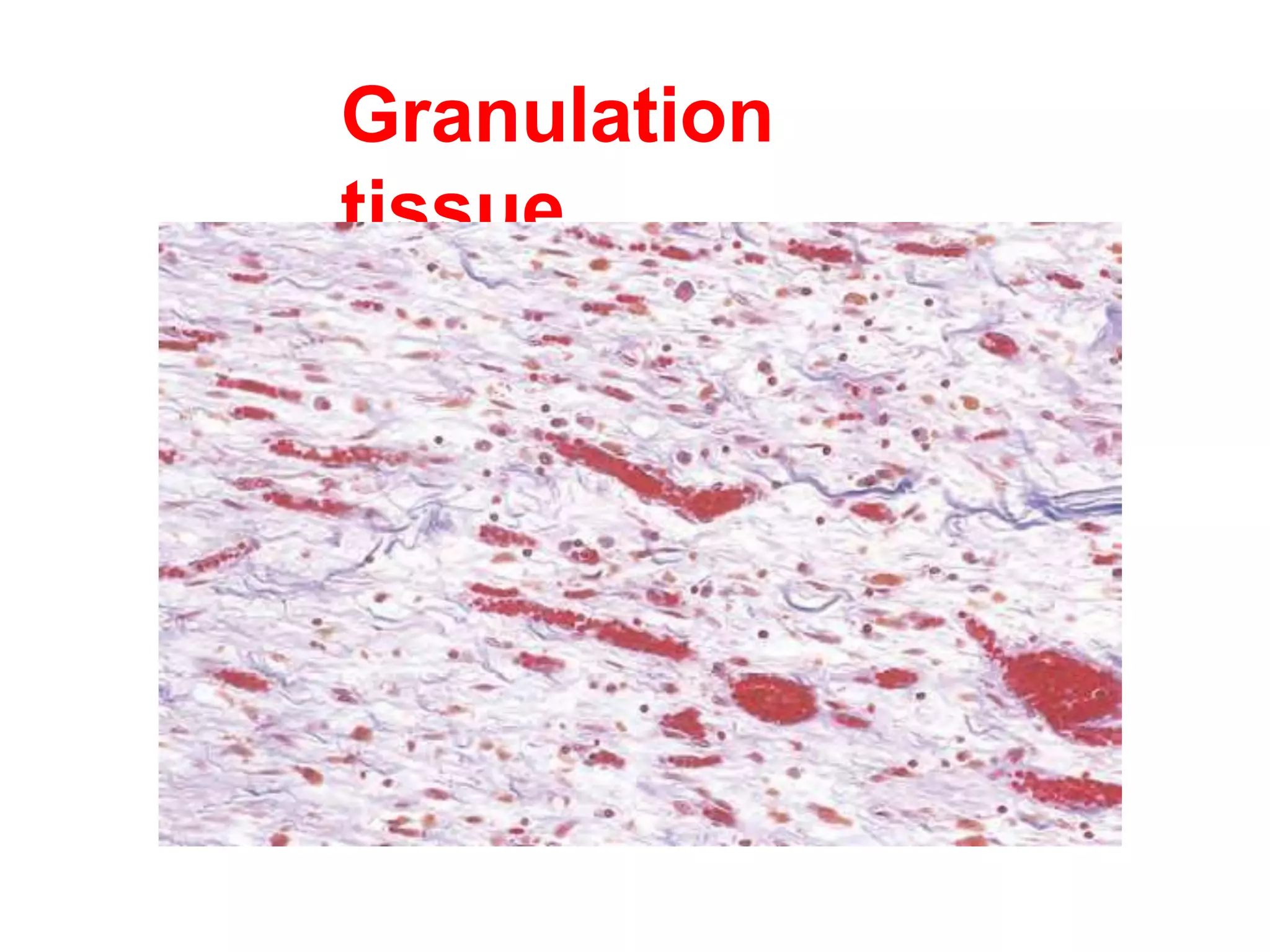
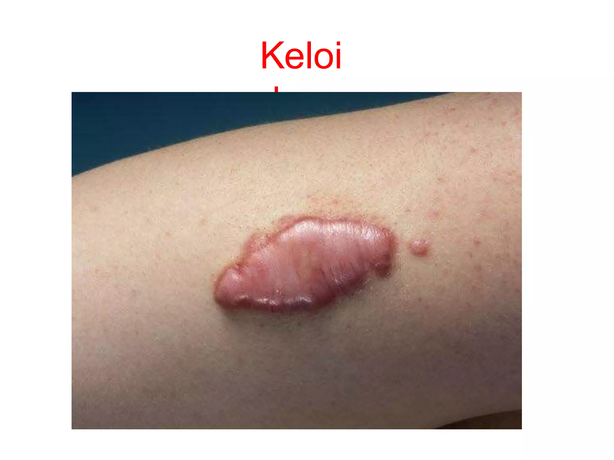
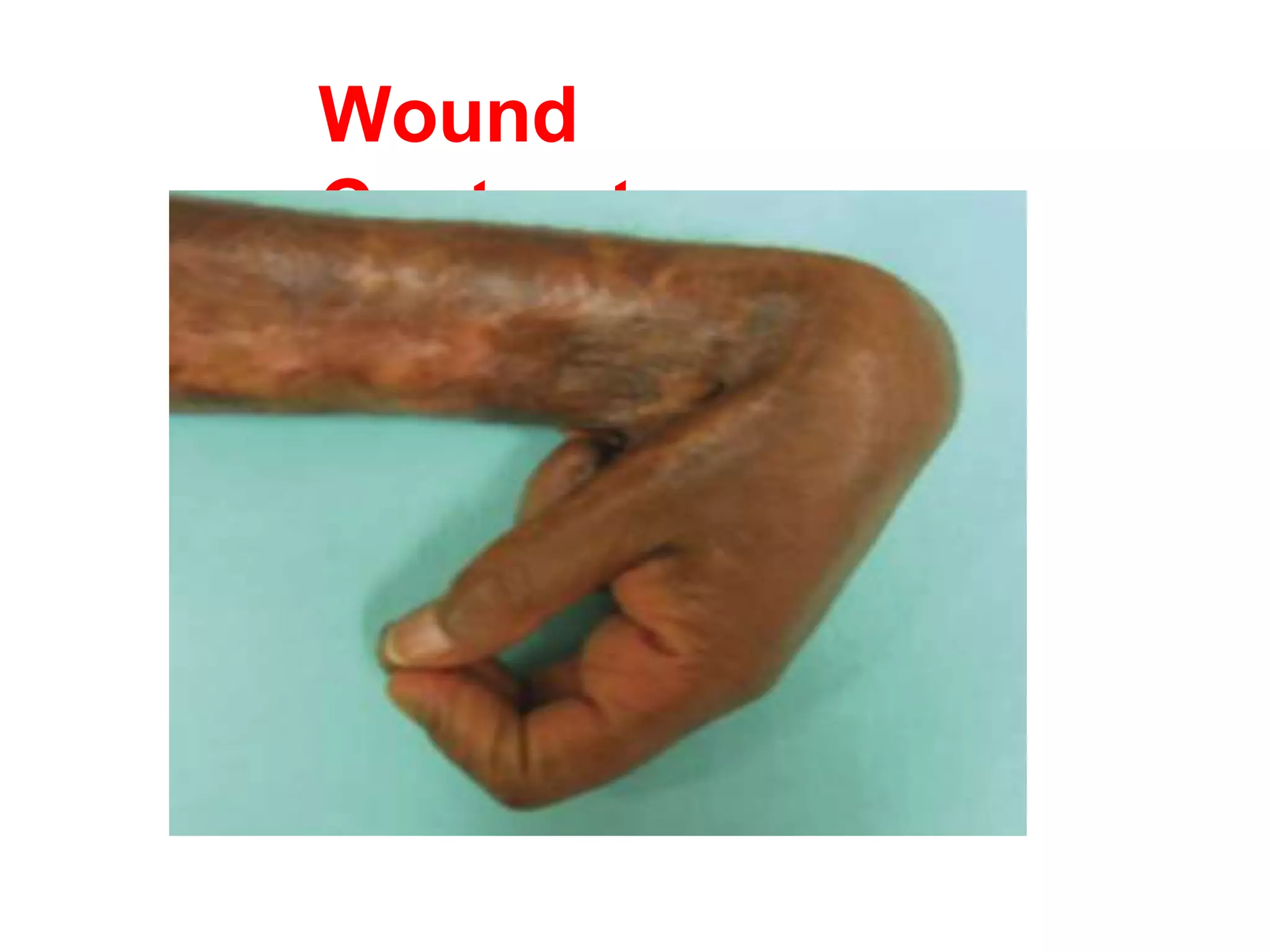
Tissue Repair
Dr. Saket Kumar
Tissue
Repair/Healing
• Repair, sometimes called healing refers to the
restoration of tissue architecture and function
after an injury.
• Repair of damaged tissues occurs by two
types of reactions:
1. Regeneration.
2. Connective tissue deposition (scar
formation).
Tissue
Repair/Healing
• Regeneration: is complete restitution of lost
tissue components identical to those
removed or killed by proliferation of residual
(uninjured) cells and maturation of tissue
stem cells.
• Connective tissue deposition (scar formation):
If the injured tissues are incapable of
regeneration, or if the the tissue is severely
damaged, repair occurs by the deposition of
connective tissue.
Tissue
Repair/Healing
• Regeneration occurs for example, in the
rapidly dividing epithelia of the skin and
intestines, and in some parenchymal
organs, notably the liver.
• Scar formation takes place in fibrosis of
the lungs, liver, kidney and in Myocardial
Infarction.
Tissue
Repair/Healing
Components of wound
healing 1. Inflammation.

2. Proliferation and migration of
parenchymal and
connective tissue cells.

3. Formation of granulation tissue.

4. Wound contraction.

5. Acquisition of wound strength.
Tissue
Repair/Healing
Tissue
Regeneration
• Both regeneration and scar formation
contribute in varying degrees to the
ultimate repair.
• Both processes involve the proliferation of
various cells, and close interactions
between cells and the extracellular matrix
(ECM).
• The cell proliferation in regeneration and
Tissue proliferative
activity
1.Continually dividing cells (labile
tissues): These cells proliferate
through-out life.
Epithelium of the skin, GIT and cells of bone
marrow. 2.Quiescent cells (Stable tissue):
Have a low level of replication. Cells from
these tissue can undergo rapid division in
response to stimuli. parenchymal cells of
liver and kidneys.
3.Non-dividing (permanent cells):
Cells that can not undergo mitotic division in
Granulation
tissue
• Granulation tissue is the hallmark of
healing. Granulation tissue progressively
invades the site of injury.
• The term granulation tissue derives from
its pink, soft, granular appearance seen in
area of ongoing wound healing.
Granulation
tissue
• Granulation tissue is:
i. the proliferation of fibroblasts
ii. new thin-walled, delicate
capillaries (angiogenesis), in a
loose extracellular matrix.
iii. admixed inflammatory cells,
mainly macrophages.
Granulation
tissue
Healing of skin
wound
• Healing of skin wound is a process that
involves both epithelial regeneration and
the formation of connective tissue scar.
• Based on the nature and size of the
wound, the healing of skin wounds
occurs by first or second intention.
Healing of skin
wound
Healing by first intension
• When the injury involves only the epithelial
layer and the principal mechanism of
repair is epithelial regeneration it is called
primary union or healing by first intention.
• It is the process of healing of a clean,
uninfected surgical incisions
approximated by sutures.
Healing by second
intension
• Healing by second intention is also
known as healing by secondary union.
• When cell or tissue loss is more extensive
like in large wounds, abscesses,
ulceration, and ischemic necrosis
(infarction) in parenchymal organs.
• The repair process in second intension
involves a combination of regeneration
and scar formation.
Difference
between
Primary intension
1. Inflammatory
reaction is less
marked.
2. No wound
contraction.
3. Margins are
apposed
together.
4. Minimal tissue
damage
5. Minimal bleeding
6. Heals quickly
Secondary intension
1. Intense
inflammation.
2. Much granulation
tissue formation.
3. Gap persists
between margins.
4. Marked damage
of tissues.
5. More bleeding.
6. Takes time to heal.
7. Large scar
Healing by first VS second
intension
Angiogenesis
New blood vessel development from existing vessel
Steps:
- Vasodilation
- Separation of pericytes and disintegration of BM
- Migration of endothelial cells
- Proliferation of endothelial cells behind leading tip
- Recruitment of periendothelial cells
- Suppression of endothelial proliferation and deposition of
BM
Growth factors – VEGF, FGF, PDGF, TGF beta
Notch signalling – crosstalk with VEGF
Regulates sprouting and branching of
vessel
Factors influencing wound
healing
Systemic factors:
1.Nutritional status: Protein deficiency and
vitamin C deficiency inhibit collagen synthesis
and delay wound healing.
2.Metabolic status: DM is associated delay in
wound healing.
3.Circulatory status, poor perfusion: Inadequate
blood supply impair wound healing.
4.Hormones: Glucocorticoids have anti-
inflammatory effects and inhibits collagen
synthesis causing delay.
Factors influencing wound
healing
Local factors:
1.Infection (one of the most important cause of
delay in healing).
2.Mechanical factors (increased local
pressure, high mobility delays healing).
3.Foreign bodies (splinter, steel, glass delays
healing).
4.Size and shape of wound (clear cut surgical
wound heals quickly).
5.Location of the wound (wound in the face
heals quickly)
Complications of wound healing
Complications of wound healing are
grouped into three categories:
i. Deficient scar formation.
ii. Excessive scar formation.
iii.Exuberant granulation tissue
formation
iv.Formation of contractures.
Complications of wound healing
Deficient scar formation can lead to two
types of complications:
1.Dehiscence or rupture of a wound.
2. Ulceration.
Wound dehiscence is common after
abdominal surgery and is due to
increased abdominal pressure.
Wounds ulceration is due to inadequate
vascularization. Example lower extremity
wounds in with atherosclerosis.
Complications of wound healing
Excessive scar formation can give rise to:
(1) hypertrophic scar
(2)keloids.
• The accumulation of excessive
amounts of collagen give rise to a
hypertrophic scar.
• If the scar tissue grows beyond the
boundaries of original wound and does
not regress, it is called keloid.
Keloi
d
Exuberant granulation
tissue
• Exuberant granulation is the formation of
excessive amounts of granulation tissue,
which protrudes above the level of the
surrounding skin and blocks re-
epithelialization (proud flesh).
• Excessive granulation must be removed by
cautery or surgical excision to allow re-
epithelialization.
Wound
Contracture
• An exaggeration of contraction of wound
gives rise to contracture and results in
deformities of the wound and the
surrounding tissues.
• Common areas affected palms, the soles
and the anterior aspect of the thorax.
• Contractures are commonly seen after
serious burns which compromise
Wound
Contracture
Remodeling of Connective
Tissue
• The connective tissue in the scar
continues to be modified and remodeled.
• To increase the strength
- cross linking of collagen
- Change of collagen from type III to type I
Thank you

Tissue repair.pptx

  • 1.
  • 2.
    Tissue Repair/Healing • Repair, sometimescalled healing refers to the restoration of tissue architecture and function after an injury. • Repair of damaged tissues occurs by two types of reactions: 1. Regeneration. 2. Connective tissue deposition (scar formation).
  • 3.
    Tissue Repair/Healing • Regeneration: iscomplete restitution of lost tissue components identical to those removed or killed by proliferation of residual (uninjured) cells and maturation of tissue stem cells. • Connective tissue deposition (scar formation): If the injured tissues are incapable of regeneration, or if the the tissue is severely damaged, repair occurs by the deposition of connective tissue.
  • 4.
    Tissue Repair/Healing • Regeneration occursfor example, in the rapidly dividing epithelia of the skin and intestines, and in some parenchymal organs, notably the liver. • Scar formation takes place in fibrosis of the lungs, liver, kidney and in Myocardial Infarction.
  • 5.
  • 6.
    Components of wound healing1. Inflammation.  2. Proliferation and migration of parenchymal and connective tissue cells.  3. Formation of granulation tissue.  4. Wound contraction.  5. Acquisition of wound strength.
  • 7.
  • 8.
    Tissue Regeneration • Both regenerationand scar formation contribute in varying degrees to the ultimate repair. • Both processes involve the proliferation of various cells, and close interactions between cells and the extracellular matrix (ECM). • The cell proliferation in regeneration and
  • 9.
    Tissue proliferative activity 1.Continually dividingcells (labile tissues): These cells proliferate through-out life. Epithelium of the skin, GIT and cells of bone marrow. 2.Quiescent cells (Stable tissue): Have a low level of replication. Cells from these tissue can undergo rapid division in response to stimuli. parenchymal cells of liver and kidneys. 3.Non-dividing (permanent cells): Cells that can not undergo mitotic division in
  • 10.
    Granulation tissue • Granulation tissueis the hallmark of healing. Granulation tissue progressively invades the site of injury. • The term granulation tissue derives from its pink, soft, granular appearance seen in area of ongoing wound healing.
  • 11.
    Granulation tissue • Granulation tissueis: i. the proliferation of fibroblasts ii. new thin-walled, delicate capillaries (angiogenesis), in a loose extracellular matrix. iii. admixed inflammatory cells, mainly macrophages.
  • 12.
  • 13.
    Healing of skin wound •Healing of skin wound is a process that involves both epithelial regeneration and the formation of connective tissue scar. • Based on the nature and size of the wound, the healing of skin wounds occurs by first or second intention.
  • 14.
    Healing of skin wound Healingby first intension • When the injury involves only the epithelial layer and the principal mechanism of repair is epithelial regeneration it is called primary union or healing by first intention. • It is the process of healing of a clean, uninfected surgical incisions approximated by sutures.
  • 15.
    Healing by second intension •Healing by second intention is also known as healing by secondary union. • When cell or tissue loss is more extensive like in large wounds, abscesses, ulceration, and ischemic necrosis (infarction) in parenchymal organs. • The repair process in second intension involves a combination of regeneration and scar formation.
  • 16.
    Difference between Primary intension 1. Inflammatory reactionis less marked. 2. No wound contraction. 3. Margins are apposed together. 4. Minimal tissue damage 5. Minimal bleeding 6. Heals quickly Secondary intension 1. Intense inflammation. 2. Much granulation tissue formation. 3. Gap persists between margins. 4. Marked damage of tissues. 5. More bleeding. 6. Takes time to heal. 7. Large scar
  • 17.
    Healing by firstVS second intension
  • 18.
    Angiogenesis New blood vesseldevelopment from existing vessel Steps: - Vasodilation - Separation of pericytes and disintegration of BM - Migration of endothelial cells - Proliferation of endothelial cells behind leading tip - Recruitment of periendothelial cells - Suppression of endothelial proliferation and deposition of BM
  • 20.
    Growth factors –VEGF, FGF, PDGF, TGF beta Notch signalling – crosstalk with VEGF Regulates sprouting and branching of vessel
  • 21.
    Factors influencing wound healing Systemicfactors: 1.Nutritional status: Protein deficiency and vitamin C deficiency inhibit collagen synthesis and delay wound healing. 2.Metabolic status: DM is associated delay in wound healing. 3.Circulatory status, poor perfusion: Inadequate blood supply impair wound healing. 4.Hormones: Glucocorticoids have anti- inflammatory effects and inhibits collagen synthesis causing delay.
  • 22.
    Factors influencing wound healing Localfactors: 1.Infection (one of the most important cause of delay in healing). 2.Mechanical factors (increased local pressure, high mobility delays healing). 3.Foreign bodies (splinter, steel, glass delays healing). 4.Size and shape of wound (clear cut surgical wound heals quickly). 5.Location of the wound (wound in the face heals quickly)
  • 23.
    Complications of woundhealing Complications of wound healing are grouped into three categories: i. Deficient scar formation. ii. Excessive scar formation. iii.Exuberant granulation tissue formation iv.Formation of contractures.
  • 24.
    Complications of woundhealing Deficient scar formation can lead to two types of complications: 1.Dehiscence or rupture of a wound. 2. Ulceration. Wound dehiscence is common after abdominal surgery and is due to increased abdominal pressure. Wounds ulceration is due to inadequate vascularization. Example lower extremity wounds in with atherosclerosis.
  • 25.
    Complications of woundhealing Excessive scar formation can give rise to: (1) hypertrophic scar (2)keloids. • The accumulation of excessive amounts of collagen give rise to a hypertrophic scar. • If the scar tissue grows beyond the boundaries of original wound and does not regress, it is called keloid.
  • 26.
  • 27.
    Exuberant granulation tissue • Exuberantgranulation is the formation of excessive amounts of granulation tissue, which protrudes above the level of the surrounding skin and blocks re- epithelialization (proud flesh). • Excessive granulation must be removed by cautery or surgical excision to allow re- epithelialization.
  • 28.
    Wound Contracture • An exaggerationof contraction of wound gives rise to contracture and results in deformities of the wound and the surrounding tissues. • Common areas affected palms, the soles and the anterior aspect of the thorax. • Contractures are commonly seen after serious burns which compromise
  • 29.
  • 30.
    Remodeling of Connective Tissue •The connective tissue in the scar continues to be modified and remodeled. • To increase the strength - cross linking of collagen - Change of collagen from type III to type I
  • 31.