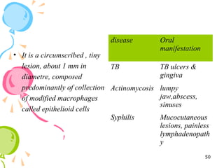
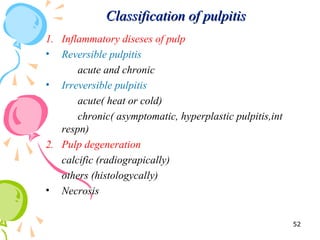
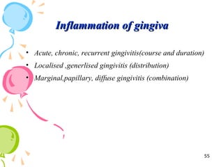
This document defines inflammation and discusses the various types. Inflammation is the body's response to injury or infection and involves increased blood flow, immune cell infiltration, and release of chemical mediators. There are two main types - acute inflammation, which lasts minutes to days and resolves on its own, involving redness, swelling, heat and pain. Chronic inflammation persists longer and involves tissue destruction alongside inflammation. Specific types of chronic inflammation include granulomatous inflammation, which features collections of immune cells called granulomas. The document also discusses inflammation of different oral tissues like the pulp, periodontium and gingiva.
INTRODUCTION
HISTORY
CAUSES OF INFLAMMATION
CLASSIFICATION
ACUTE INFLAMMATION
CHEMICAL MEDIATORS OF INFLAMMATION
OUTCOMES OF ACUTE INFLAMMATION
CHRONIC INFLAMMATION
INFLAMMATORY DISEASES
REFERENCES
INTRODUCTION
HISTORY
CAUSES OF INFLAMMATION
CLASSIFICATION
ACUTE INFLAMMATION
CHEMICAL MEDIATORS OF INFLAMMATION
OUTCOMES OF ACUTE INFLAMMATION
CHRONIC INFLAMMATION
INFLAMMATORY DISEASES
REFERENCES
Inflammation- General Pathology seminar PG 1st yearDr. Ritu Gupta
this seminar includes general inflammation, its etiology, acute inflammation, features, events, fate, chronic inflammation, causes, features, types, granulomatous inflammation, acute v/s chronic inflammation, inflammatory disorders of pulp and periradicular tissues
“Inflame” redirects here. For the 2017 Turkish film, see
Inflame (film).
Toes inflamed by chilblains
Inflammation (from Latin inflammatio) is part of the
complex biological response of body tissues to harmful
stimuli, such as pathogens, damaged cells, or irritants,[1]
and is a protective response involving immune cells,
blood vessels, and molecular mediators. The function of
inflammation is to eliminate the initial cause of cell injury,
clear out necrotic cells and tissues damaged from
the original insult and the inflammatory process, and to
initiate tissue repair.
The classical signs of inflammation are heat, pain, redness,
swelling, and loss of function. Inflammation is a
generic response, and therefore it is considered as a mechanism
of innate immunity, as compared to adaptive immunity,
which is specific for each pathogen.[2] Too little
inflammation could lead to progressive tissue destruction
by the harmful stimulus (e.g. bacteria) and compromise
the survival of the organism. In contrast, chronic
inflammation may lead to a host of diseases, such as hay
fever, periodontitis, atherosclerosis, rheumatoid arthritis,
and even cancer (e.g., gallbladder carcinoma). Inflammation
is therefore normally closely regulated by the body.
Inflammation can be classified as either acute or chronic.
Acute inflammation is the initial response of the body to
harmful stimuli and is achieved by the increased movement
of plasma and leukocytes (especially granulocytes)
from the blood into the injured tissues. A series of biochemical
events propagates and matures the inflammatory
response, involving the local vascular system, the
immune system, and various cells within the injured tissue.
Prolonged inflammation, known as chronic inflammation,
leads to a progressive shift in the type of cells
present at the site of inflammation, such as mononuclear
cells, and is characterized by simultaneous destruction
and healing of the tissue from the inflammatory process.
Inflammation is not a synonym for infection. Infection
describes the interaction between the action of microbial
invasion and the reaction of the body’s inflammatory response
— the two components are considered together
when discussing an infection, and the word is used to imply
a microbial invasive cause for the observed inflammatory
reaction. Inflammation on the other hand describes
purely the body’s immunovascular response, whatever the
cause may be. But because of how often the two are
correlated, words ending in the suffix -itis (which refers
to inflammation) are sometimes informally described as
referring to infection. For example, the word urethritis
strictly means only “urethral inflammation”, but clinical
health care providers usually
inflammation is the body's immune system's response to an irritant. The irritant might be a germ, but it could also be a foreign object, such as a splinter in your finger.
aetiology of inflammation; types of inflammation; how inflammation occur; cells involve in inflammation; role of wbc in inflammation; outcome of inflammation; how inflammation associated with immunity, clotting system, complementary system kinin system, how inflammation is associated with oral cavity; disease associated with inflammatory system
NVBDCP.pptx Nation vector borne disease control programSapna Thakur
NVBDCP was launched in 2003-2004 . Vector-Borne Disease: Disease that results from an infection transmitted to humans and other animals by blood-feeding arthropods, such as mosquitoes, ticks, and fleas. Examples of vector-borne diseases include Dengue fever, West Nile Virus, Lyme disease, and malaria.
- Video recording of this lecture in English language: https://youtu.be/lK81BzxMqdo
- Video recording of this lecture in Arabic language: https://youtu.be/Ve4P0COk9OI
- Link to download the book free: https://nephrotube.blogspot.com/p/nephrotube-nephrology-books.html
- Link to NephroTube website: www.NephroTube.com
- Link to NephroTube social media accounts: https://nephrotube.blogspot.com/p/join-nephrotube-on-social-media.html
Title: Sense of Smell
Presenter: Dr. Faiza, Assistant Professor of Physiology
Qualifications:
MBBS (Best Graduate, AIMC Lahore)
FCPS Physiology
ICMT, CHPE, DHPE (STMU)
MPH (GC University, Faisalabad)
MBA (Virtual University of Pakistan)
Learning Objectives:
Describe the primary categories of smells and the concept of odor blindness.
Explain the structure and location of the olfactory membrane and mucosa, including the types and roles of cells involved in olfaction.
Describe the pathway and mechanisms of olfactory signal transmission from the olfactory receptors to the brain.
Illustrate the biochemical cascade triggered by odorant binding to olfactory receptors, including the role of G-proteins and second messengers in generating an action potential.
Identify different types of olfactory disorders such as anosmia, hyposmia, hyperosmia, and dysosmia, including their potential causes.
Key Topics:
Olfactory Genes:
3% of the human genome accounts for olfactory genes.
400 genes for odorant receptors.
Olfactory Membrane:
Located in the superior part of the nasal cavity.
Medially: Folds downward along the superior septum.
Laterally: Folds over the superior turbinate and upper surface of the middle turbinate.
Total surface area: 5-10 square centimeters.
Olfactory Mucosa:
Olfactory Cells: Bipolar nerve cells derived from the CNS (100 million), with 4-25 olfactory cilia per cell.
Sustentacular Cells: Produce mucus and maintain ionic and molecular environment.
Basal Cells: Replace worn-out olfactory cells with an average lifespan of 1-2 months.
Bowman’s Gland: Secretes mucus.
Stimulation of Olfactory Cells:
Odorant dissolves in mucus and attaches to receptors on olfactory cilia.
Involves a cascade effect through G-proteins and second messengers, leading to depolarization and action potential generation in the olfactory nerve.
Quality of a Good Odorant:
Small (3-20 Carbon atoms), volatile, water-soluble, and lipid-soluble.
Facilitated by odorant-binding proteins in mucus.
Membrane Potential and Action Potential:
Resting membrane potential: -55mV.
Action potential frequency in the olfactory nerve increases with odorant strength.
Adaptation Towards the Sense of Smell:
Rapid adaptation within the first second, with further slow adaptation.
Psychological adaptation greater than receptor adaptation, involving feedback inhibition from the central nervous system.
Primary Sensations of Smell:
Camphoraceous, Musky, Floral, Pepperminty, Ethereal, Pungent, Putrid.
Odor Detection Threshold:
Examples: Hydrogen sulfide (0.0005 ppm), Methyl-mercaptan (0.002 ppm).
Some toxic substances are odorless at lethal concentrations.
Characteristics of Smell:
Odor blindness for single substances due to lack of appropriate receptor protein.
Behavioral and emotional influences of smell.
Transmission of Olfactory Signals:
From olfactory cells to glomeruli in the olfactory bulb, involving lateral inhibition.
Primitive, less old, and new olfactory systems with different path
New Drug Discovery and Development .....NEHA GUPTA
The "New Drug Discovery and Development" process involves the identification, design, testing, and manufacturing of novel pharmaceutical compounds with the aim of introducing new and improved treatments for various medical conditions. This comprehensive endeavor encompasses various stages, including target identification, preclinical studies, clinical trials, regulatory approval, and post-market surveillance. It involves multidisciplinary collaboration among scientists, researchers, clinicians, regulatory experts, and pharmaceutical companies to bring innovative therapies to market and address unmet medical needs.
Couples presenting to the infertility clinic- Do they really have infertility...Sujoy Dasgupta
Dr Sujoy Dasgupta presented the study on "Couples presenting to the infertility clinic- Do they really have infertility? – The unexplored stories of non-consummation" in the 13th Congress of the Asia Pacific Initiative on Reproduction (ASPIRE 2024) at Manila on 24 May, 2024.
Acute scrotum is a general term referring to an emergency condition affecting the contents or the wall of the scrotum.
There are a number of conditions that present acutely, predominantly with pain and/or swelling
A careful and detailed history and examination, and in some cases, investigations allow differentiation between these diagnoses. A prompt diagnosis is essential as the patient may require urgent surgical intervention
Testicular torsion refers to twisting of the spermatic cord, causing ischaemia of the testicle.
Testicular torsion results from inadequate fixation of the testis to the tunica vaginalis producing ischemia from reduced arterial inflow and venous outflow obstruction.
The prevalence of testicular torsion in adult patients hospitalized with acute scrotal pain is approximately 25 to 50 percent
These simplified slides by Dr. Sidra Arshad present an overview of the non-respiratory functions of the respiratory tract.
Learning objectives:
1. Enlist the non-respiratory functions of the respiratory tract
2. Briefly explain how these functions are carried out
3. Discuss the significance of dead space
4. Differentiate between minute ventilation and alveolar ventilation
5. Describe the cough and sneeze reflexes
Study Resources:
1. Chapter 39, Guyton and Hall Textbook of Medical Physiology, 14th edition
2. Chapter 34, Ganong’s Review of Medical Physiology, 26th edition
3. Chapter 17, Human Physiology by Lauralee Sherwood, 9th edition
4. Non-respiratory functions of the lungs https://academic.oup.com/bjaed/article/13/3/98/278874
Report Back from SGO 2024: What’s the Latest in Cervical Cancer?bkling
Are you curious about what’s new in cervical cancer research or unsure what the findings mean? Join Dr. Emily Ko, a gynecologic oncologist at Penn Medicine, to learn about the latest updates from the Society of Gynecologic Oncology (SGO) 2024 Annual Meeting on Women’s Cancer. Dr. Ko will discuss what the research presented at the conference means for you and answer your questions about the new developments.
4. Inflammation:
1. To eliminate the cause of the tissue damage,
2. To repair injured and damaged tissue.
• Inflammation is necessary for the survival of the host.
• In the absence of inflammation the body would be unable
to kill and eliminate infectious agents.
• One of the innate defense mechanisms of the body.
Introduction
4
5. DefinationDefination
• Inflammation is defined as complex series of events that
occurs in vascularized living tissues in response to local
injury or tissue damage.
• Inflammation is a programmed local tissue response
peculiar to vascularized living tissues.
5
6. Causes of inflammationCauses of inflammation
• Physical agents –
heat
cold
radiation
mechanical trauma
• Chemical agents –
organic and inorganic poisons
• Infective agents –
bacteria and virus
• Immunological agents –
cell mediated
antigen-antibody reactions
6
7. Inflammation and infection..Inflammation and infection..
• Inflammation protective response by the body
• Infection invasion into body by harmful microbes
and their resultant ill-effects by toxins.
7
8. Signs of inflammationSigns of inflammation
Rubor (redness)
Tumor (swelling)
Calor (heat)
Dolor (pain)
Functio lasea (loss of function)
8
9. English Greek/Latin Caused By
Redness Rubor Hyperemia
Warmth Calor Hyperemia
Swelling Tumor Increased vascular
permeability
Pain Dolor Low pH
Loss of function Functio laesa Pain, swelling
9
14. Vascular events in acute inflammationVascular events in acute inflammation
Haemodynamic changes
Changes in vascular permeability
14
15. 1) Haemodynamic changes1) Haemodynamic changes
Persistent progressive vasodilation –
1. Affects venules & capillaries
2. in blood vol redness and warmth at site of
inflammation.
3. Transudation of fluid in extracellular space
4. Swelling at local site of acute inflammation
15
17. Cellular events in acute inflammationCellular events in acute inflammation
• Cellular phase of inflammation consists of 2 process
Exudation of leukocytes phagocytosis
17
18. 1) Exudation of leukocytes1) Exudation of leukocytes
• The changes leading to migration of leucocytes are:
Changes in the formed elements of blood
Rolling and adhesion
Emigration
Chemotaxis
18
19. 1)changes in the formed elements:
- Rate of blood flow is increased
- Stasis(change in normal axial flow of blood)
widening
narrowing
Central stream
plasma
19
20. 2)Rolling and adhesion
- PMS’s roll over the endothelial cells.
- Bond btn leukocytes and endothelial cells.
20
21. 3) Emigration and diapedesis –
neutrophils throw cytoplasmic pseudopods
cross the basement membrane
21
22. • The chemotactic factor-mediated
transmigration of leukocytes after crossing
several barriers to reach the interstitial tissues
is called chemotaxis
• Chemotactic substances – chemokines
eg – leukotrines, platelet
factor,cytokines, etc
4)Chemotaxis –
22
25. 2) Phagocytosis2) Phagocytosis
• Phagocytosis is defined as the process of engulfment of
solid particulate material by the cells
• Phagocytes (cell eating cells)
• cells
microphages macrophages
25
26. Steps in phagocytosis
Recognition and attachment stage
(opsonisation)
Engulfment stage
Secretion (degranulation) stage
Digestion or degradation stage
26
27. • Recognition and attachment
stage:
To bond the bacteria & the
cell membrane of the
phagocytic cell, the
microorganisms get coated
with opsonins which are
naturally occurring factors
in the serum.
The main opsonins present
in the serum are IgG
opsonin , C3b opsonin &
lectins
27
28. Engulfment stage
Cytoplasmic pseudopods
are formed around the
particle, enveloping it in
a phagocytic vacuole.
Eventually the plasma
membrane enclosing the
phagocytic vacuole
breaks the cell surface
The lysosomes of the cell
fuse with the vacuole and
form phagolysosome or
phagosome.
28
29. Degranulation stage :
preformed granule
products of PMN’s are
discharged.
Specific or secondary
granules of PMN’s are
released with
interlukin2,TNF,superox
ide oxygen.
29
30. Killing/degradation stage:
Killing & digestion of the
microorganisms by the phagocytes as
scavenger cells is done.
The microorganisms are degraded by
the hydrolytic enzymes.
30
37. Morphology of acute inflammationMorphology of acute inflammation
1.Pseudomembranous
inflammation-
• it’s a inflammatory response
of the mucous surface to
toxins of diphtheria or irritant
gases.
• denudation of the epithelium
• plasma exudes on the surface
where it coagulates &
together with the necrosed
epithelium forms a false
membrane.
37
38. 2.Ulcer-
• Ulcers are local defects on
the surface of an organ
produced by inflammation.
38
39. 3.Suppuration(Abscess
formation)-
• neutrophilic infiltrate in the
inflamed tissue results in
tissue necrosis.
• A cavity is formed which is
called an abscess & contains
a purulent exudate
• Boil or Furuncle is an acute
inflammation of the hair
follicles in the dermal tissues.
39
40. 4.Cellulitits-
it’s a diffuse
inflammation of soft tissues
resulting from spreading
effects of substances like
hyaluronidase released by
some bacteria.
5.Bacterial infection of the
blood-
a) Bacteraemia
b) Septicemia
c) Pyaemia
40
41. The systemic effects of inflammationThe systemic effects of inflammation
1.fever
2.Leucocytosis
3.Lymphadenitis
4.Shock
41
42. Fate of acute inflammationFate of acute inflammation
1. Resolution
2. Healing by scarring
3. Progression to suppuration
4. Progression to Chronic inflammation.
42
44. Chronic inflammation can be defined as a prolonged
process in which tissue destruction and
inflammation occur at the same time.
44
45. Causes of Chronic inflammation
1.Chronic inflammation following Acute inflammation
2.Recurrrent attacks of acute inflammation
3.Chronic inflammation starting de novo
45
46. General features of chronic inflammationGeneral features of chronic inflammation
1.Mononuclear cell infiltration-
phagocytes, circulating
monocytes, macrophages &
giant cells.
2.Tissue destruction or necrosis.
3.Proliferative changes- small
blood vessels & fibroblasts
46
50. • It is a circumscribed , tiny
lesion, about 1 mm in
diametre, composed
predominantly of collection
of modified macrophages
called epithelioid cells
disease Oral
manifestation
TB TB ulcers &
gingiva
Actinomycosis lumpy
jaw,abscess,
sinuses
Syphilis Mucocutaneous
lesions, painless
lymphadenopath
y
50