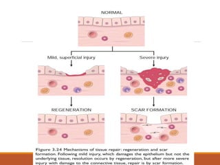
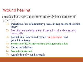
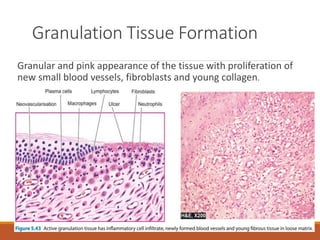
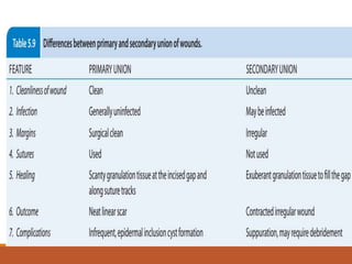
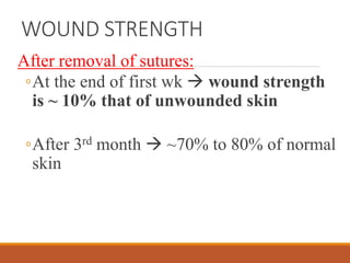
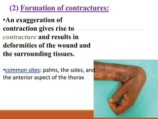
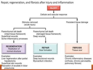
TISSUE REPAIR
(WOUND HEALING)
DR. RAM THAPA
Course Content
Types, etiopathogenesis
Factors affecting Healings – Promoting &
Delaying
Mechanism of healing – Primary and secondary
intention
Definitions
Repair, also called healing, refers to the
restoration of tissue architecture and
function after an injury.
Repair is used for parenchymal and
connective tissues
Healing for surface epithelia,
Definitions:
Regeneration: refers to growth of cells and tissues to
replace lost structures
Healingis usually a tissue response
◦to a wound (commonly in the skin),
◦to inflammatory processes in internal organs, or
◦to cell necrosis in organs incapable of
regeneration.
Healing
Regeneration scar formation
When healing can not be accomplished by
regeneration, the main healing process is
repair by deposition of collagen and other
ECM components, causing the formation of
a scar.
1. Labile Cells: epithelial cells,haematopoietic cells
of bone marrow
2.Stable cells:-Parenchymal cells of organs like liver,
pancreas, kidneys, adrenal and thyroid;
mesenchymal cells like smooth muscle cells,
fibroblasts, vascular endothelium
3.Permanent cells:- neurons of nervous system,
skeletal muscle and cardiac muscle cells
wound healing
Inflammation
Angiogenesis
Migration and proliferation of fibroblasts
Scar formation
Connective tissue remodelling
Wound healing
complex but orderly phenomenon involving a number of
processes:
1) Induction of an inflammatory process in response to the initial
injury
2) Proliferation and migration of parenchymal and connective
tissue cells
3) Formation of new blood vessels (angiogenesis) and
granulation tissue
4) Synthesis of ECM proteins and collagen deposition
5) Tissue remodelling
6) Wound contraction
7) Acquisition of wound strength
Cutaneous wound healing is generally divided
into three phases:
(1) inflammation (early and late);
(2) granulation tissue formation and re-epithelialization
(3) wound contraction, ECM deposition, and remodelling
Granulation Tissue Formation
Granular and pink appearance of the tissue with proliferation of
new small blood vessels, fibroblasts and young collagen.
Phases of cutaneous wound healing
PROCESS OF WOUND HEALING
1. Primary intention
2. Secondary intention
WOUND HEALING BY PRIMARY INTENTION
The healing of a clean, uninfected surgical
incision approximated by surgical sutures
Such healing is referred to as primary union
or healing by first intention
WOUND HEALING BY PRIMARY
INTENTION
Defined as healing of a wound which has the
following characteristics:
i) clean and uninfected;
ii) surgically incised;
iii) without much loss of cells and tissue; and
iv) edges of wound are approximated by surgical
sutures.
Healing process follows a series of
sequential steps:
Within 24 hours:
◦neutrophils appear at the margins of the
incision, moving toward the fibrin clot.
In 24 to 48 hours:
◦epithelial cells move from the wound edges
(with little cell proliferation) along the cut
margins of the dermis
◦Deposition of basement membrane components
as they move
By day 3:
◦neutrophils replaced by macrophages
◦Granulation tissue progressively invades the
incision space.
◦Collagen fibers are now present in the margins of
the incision, but at first these are vertically oriented
and do not bridge the incision.
◦Epithelial cell proliferation thickens the epidermal
layer
By day 5:
◦the incisional space is filled with granulation tissue.
◦Neovascularization is maximal.
◦Collagen fibrils  more abundant and begin to
bridge the incision.
◦differentiation of surface cells yields a mature
epidermal architecture with surface keratinization.
During the second week:
collagen +++
proliferation of fibroblasts ++
leukocytic infiltrate, edema, and increased
vascularity largely disappear
the long process of blanching begins,
accomplished by the increased accumulation of
collagen within the incisional scar, accompanied
by regression of vascular channels.
By the end of the first month:
◦Scar is made up of a cellular connective tissue
devoid of inflammatory infiltrate, covered now
by intact epidermis.
◦The dermal appendages  permanently lost.
◦Tensile strength of the wound increases
WOUND HEALING BY SECONDARY INTENTION
Occurs when there is
◦ more extensive loss of cells and tissue
◦ large defects in surface wounds
The reparative process is more complicated
Abundant granulation tissue grows in from the margin to
complete the repair.
referred to as secondary union or healing by second intention
HEALING BY SECOND INTENTION
Defined as healing of a wound having the following
characteristics:
i) open with a large tissue defect, at times infected;
ii) having extensive loss of cells and tissues;
iii) the wound is not approximated by surgical
sutures but is left open.
Differs from healing by primary intention by
the following points:
1. Large tissue defects  a larger fibrin clot that fills the
defect more necrotic debris and exudates that must be
removed. Inflammatory reaction is more intense.
2. Much larger amounts of granulation tissue are formed.
3. wound contraction
4. scar formation and thinning of the epidermis.
Development of fibrosis in chronic inflammation
HEALING OF SKIN ULCER
(A)Inflammation
(B)Proliferation of epithelial
cells; formation of
granulation tissue by vessel
growth proliferating
fibroblasts.
(C) Remodeling to produce the
fibrous scar.
Steps in repair by scar formation: wound healing in
the skin.
WOUND STRENGTH
After removal of sutures:
◦At the end of first wk  wound strength
is ~ 10% that of unwounded skin
◦After 3rd month  ~70% to 80% of normal
skin
FACTORS THAT INFLUENCE WOUND HEALING:
SYSTEMIC:
1. Nutrition Protein deficiency vitamin C
deficiency  inhibit collagen synthesis  retard
healing
2.Metabolic status  eg. Diabetes mellitus  retard
healing .
3.Circulatory status  eg. Inadequate blood supply 
arteriosclerosis or venous abnormalities (e.g.,
varicose veins)  retard venous drainage, also
impairs healing
4. Hormones  eg. glucocorticoids  inhibit collagen
synthesis  impairs healing
LOCAL:
1. Infection  persistent tissue injury and
inflammation.
2. Mechanical factors  early motion of wounds 
compressing blood vessels and separating the
edges of the wound.
3. Foreign bodies  unnecessary sutures or
fragments of steel, glass, or even bone
4. Size, location & type of injury
FACTORS THAT INFLUENCE WOUND
HEALING
COMPLICATIONS OF WOUND
HEALING
(1)Wound dehiscence and ulceration
◦ Inadequate formation of granulation
tissue or assembly of a scar
◦ inadequate vascularization
◦ diabetic peripheral neuropathy
(2) Formation of contractures:
•An exaggeration of
contraction gives rise to
contracture and results in
deformities of the wound and
the surrounding tissues.
•common sites: palms, the soles, and
the anterior aspect of the thorax
(3) excessive formation of the repair
components
◦ hypertrophic scar
◦ keloid
◦ exuberant granulation
◦ desmoids, or aggressive fibromatoses
Repair, regeneration, and fibrosis after injury and inflammation
4.Wound Healing.pptx

4.Wound Healing.pptx

  • 1.
  • 2.
    Course Content Types, etiopathogenesis Factorsaffecting Healings – Promoting & Delaying Mechanism of healing – Primary and secondary intention
  • 3.
    Definitions Repair, also calledhealing, refers to the restoration of tissue architecture and function after an injury. Repair is used for parenchymal and connective tissues Healing for surface epithelia,
  • 4.
    Definitions: Regeneration: refers togrowth of cells and tissues to replace lost structures Healingis usually a tissue response ◦to a wound (commonly in the skin), ◦to inflammatory processes in internal organs, or ◦to cell necrosis in organs incapable of regeneration.
  • 6.
  • 9.
    When healing cannot be accomplished by regeneration, the main healing process is repair by deposition of collagen and other ECM components, causing the formation of a scar.
  • 10.
    1. Labile Cells:epithelial cells,haematopoietic cells of bone marrow 2.Stable cells:-Parenchymal cells of organs like liver, pancreas, kidneys, adrenal and thyroid; mesenchymal cells like smooth muscle cells, fibroblasts, vascular endothelium 3.Permanent cells:- neurons of nervous system, skeletal muscle and cardiac muscle cells
  • 11.
    wound healing Inflammation Angiogenesis Migration andproliferation of fibroblasts Scar formation Connective tissue remodelling
  • 12.
    Wound healing complex butorderly phenomenon involving a number of processes: 1) Induction of an inflammatory process in response to the initial injury 2) Proliferation and migration of parenchymal and connective tissue cells 3) Formation of new blood vessels (angiogenesis) and granulation tissue 4) Synthesis of ECM proteins and collagen deposition 5) Tissue remodelling 6) Wound contraction 7) Acquisition of wound strength
  • 13.
    Cutaneous wound healingis generally divided into three phases: (1) inflammation (early and late); (2) granulation tissue formation and re-epithelialization (3) wound contraction, ECM deposition, and remodelling
  • 14.
    Granulation Tissue Formation Granularand pink appearance of the tissue with proliferation of new small blood vessels, fibroblasts and young collagen.
  • 15.
    Phases of cutaneouswound healing
  • 16.
    PROCESS OF WOUNDHEALING 1. Primary intention 2. Secondary intention
  • 17.
    WOUND HEALING BYPRIMARY INTENTION The healing of a clean, uninfected surgical incision approximated by surgical sutures Such healing is referred to as primary union or healing by first intention
  • 18.
    WOUND HEALING BYPRIMARY INTENTION Defined as healing of a wound which has the following characteristics: i) clean and uninfected; ii) surgically incised; iii) without much loss of cells and tissue; and iv) edges of wound are approximated by surgical sutures.
  • 19.
    Healing process followsa series of sequential steps: Within 24 hours: ◦neutrophils appear at the margins of the incision, moving toward the fibrin clot.
  • 20.
    In 24 to48 hours: ◦epithelial cells move from the wound edges (with little cell proliferation) along the cut margins of the dermis ◦Deposition of basement membrane components as they move
  • 21.
    By day 3: ◦neutrophilsreplaced by macrophages ◦Granulation tissue progressively invades the incision space. ◦Collagen fibers are now present in the margins of the incision, but at first these are vertically oriented and do not bridge the incision. ◦Epithelial cell proliferation thickens the epidermal layer
  • 22.
    By day 5: ◦theincisional space is filled with granulation tissue. ◦Neovascularization is maximal. ◦Collagen fibrils  more abundant and begin to bridge the incision. ◦differentiation of surface cells yields a mature epidermal architecture with surface keratinization.
  • 23.
    During the secondweek: collagen +++ proliferation of fibroblasts ++ leukocytic infiltrate, edema, and increased vascularity largely disappear the long process of blanching begins, accomplished by the increased accumulation of collagen within the incisional scar, accompanied by regression of vascular channels.
  • 24.
    By the endof the first month: ◦Scar is made up of a cellular connective tissue devoid of inflammatory infiltrate, covered now by intact epidermis. ◦The dermal appendages  permanently lost. ◦Tensile strength of the wound increases
  • 26.
    WOUND HEALING BYSECONDARY INTENTION Occurs when there is ◦ more extensive loss of cells and tissue ◦ large defects in surface wounds The reparative process is more complicated Abundant granulation tissue grows in from the margin to complete the repair. referred to as secondary union or healing by second intention
  • 27.
    HEALING BY SECONDINTENTION Defined as healing of a wound having the following characteristics: i) open with a large tissue defect, at times infected; ii) having extensive loss of cells and tissues; iii) the wound is not approximated by surgical sutures but is left open.
  • 28.
    Differs from healingby primary intention by the following points: 1. Large tissue defects  a larger fibrin clot that fills the defect more necrotic debris and exudates that must be removed. Inflammatory reaction is more intense. 2. Much larger amounts of granulation tissue are formed. 3. wound contraction 4. scar formation and thinning of the epidermis.
  • 31.
    Development of fibrosisin chronic inflammation
  • 32.
    HEALING OF SKINULCER (A)Inflammation (B)Proliferation of epithelial cells; formation of granulation tissue by vessel growth proliferating fibroblasts. (C) Remodeling to produce the fibrous scar. Steps in repair by scar formation: wound healing in the skin.
  • 33.
    WOUND STRENGTH After removalof sutures: ◦At the end of first wk  wound strength is ~ 10% that of unwounded skin ◦After 3rd month  ~70% to 80% of normal skin
  • 34.
    FACTORS THAT INFLUENCEWOUND HEALING: SYSTEMIC: 1. Nutrition Protein deficiency vitamin C deficiency  inhibit collagen synthesis  retard healing 2.Metabolic status  eg. Diabetes mellitus  retard healing . 3.Circulatory status  eg. Inadequate blood supply  arteriosclerosis or venous abnormalities (e.g., varicose veins)  retard venous drainage, also impairs healing 4. Hormones  eg. glucocorticoids  inhibit collagen synthesis  impairs healing
  • 35.
    LOCAL: 1. Infection persistent tissue injury and inflammation. 2. Mechanical factors  early motion of wounds  compressing blood vessels and separating the edges of the wound. 3. Foreign bodies  unnecessary sutures or fragments of steel, glass, or even bone 4. Size, location & type of injury FACTORS THAT INFLUENCE WOUND HEALING
  • 36.
    COMPLICATIONS OF WOUND HEALING (1)Wounddehiscence and ulceration ◦ Inadequate formation of granulation tissue or assembly of a scar ◦ inadequate vascularization ◦ diabetic peripheral neuropathy
  • 37.
    (2) Formation ofcontractures: •An exaggeration of contraction gives rise to contracture and results in deformities of the wound and the surrounding tissues. •common sites: palms, the soles, and the anterior aspect of the thorax
  • 38.
    (3) excessive formationof the repair components ◦ hypertrophic scar ◦ keloid ◦ exuberant granulation ◦ desmoids, or aggressive fibromatoses
  • 39.
    Repair, regeneration, andfibrosis after injury and inflammation

Editor's Notes

  • #10 body's ability to replace injured or dead cells and to repair tissues after inflammation is critical to survival