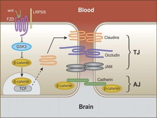
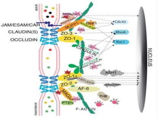
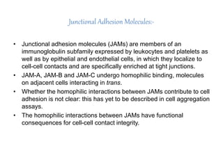
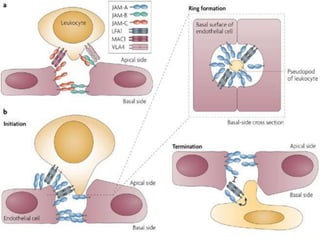
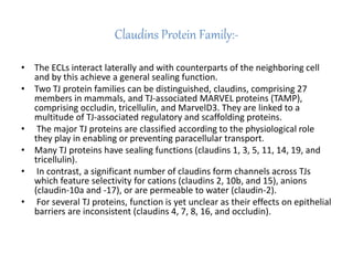
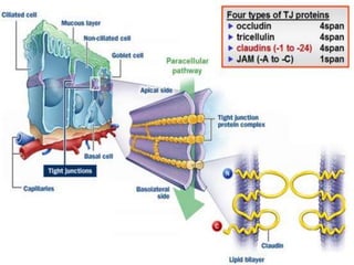
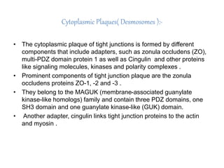
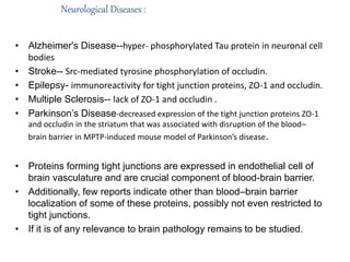
Yograj Banteria
Tight Junctions in Blood Brain Barrier:-
• Blood-brain barrier (BBB) is a dynamic interference that regulates
the nutrition and toxic substance in and out of the central nervous
system (CNS), and plays a crucial role in maintaining a stable
circumstance of the CNS.
• Tight junctions among adjacent cells form the basic structure of BBB
to limiting paracellular permeability. In the present, the constituents
of tight junction proteins are depicted, together with the regulation of
tight junction under stimulation and in pathological conditions.
Tight Junction Proteins:-
• Tight junction, one of the type of cell-cell junctions, controls the
paracellular permeability across the lateral intercellular space and
maintains the cell polarity.
• Tight junctions consist of transmembrane proteins: members of tight
junction-associated MARVEL protein (TAMP) family, claudins and
junctional adhesion molecules (JAMs), and various cytoplasmic
proteins that are necessary for the correct organization of the
integral membrane components of the junction.
Junctional Adhesion Molecules:-
• Junctional adhesion molecules (JAMs) are members of an
immunoglobulin subfamily expressed by leukocytes and platelets as
well as by epithelial and endothelial cells, in which they localize to
cell-cell contacts and are specifically enriched at tight junctions.
• JAM-A, JAM-B and JAM-C undergo homophilic binding, molecules
on adjacent cells interacting in trans.
• Whether the homophilic interactions between JAMs contribute to cell
adhesion is not clear: this has yet to be described in cell aggregation
assays.
• The homophilic interactions between JAMs have functional
consequences for cell-cell contact integrity.
Claudins Protein Family:-
• The ECLs interact laterally and with counterparts of the neighboring cell
and by this achieve a general sealing function.
• Two TJ protein families can be distinguished, claudins, comprising 27
members in mammals, and TJ-associated MARVEL proteins (TAMP),
comprising occludin, tricellulin, and MarvelD3. They are linked to a
multitude of TJ-associated regulatory and scaffolding proteins.
• The major TJ proteins are classified according to the physiological role
they play in enabling or preventing paracellular transport.
• Many TJ proteins have sealing functions (claudins 1, 3, 5, 11, 14, 19, and
tricellulin).
• In contrast, a significant number of claudins form channels across TJs
which feature selectivity for cations (claudins 2, 10b, and 15), anions
(claudin-10a and -17), or are permeable to water (claudin-2).
• For several TJ proteins, function is yet unclear as their effects on epithelial
barriers are inconsistent (claudins 4, 7, 8, 16, and occludin).
Cytoplasmic Plaques( Desmosomes ):-
• The cytoplasmic plaque of tight junctions is formed by different
components that include adapters, such as zonula occludens (ZO),
multi-PDZ domain protein 1 as well as Cingulin and other proteins
like signaling molecules, kinases and polarity complexes .
• Prominent components of tight junction plaque are the zonula
occludens proteins ZO-1, -2 and -3 .
• They belong to the MAGUK (membrane-associated guanylate
kinase-like homologs) family and contain three PDZ domains, one
SH3 domain and one guanylate kinase-like (GUK) domain.
• Another adapter, cingulin links tight junction proteins to the actin
and myosin .
Neurological Diseases :
• Alzheimer's Disease--hyper- phosphorylated Tau protein in neuronal cell
bodies
• Stroke-- Src-mediated tyrosine phosphorylation of occludin.
• Epilepsy- immunoreactivity for tight junction proteins, ZO-1 and occludin.
• Multiple Sclerosis-- lack of ZO-1 and occludin .
• Parkinson’s Disease-decreased expression of the tight junction proteins ZO-1
and occludin in the striatum that was associated with disruption of the blood–
brain barrier in MPTP-induced mouse model of Parkinson’s disease.
• Proteins forming tight junctions are expressed in endothelial cell of
brain vasculature and are crucial component of blood-brain barrier.
• Additionally, few reports indicate other than blood–brain barrier
localization of some of these proteins, possibly not even restricted to
tight junctions.
• If it is of any relevance to brain pathology remains to be studied.
Parkinson’s Disease
tight junction and blood brain barrier

tight junction and blood brain barrier

  • 1.
  • 2.
    Tight Junctions inBlood Brain Barrier:- • Blood-brain barrier (BBB) is a dynamic interference that regulates the nutrition and toxic substance in and out of the central nervous system (CNS), and plays a crucial role in maintaining a stable circumstance of the CNS. • Tight junctions among adjacent cells form the basic structure of BBB to limiting paracellular permeability. In the present, the constituents of tight junction proteins are depicted, together with the regulation of tight junction under stimulation and in pathological conditions.
  • 4.
    Tight Junction Proteins:- •Tight junction, one of the type of cell-cell junctions, controls the paracellular permeability across the lateral intercellular space and maintains the cell polarity. • Tight junctions consist of transmembrane proteins: members of tight junction-associated MARVEL protein (TAMP) family, claudins and junctional adhesion molecules (JAMs), and various cytoplasmic proteins that are necessary for the correct organization of the integral membrane components of the junction.
  • 6.
    Junctional Adhesion Molecules:- •Junctional adhesion molecules (JAMs) are members of an immunoglobulin subfamily expressed by leukocytes and platelets as well as by epithelial and endothelial cells, in which they localize to cell-cell contacts and are specifically enriched at tight junctions. • JAM-A, JAM-B and JAM-C undergo homophilic binding, molecules on adjacent cells interacting in trans. • Whether the homophilic interactions between JAMs contribute to cell adhesion is not clear: this has yet to be described in cell aggregation assays. • The homophilic interactions between JAMs have functional consequences for cell-cell contact integrity.
  • 8.
    Claudins Protein Family:- •The ECLs interact laterally and with counterparts of the neighboring cell and by this achieve a general sealing function. • Two TJ protein families can be distinguished, claudins, comprising 27 members in mammals, and TJ-associated MARVEL proteins (TAMP), comprising occludin, tricellulin, and MarvelD3. They are linked to a multitude of TJ-associated regulatory and scaffolding proteins. • The major TJ proteins are classified according to the physiological role they play in enabling or preventing paracellular transport. • Many TJ proteins have sealing functions (claudins 1, 3, 5, 11, 14, 19, and tricellulin). • In contrast, a significant number of claudins form channels across TJs which feature selectivity for cations (claudins 2, 10b, and 15), anions (claudin-10a and -17), or are permeable to water (claudin-2). • For several TJ proteins, function is yet unclear as their effects on epithelial barriers are inconsistent (claudins 4, 7, 8, 16, and occludin).
  • 10.
    Cytoplasmic Plaques( Desmosomes):- • The cytoplasmic plaque of tight junctions is formed by different components that include adapters, such as zonula occludens (ZO), multi-PDZ domain protein 1 as well as Cingulin and other proteins like signaling molecules, kinases and polarity complexes . • Prominent components of tight junction plaque are the zonula occludens proteins ZO-1, -2 and -3 . • They belong to the MAGUK (membrane-associated guanylate kinase-like homologs) family and contain three PDZ domains, one SH3 domain and one guanylate kinase-like (GUK) domain. • Another adapter, cingulin links tight junction proteins to the actin and myosin .
  • 12.
    Neurological Diseases : •Alzheimer's Disease--hyper- phosphorylated Tau protein in neuronal cell bodies • Stroke-- Src-mediated tyrosine phosphorylation of occludin. • Epilepsy- immunoreactivity for tight junction proteins, ZO-1 and occludin. • Multiple Sclerosis-- lack of ZO-1 and occludin . • Parkinson’s Disease-decreased expression of the tight junction proteins ZO-1 and occludin in the striatum that was associated with disruption of the blood– brain barrier in MPTP-induced mouse model of Parkinson’s disease. • Proteins forming tight junctions are expressed in endothelial cell of brain vasculature and are crucial component of blood-brain barrier. • Additionally, few reports indicate other than blood–brain barrier localization of some of these proteins, possibly not even restricted to tight junctions. • If it is of any relevance to brain pathology remains to be studied.
  • 13.