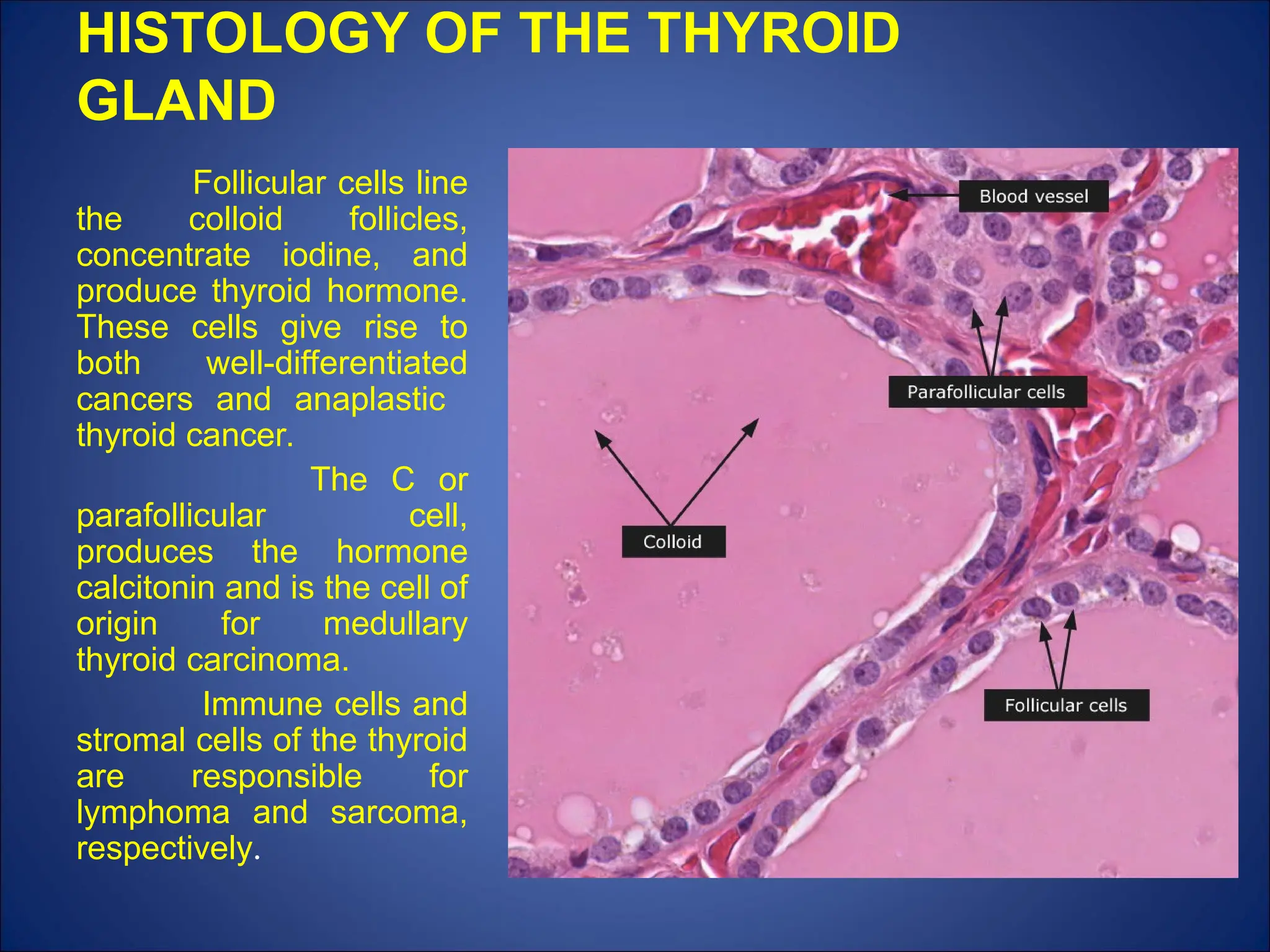
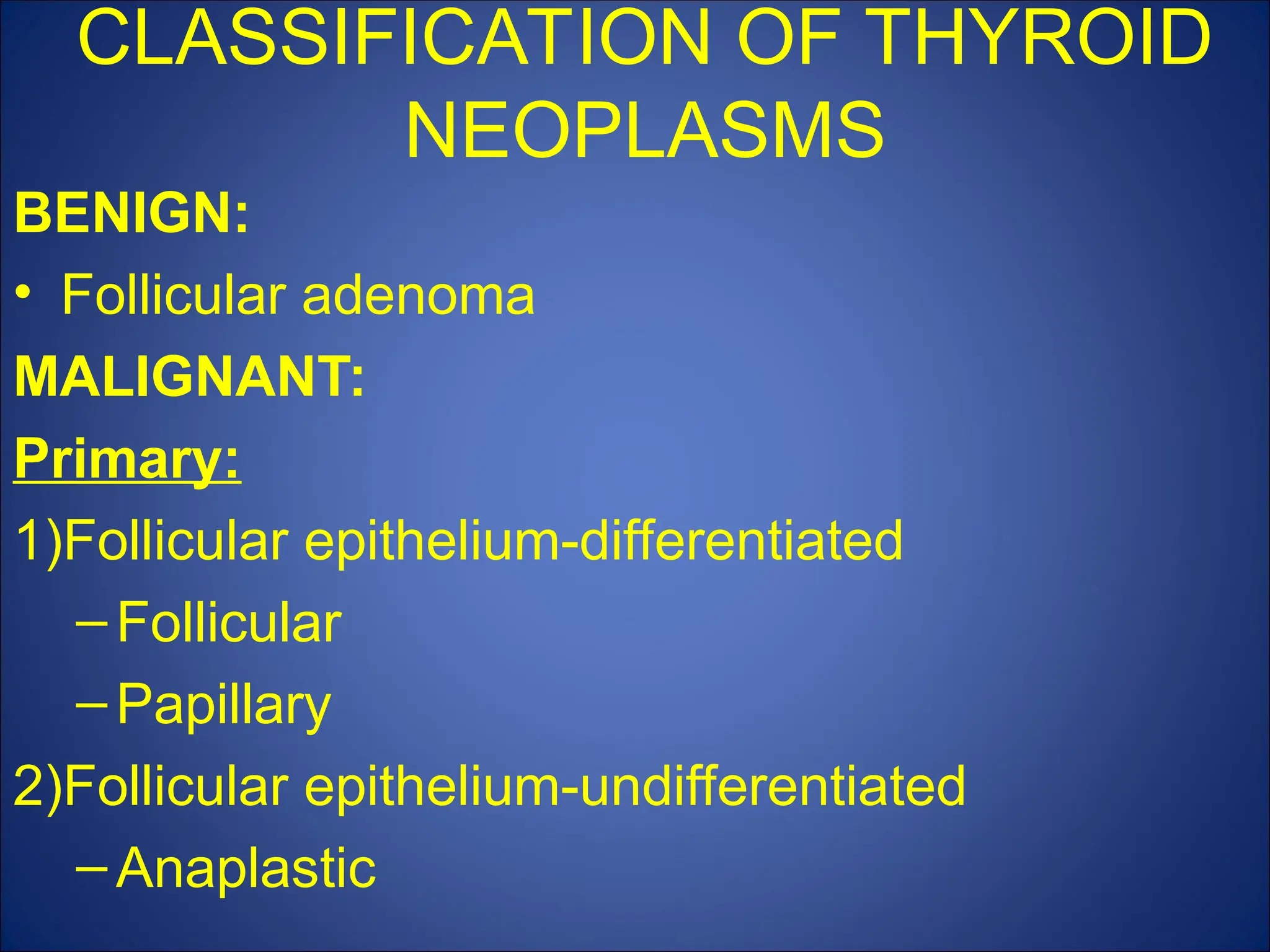
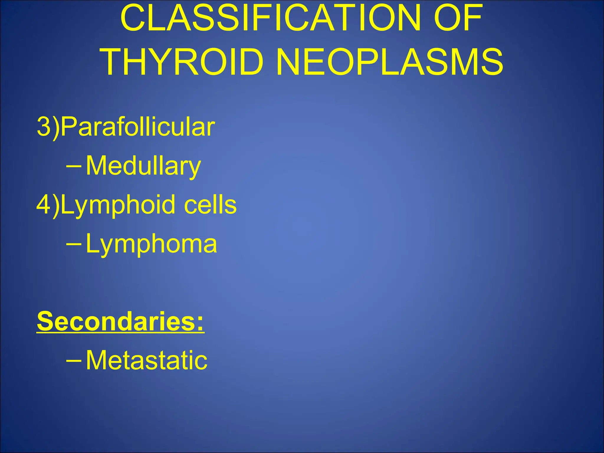
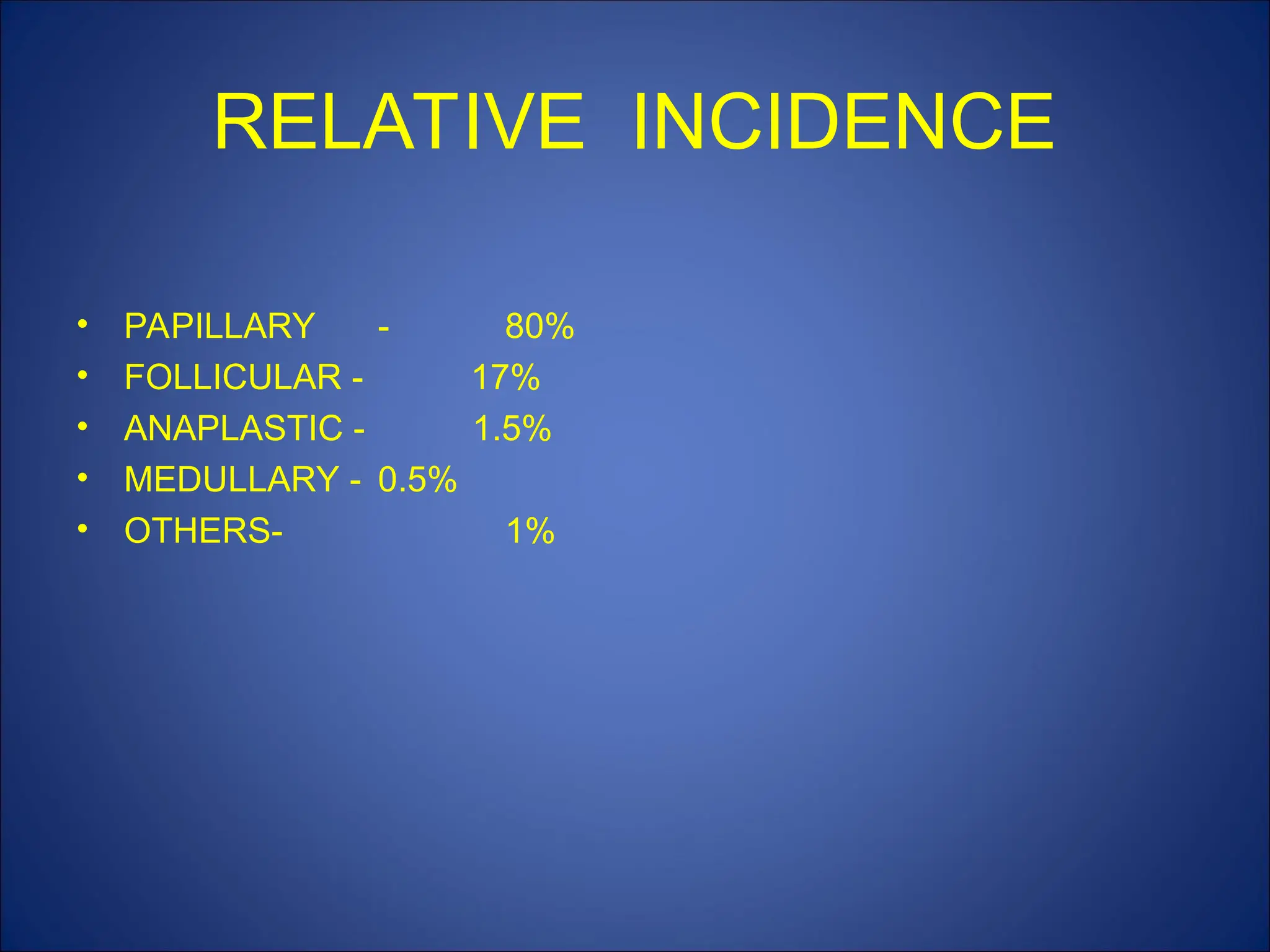
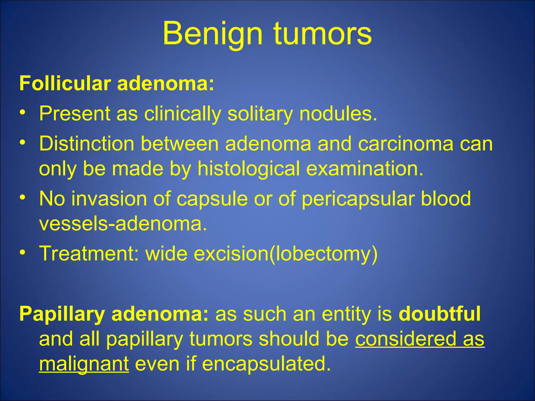
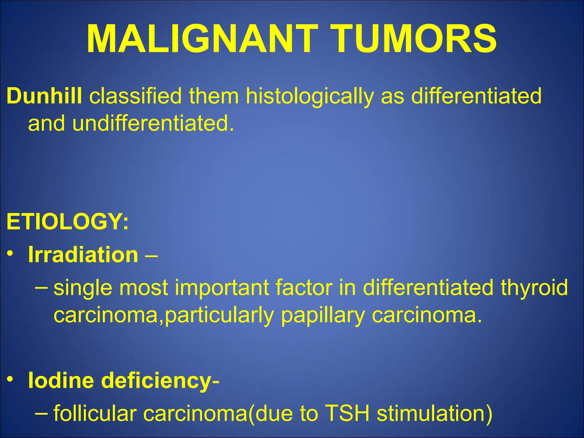
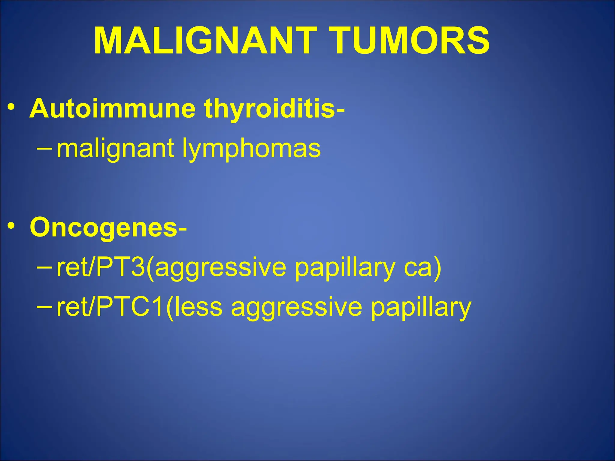
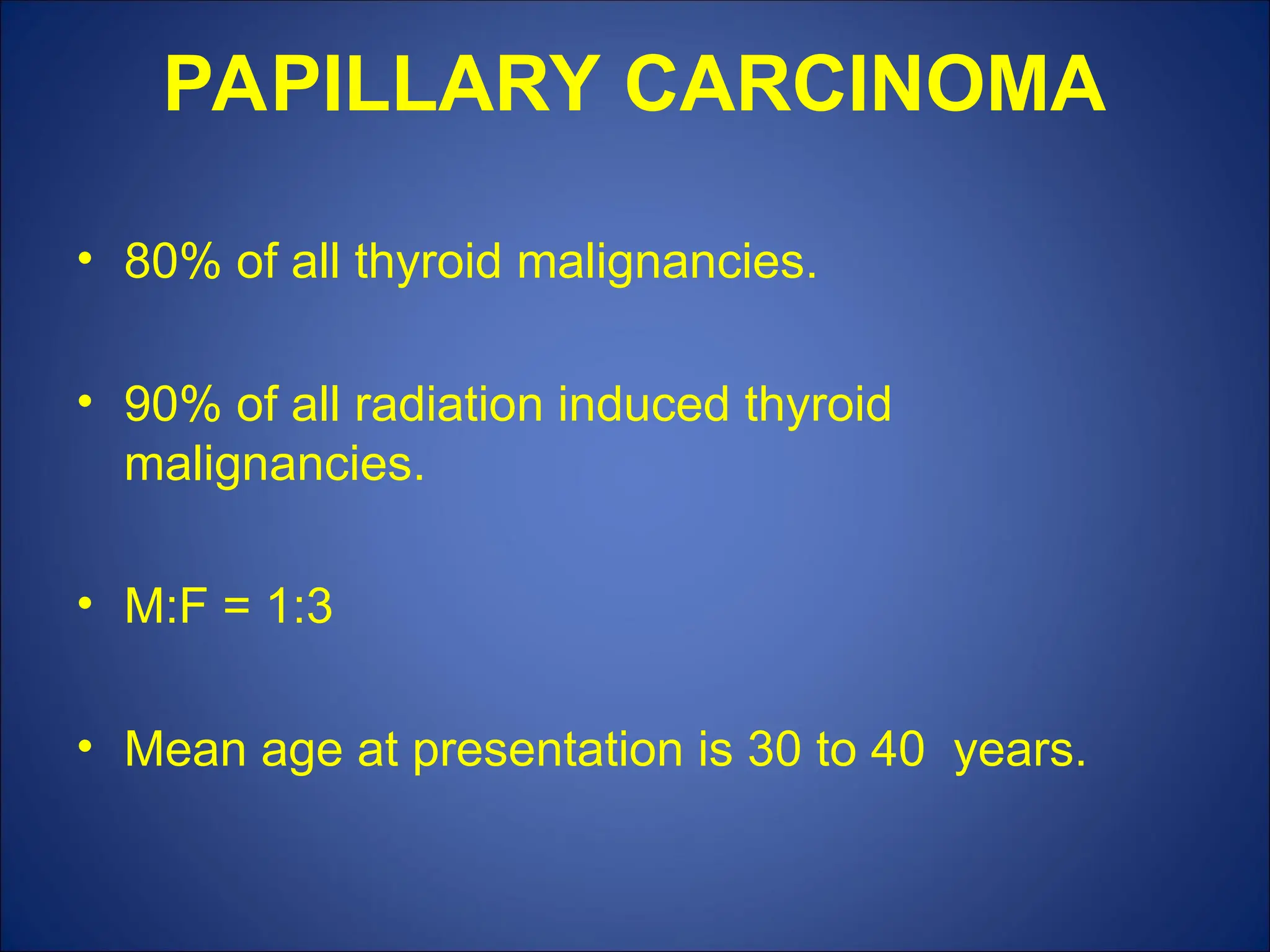
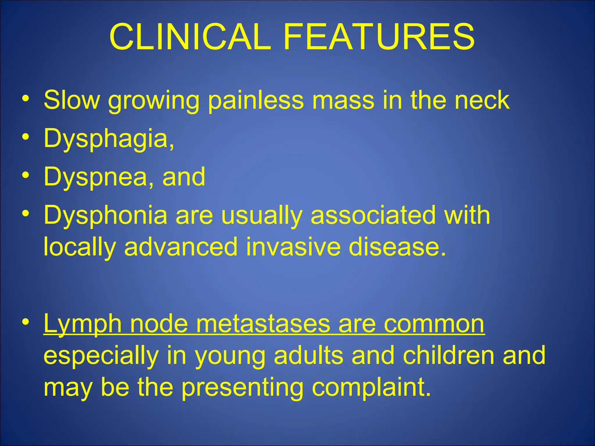
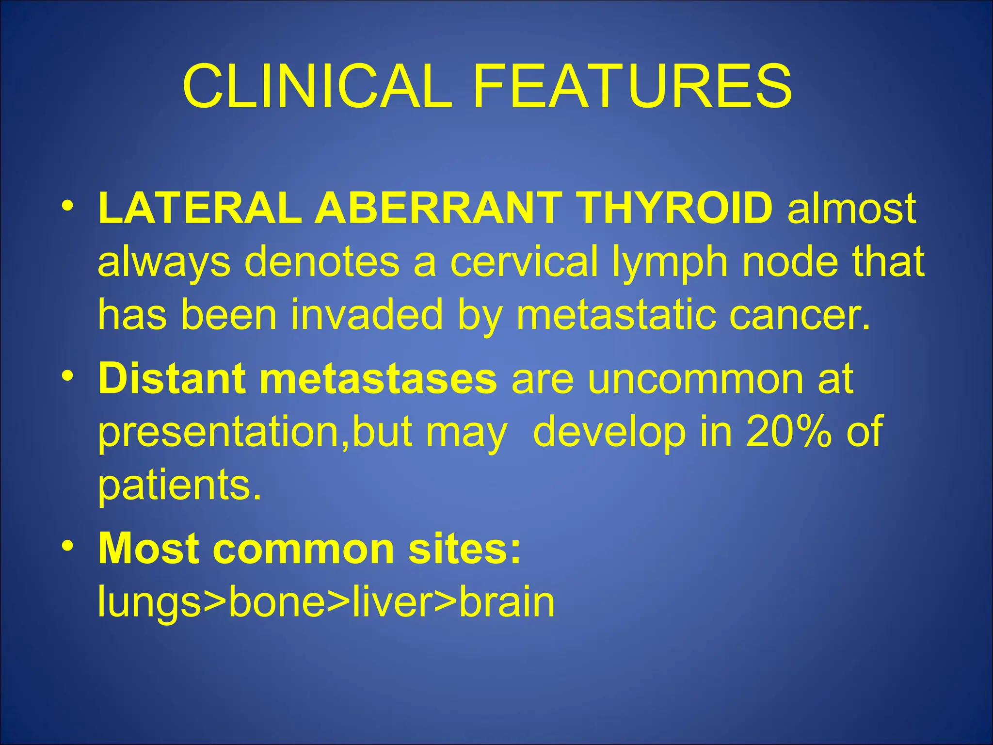
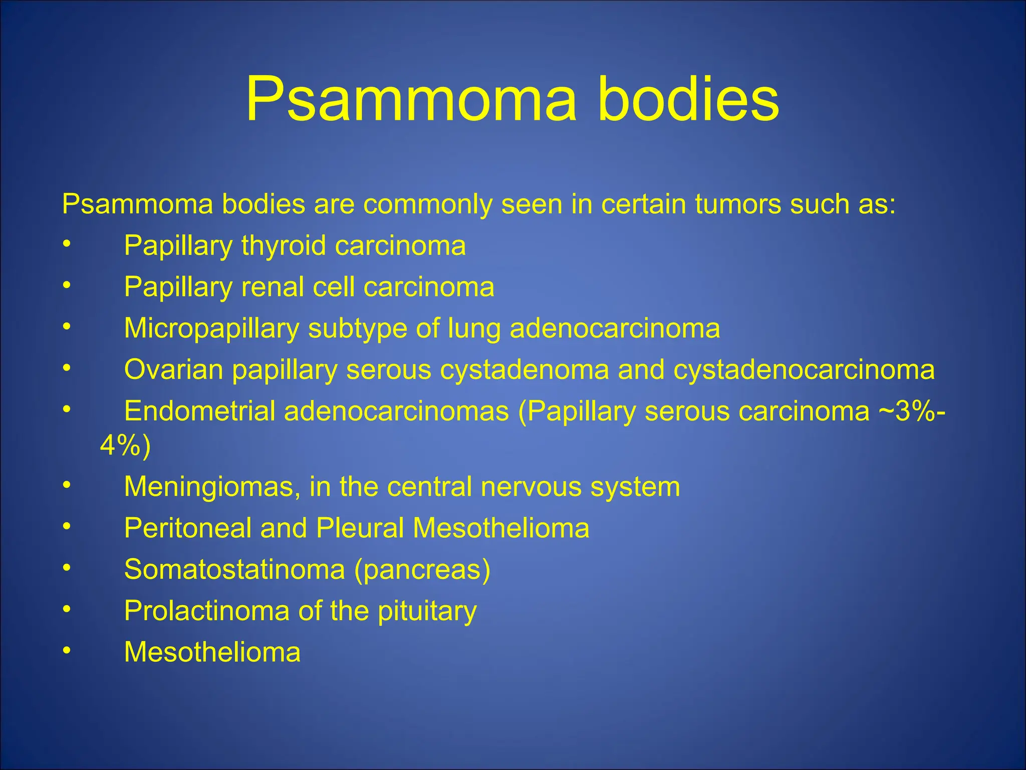
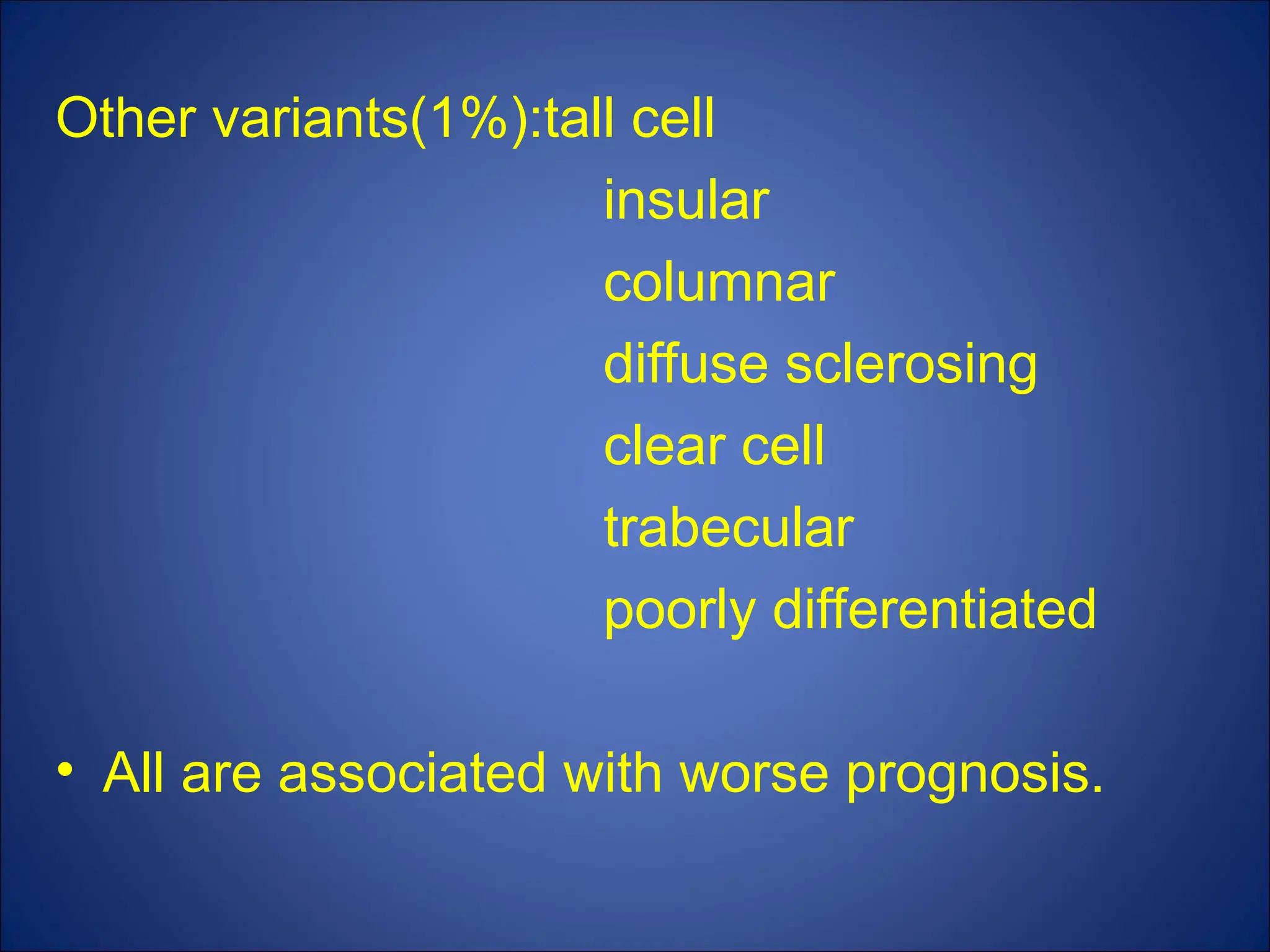
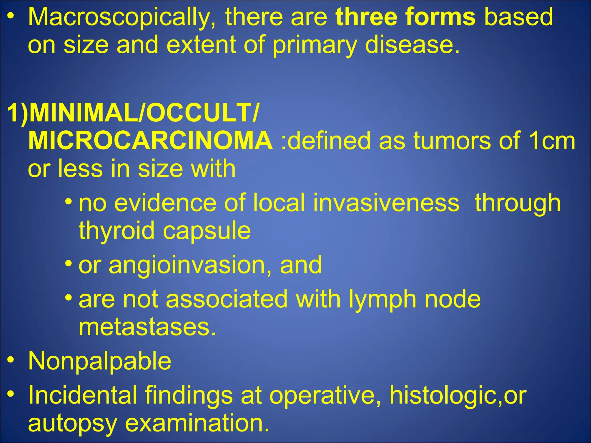
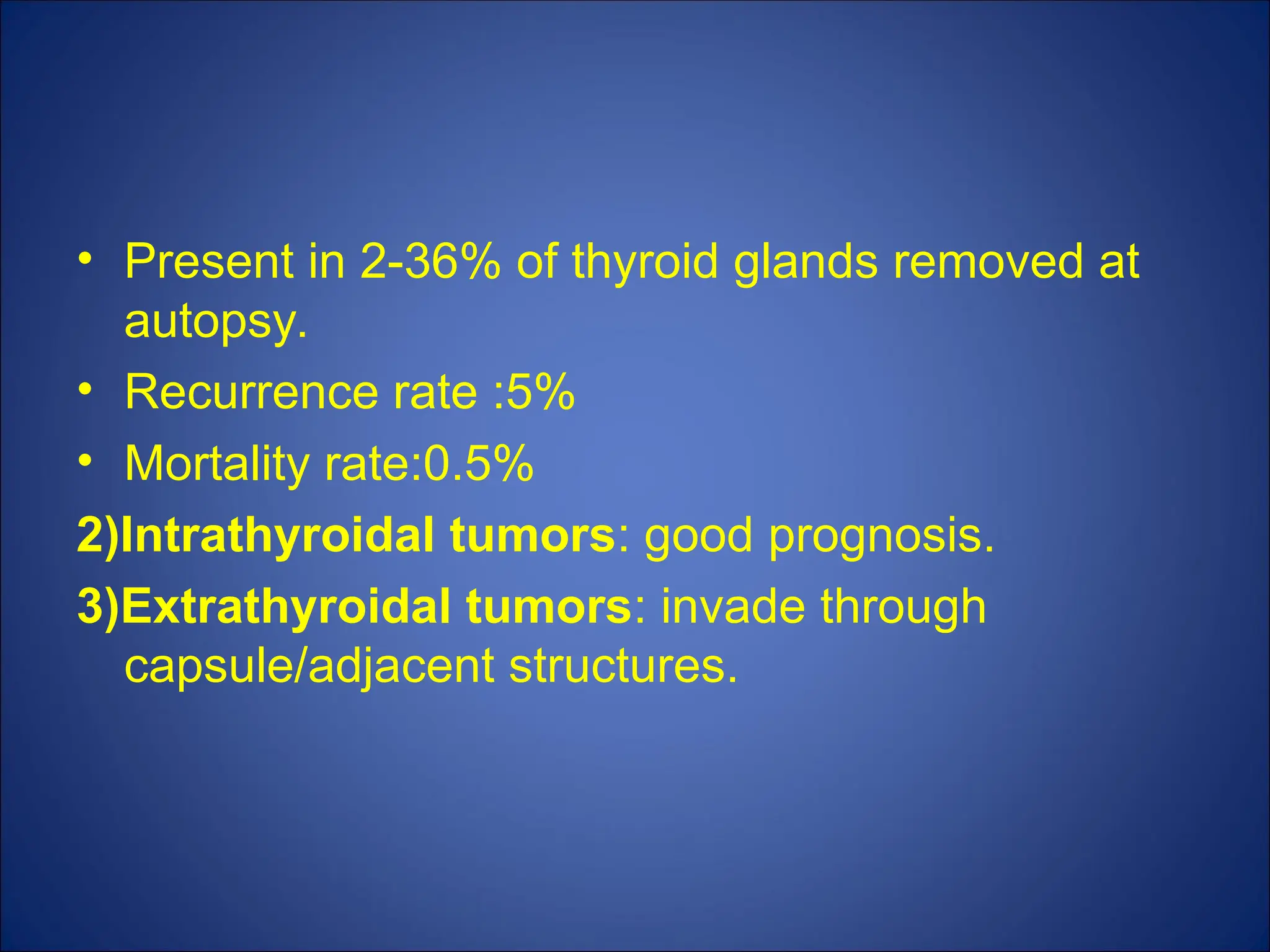
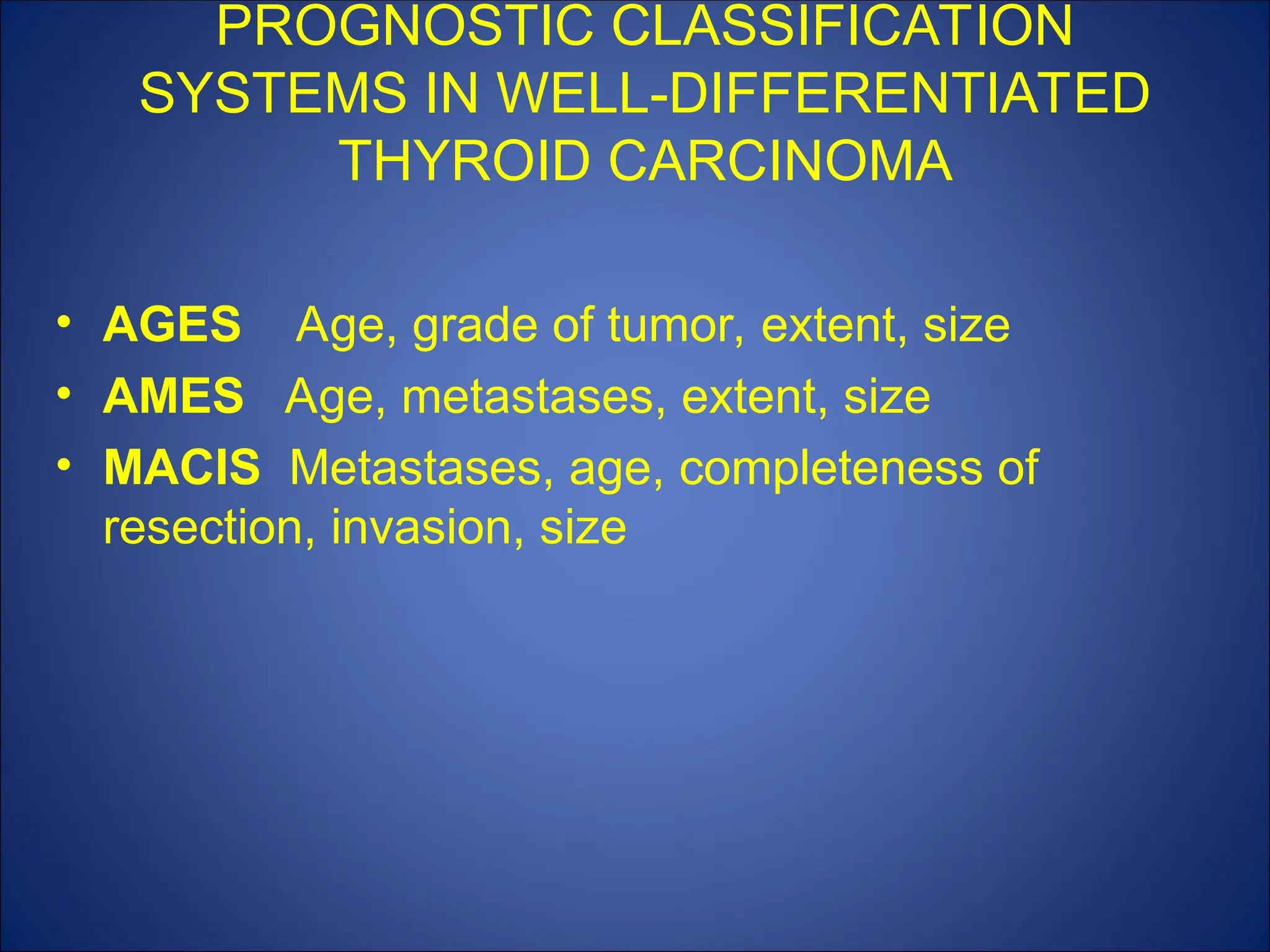
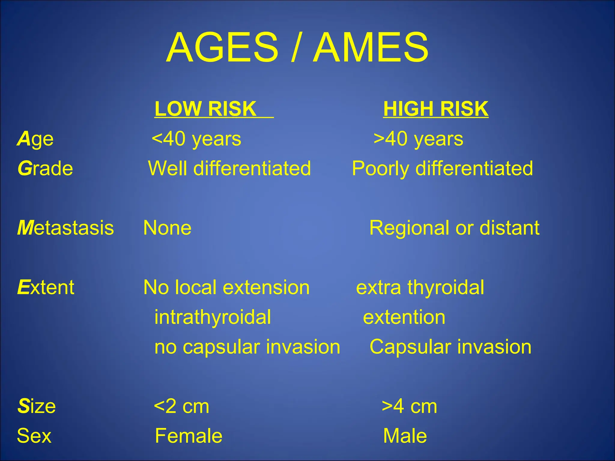
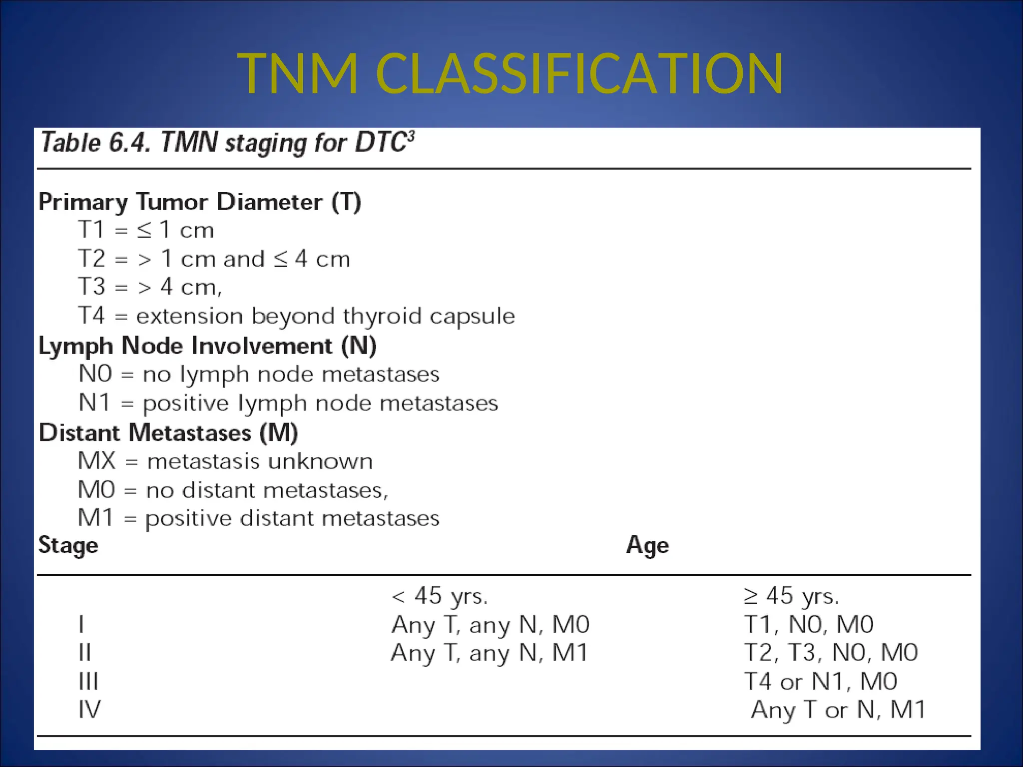
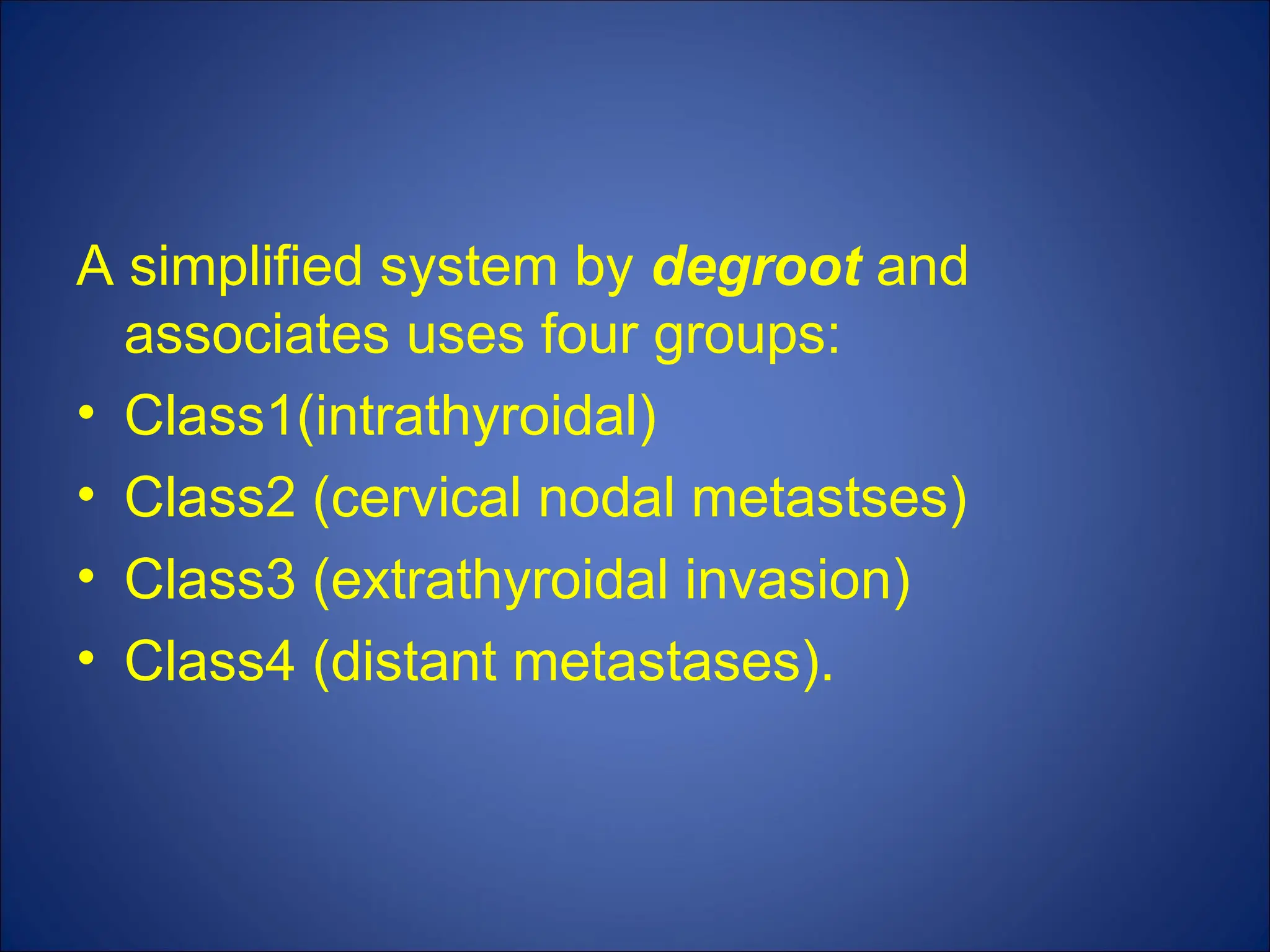
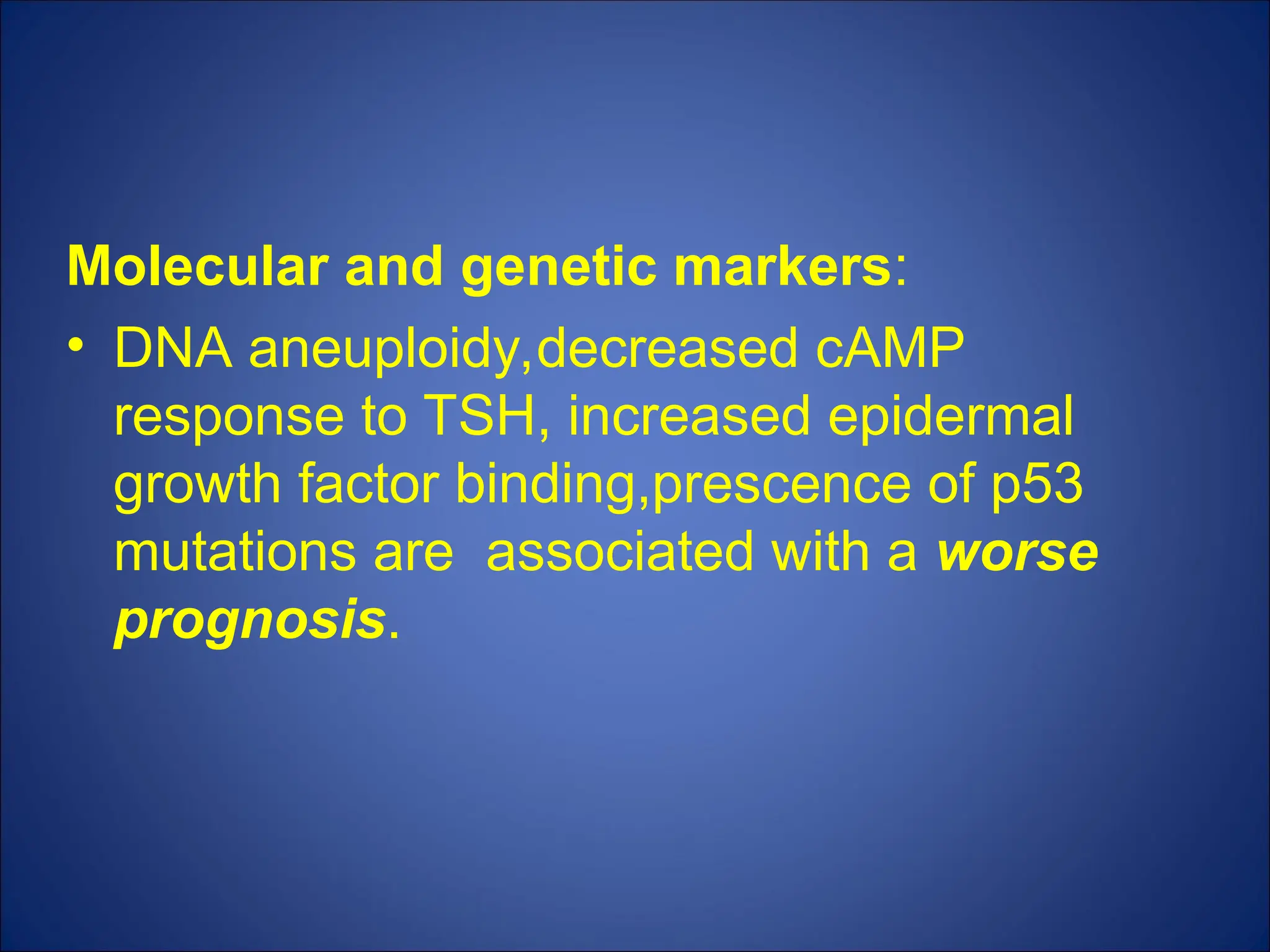
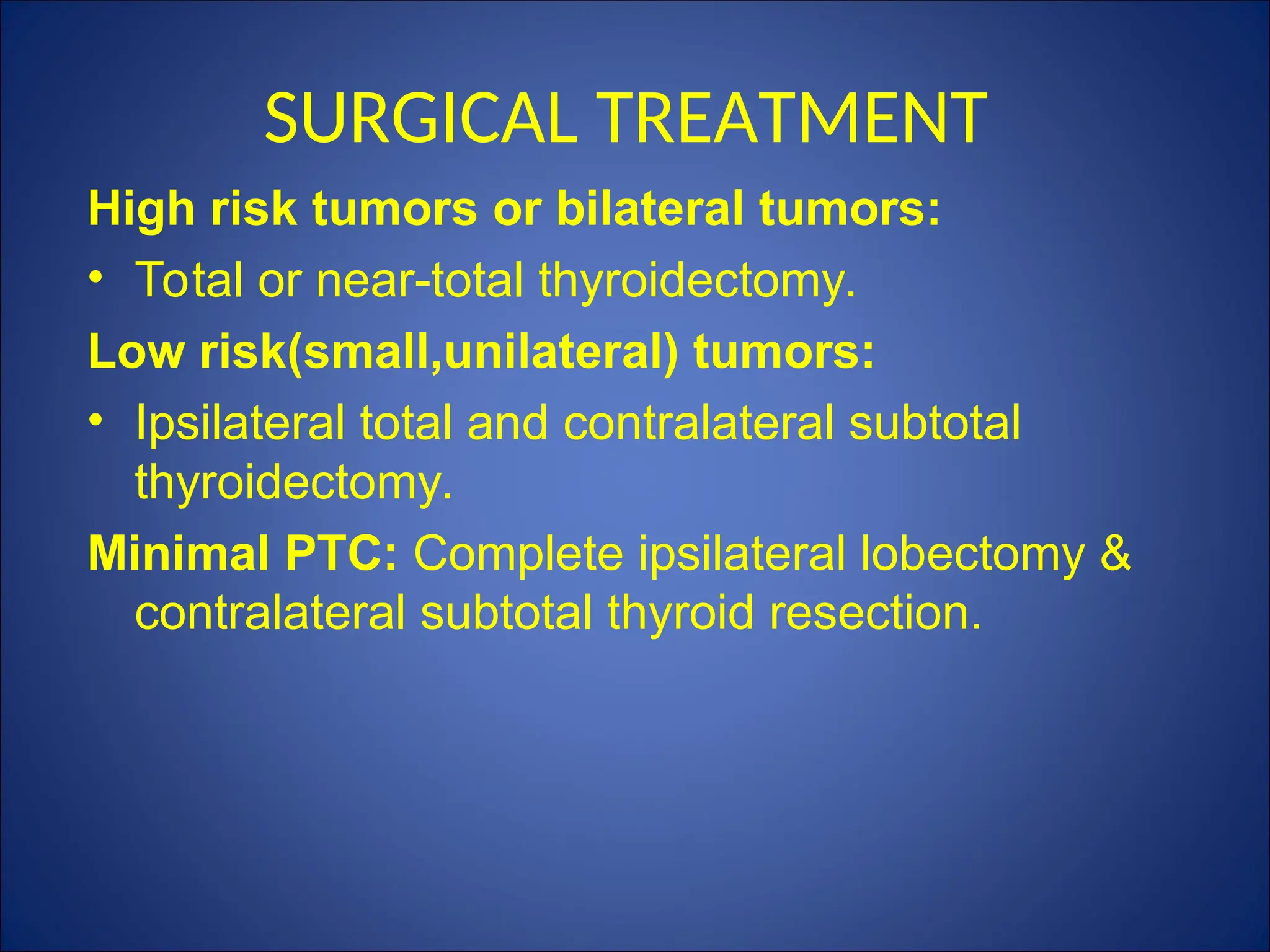
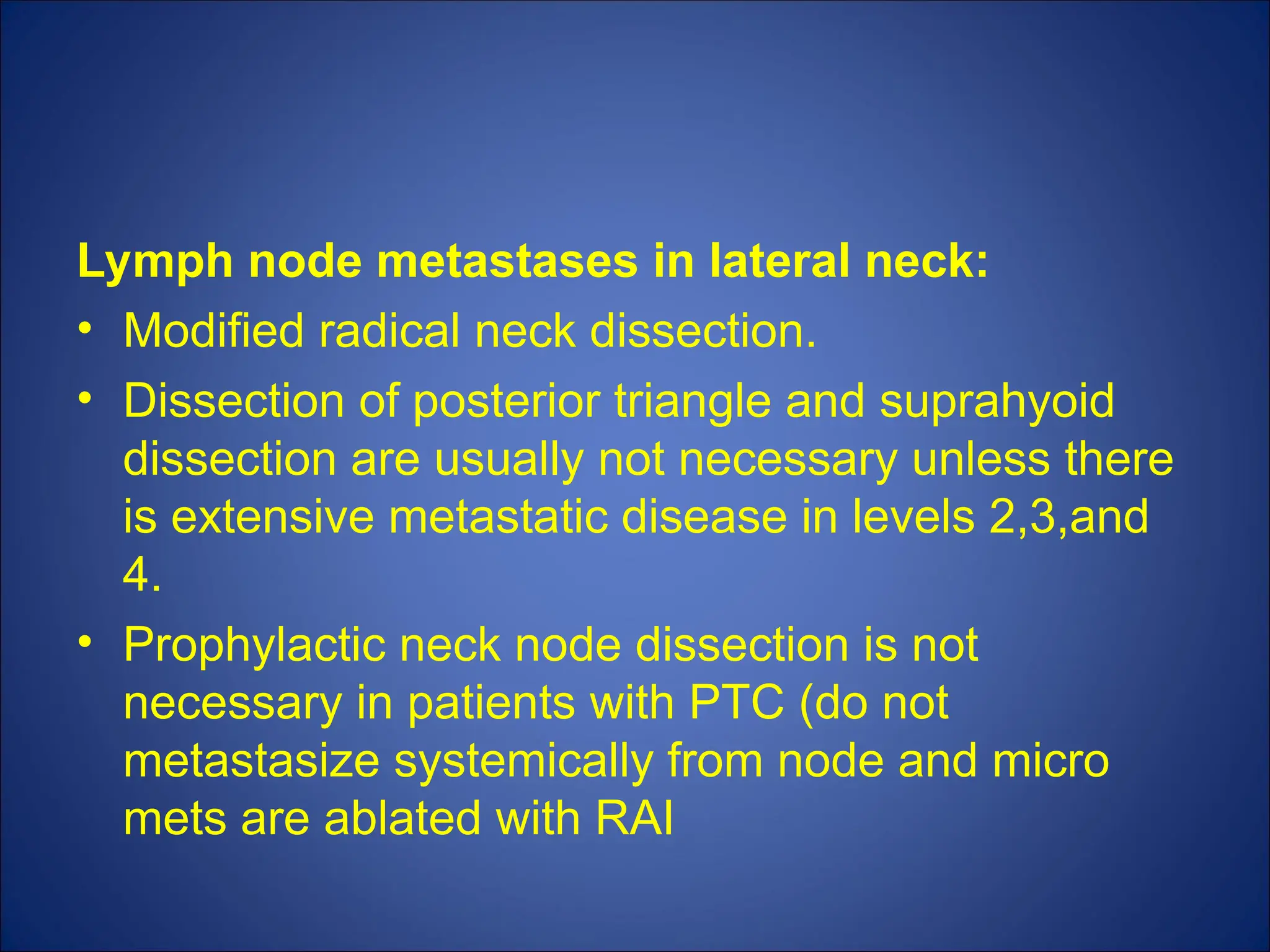
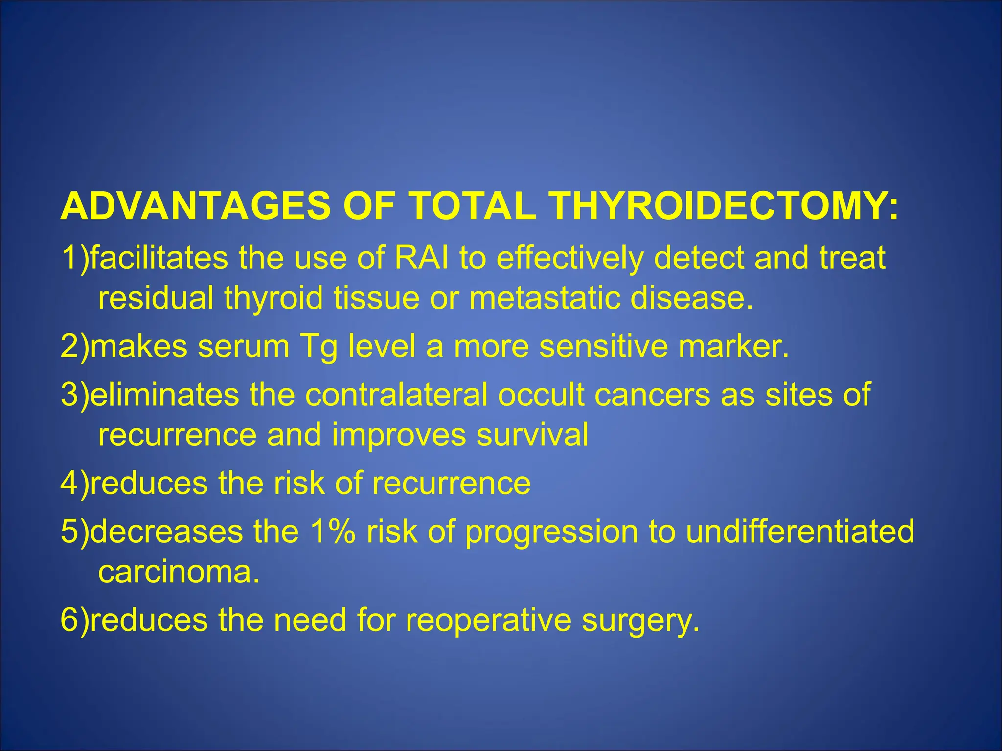
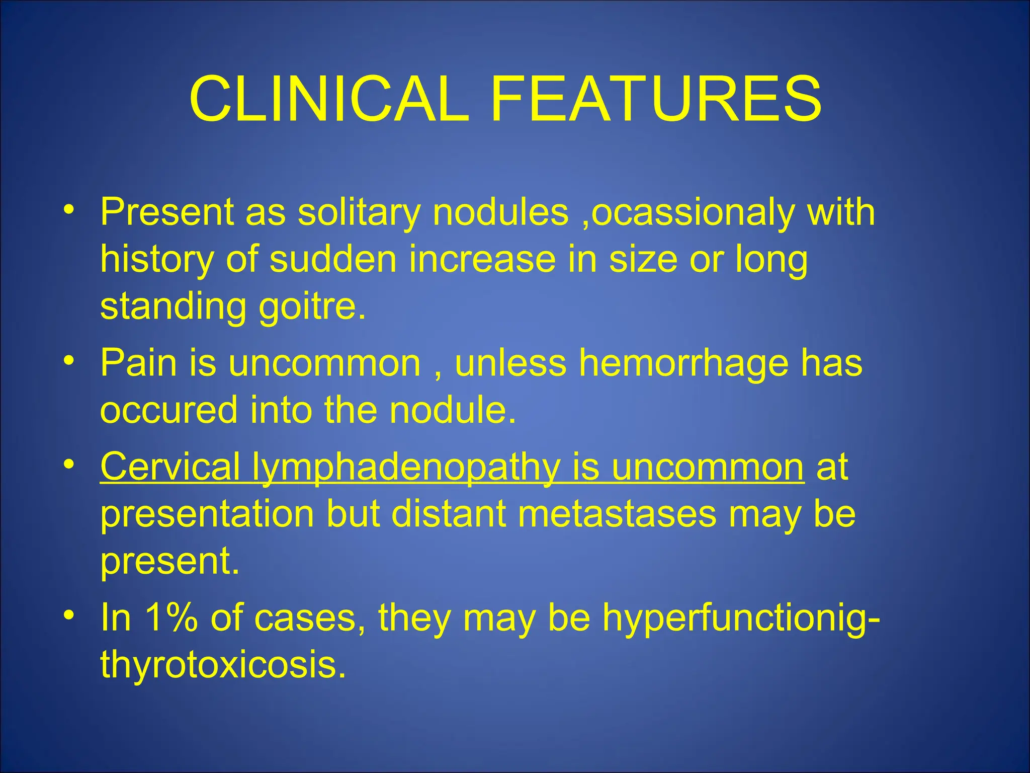
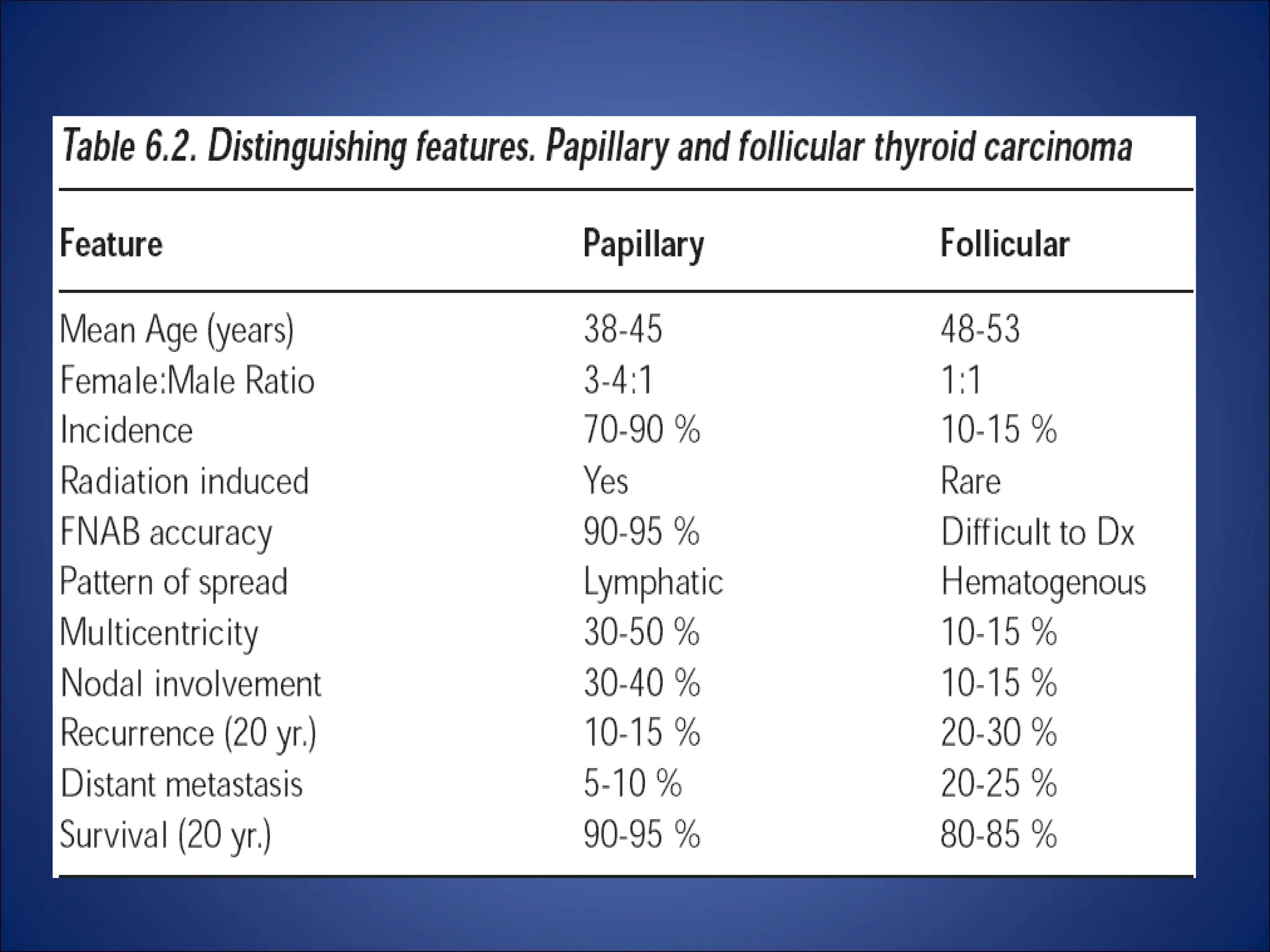
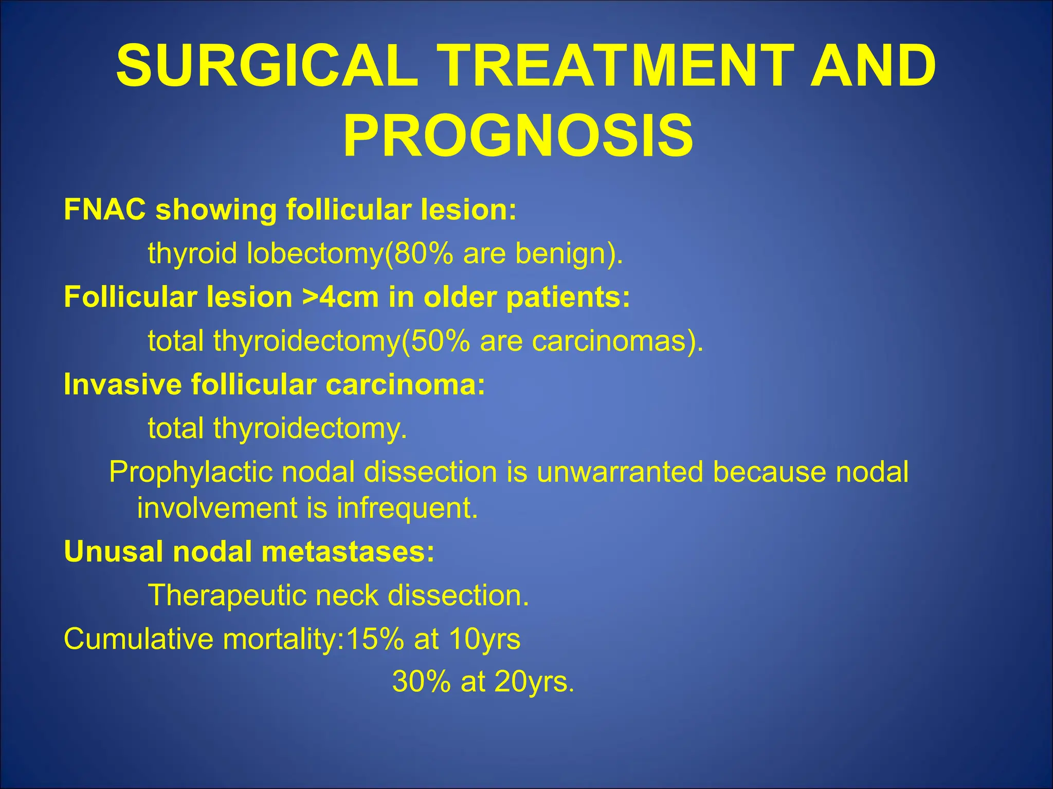
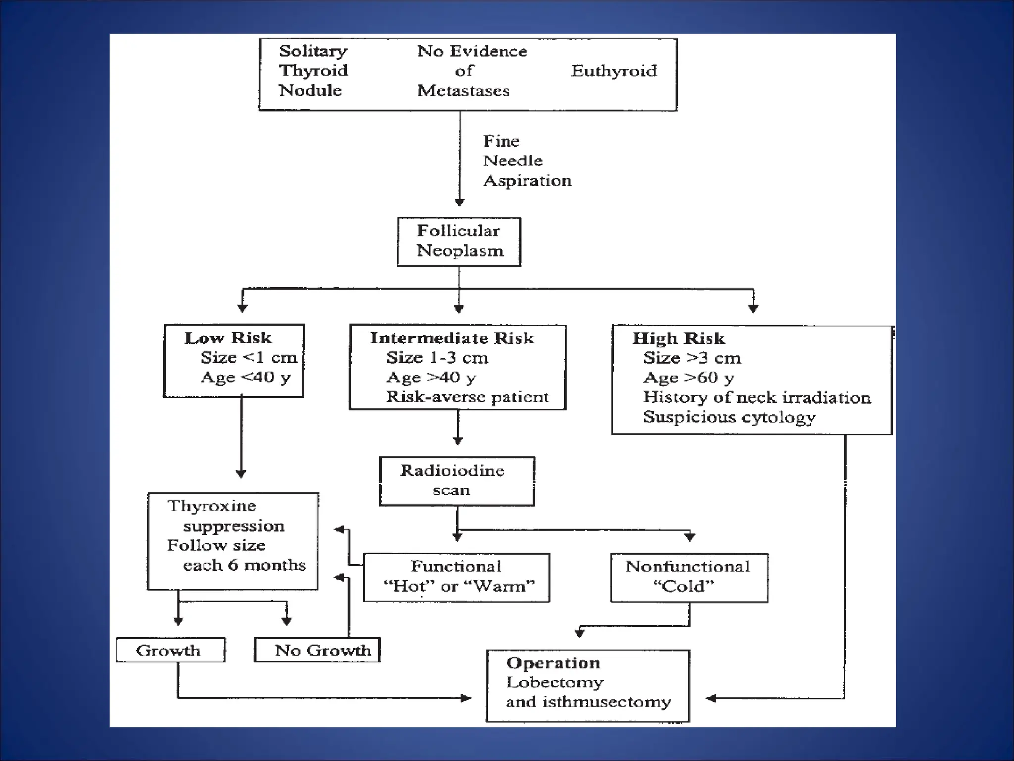
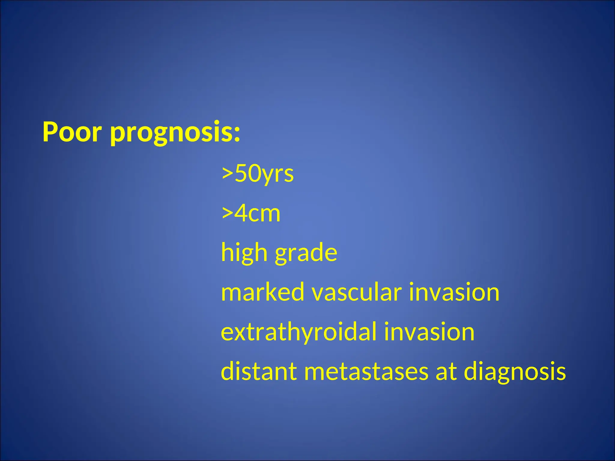
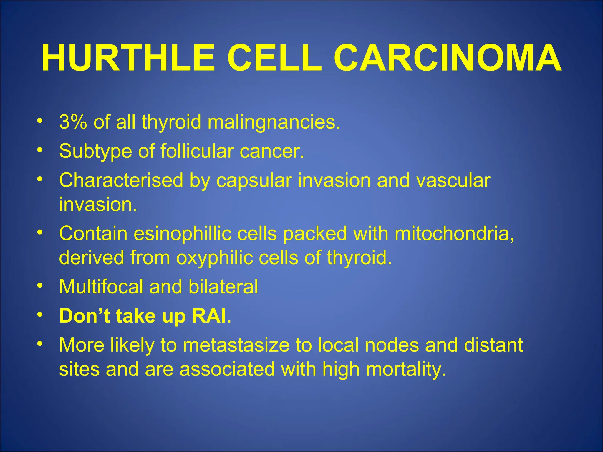
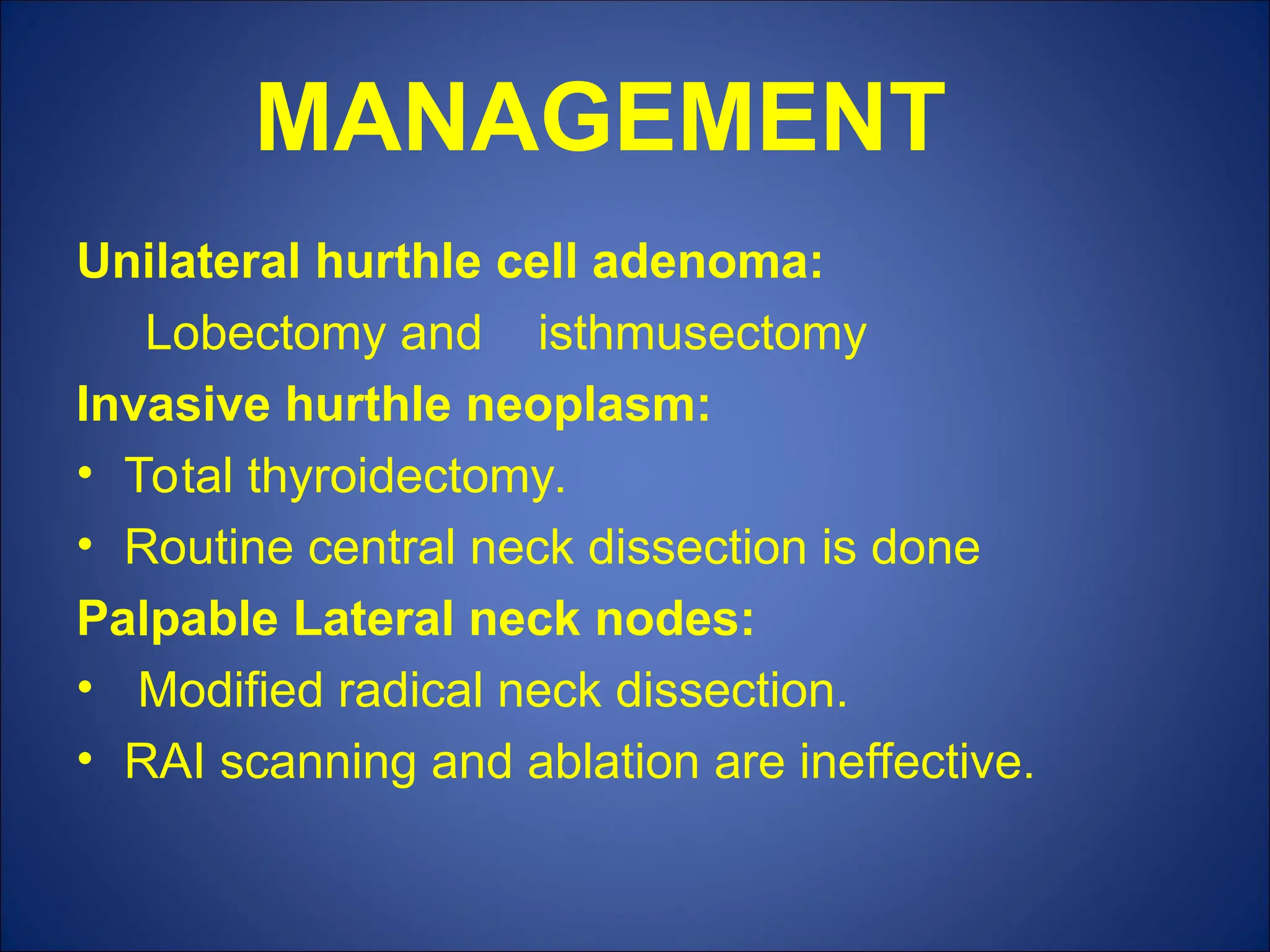
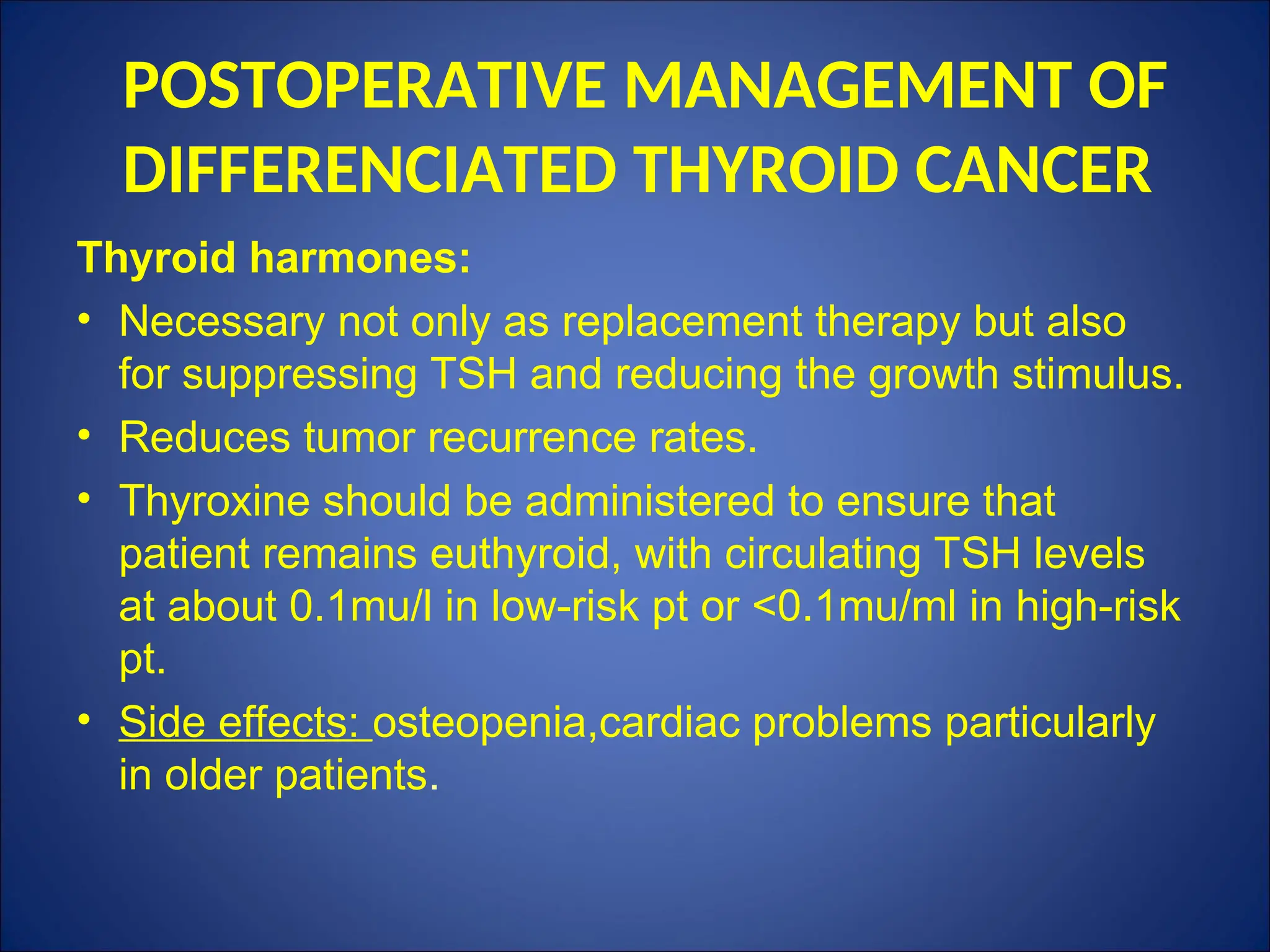
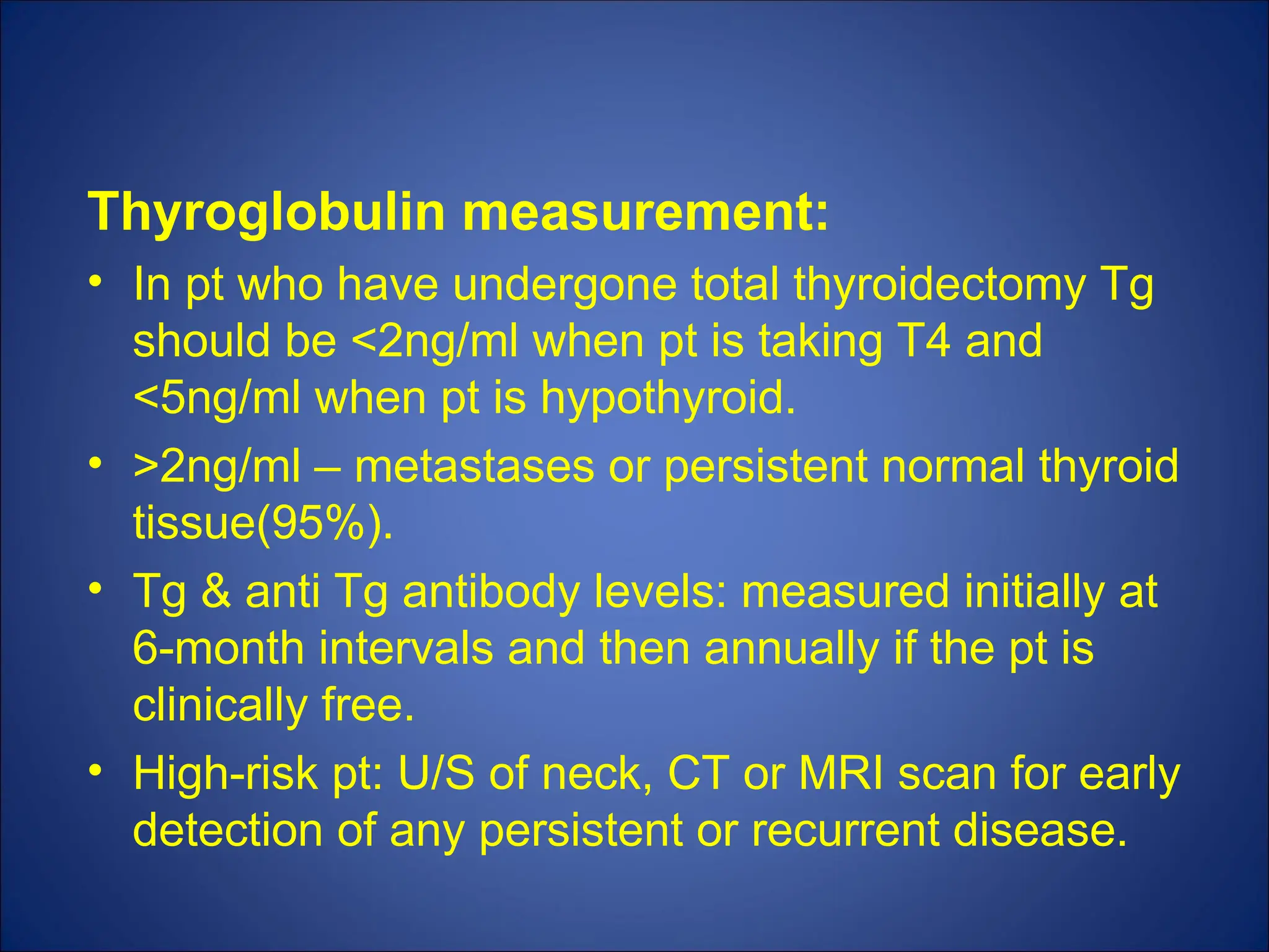
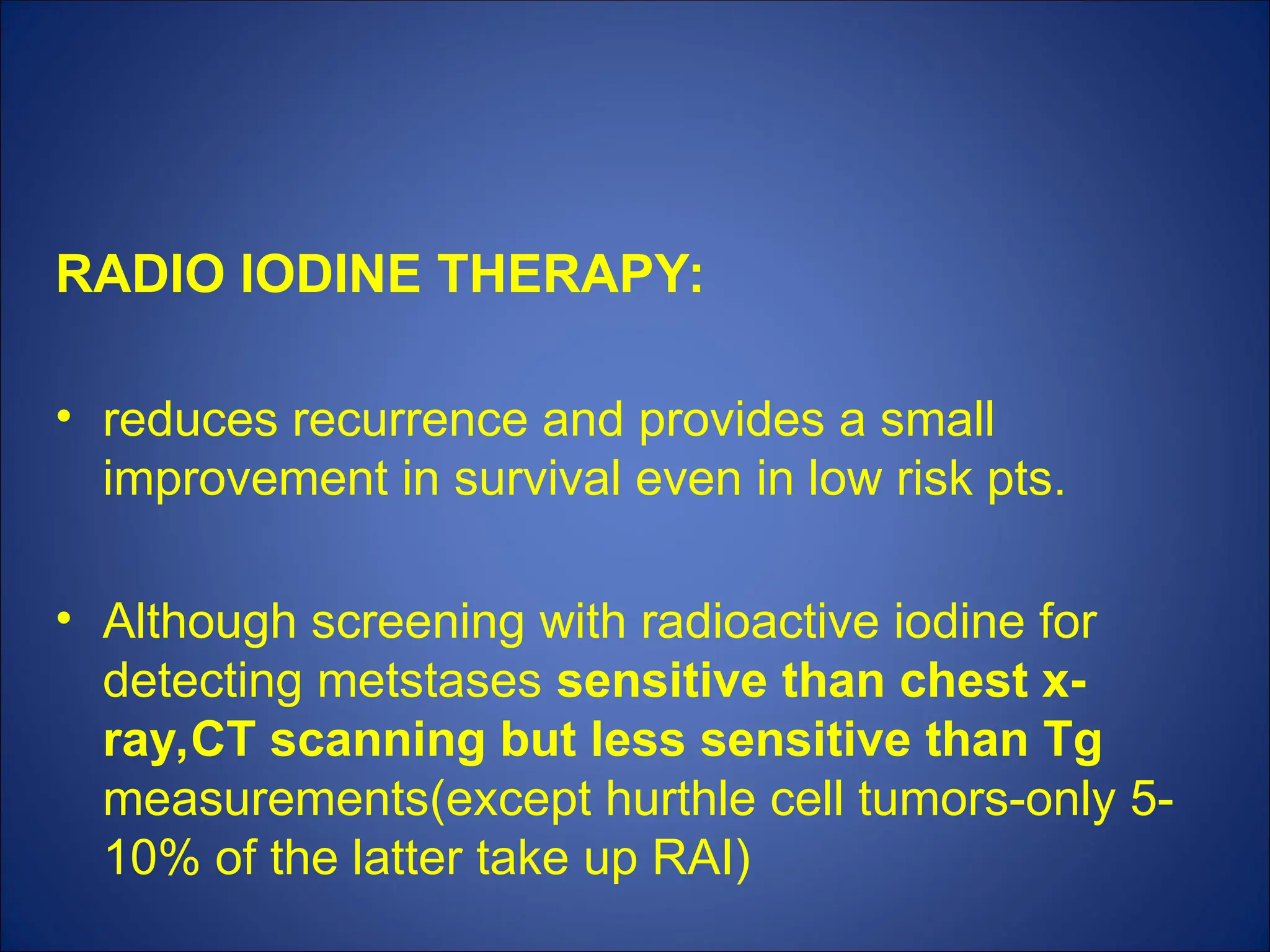
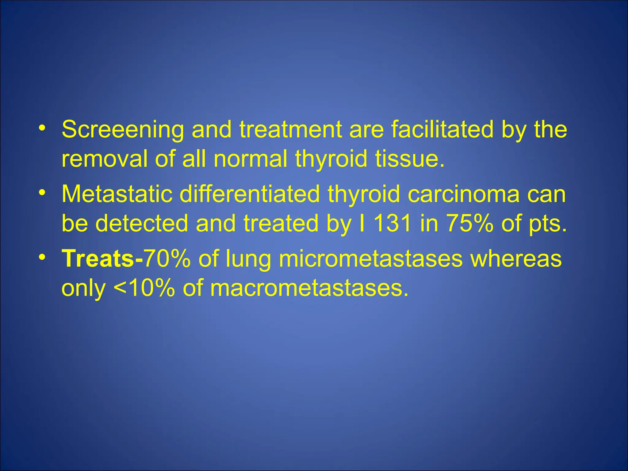
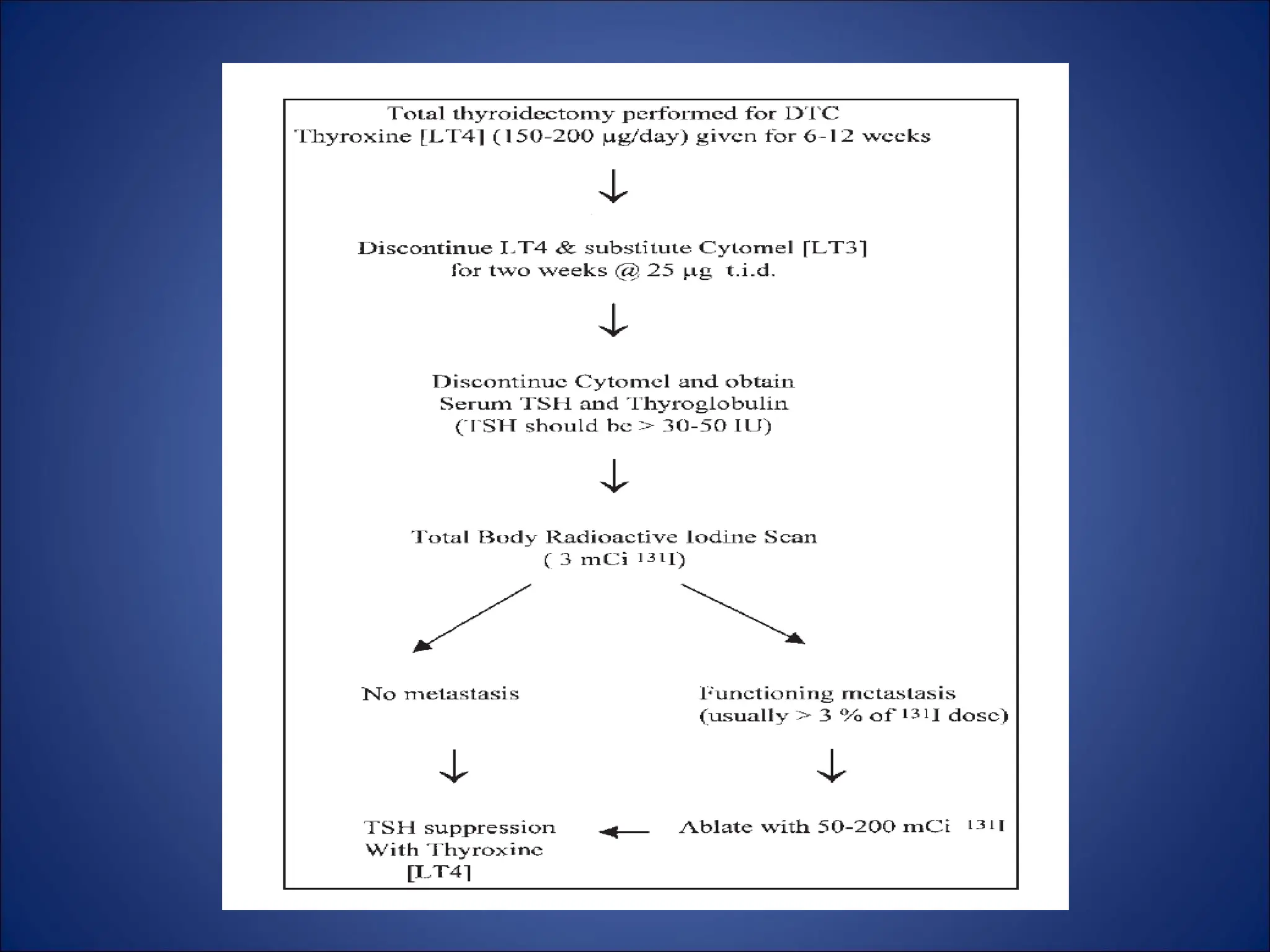
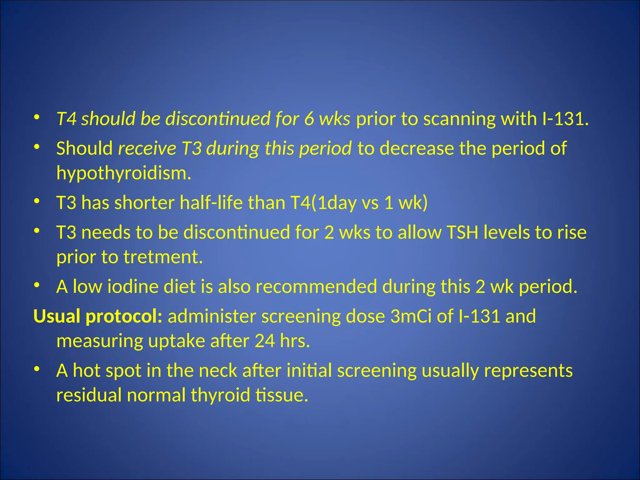
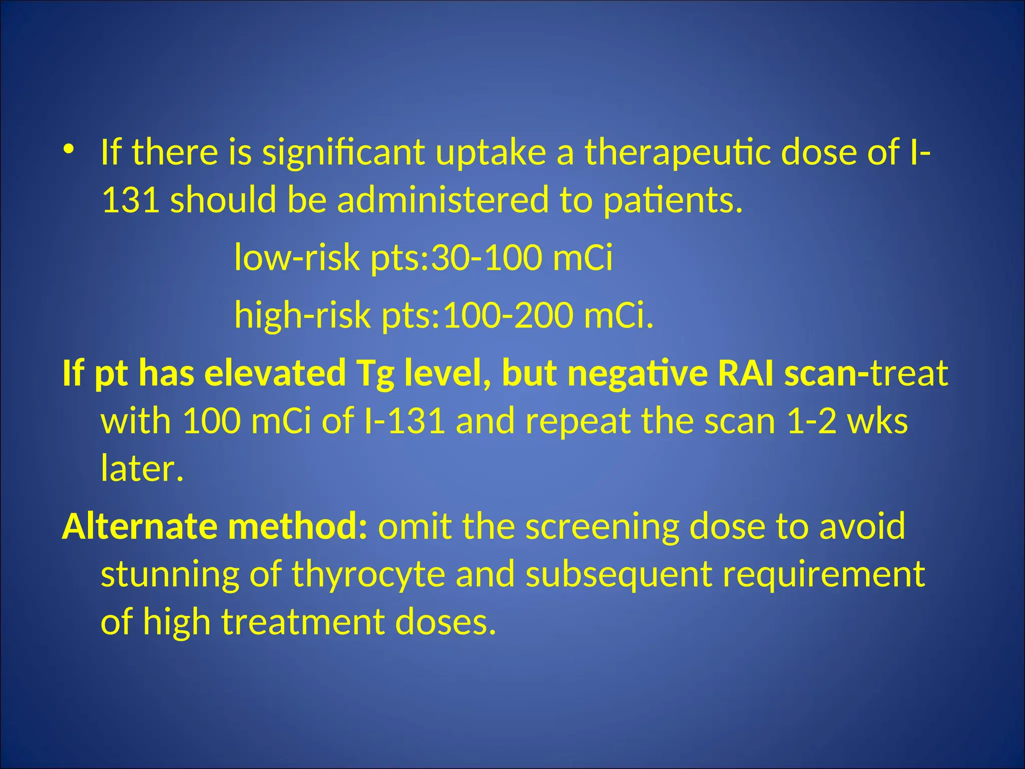
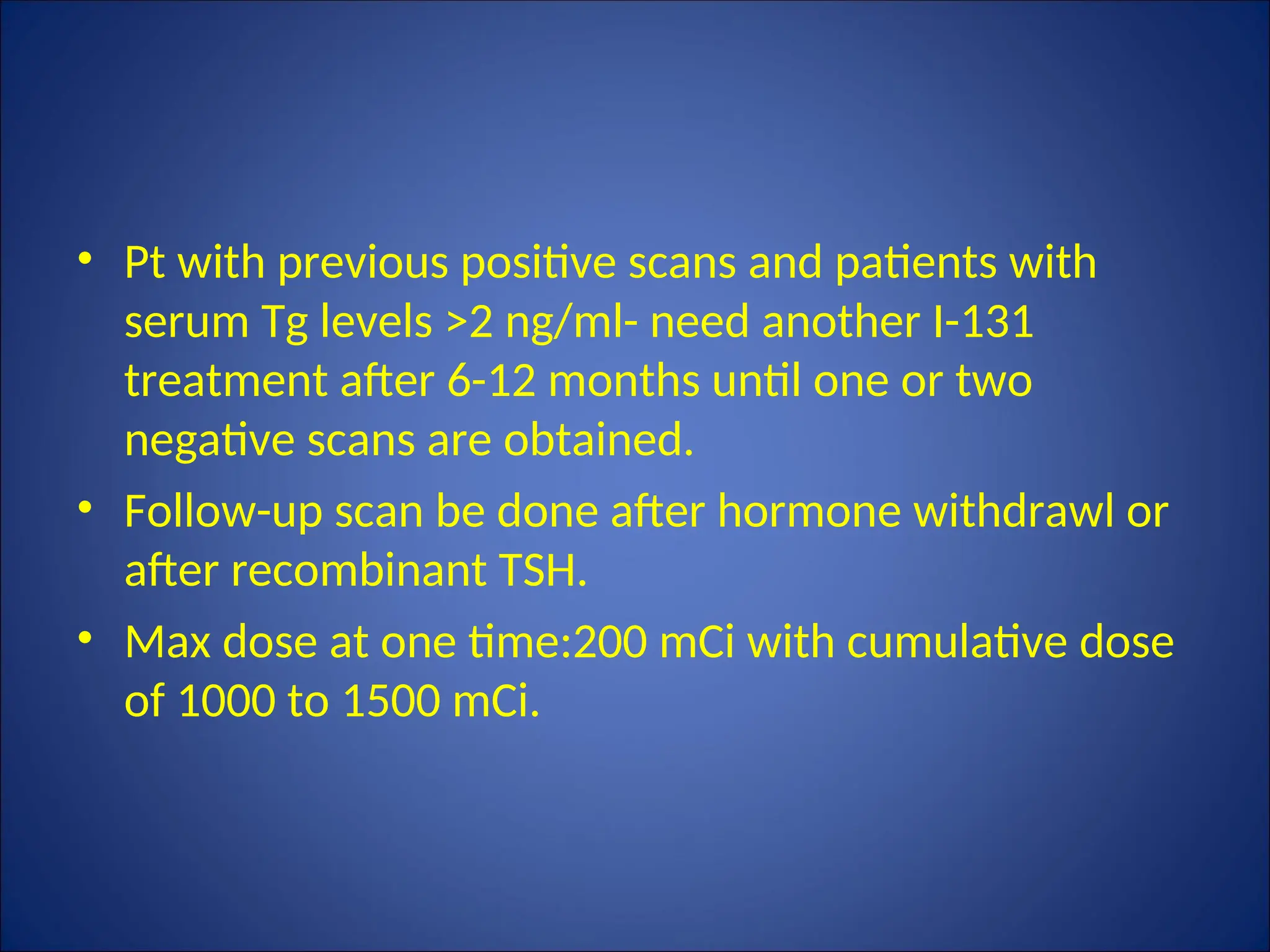
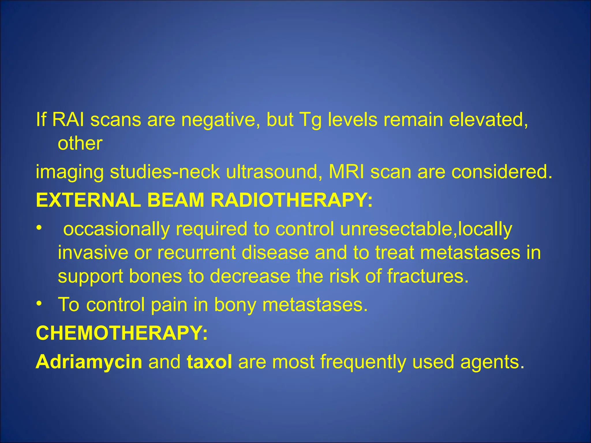
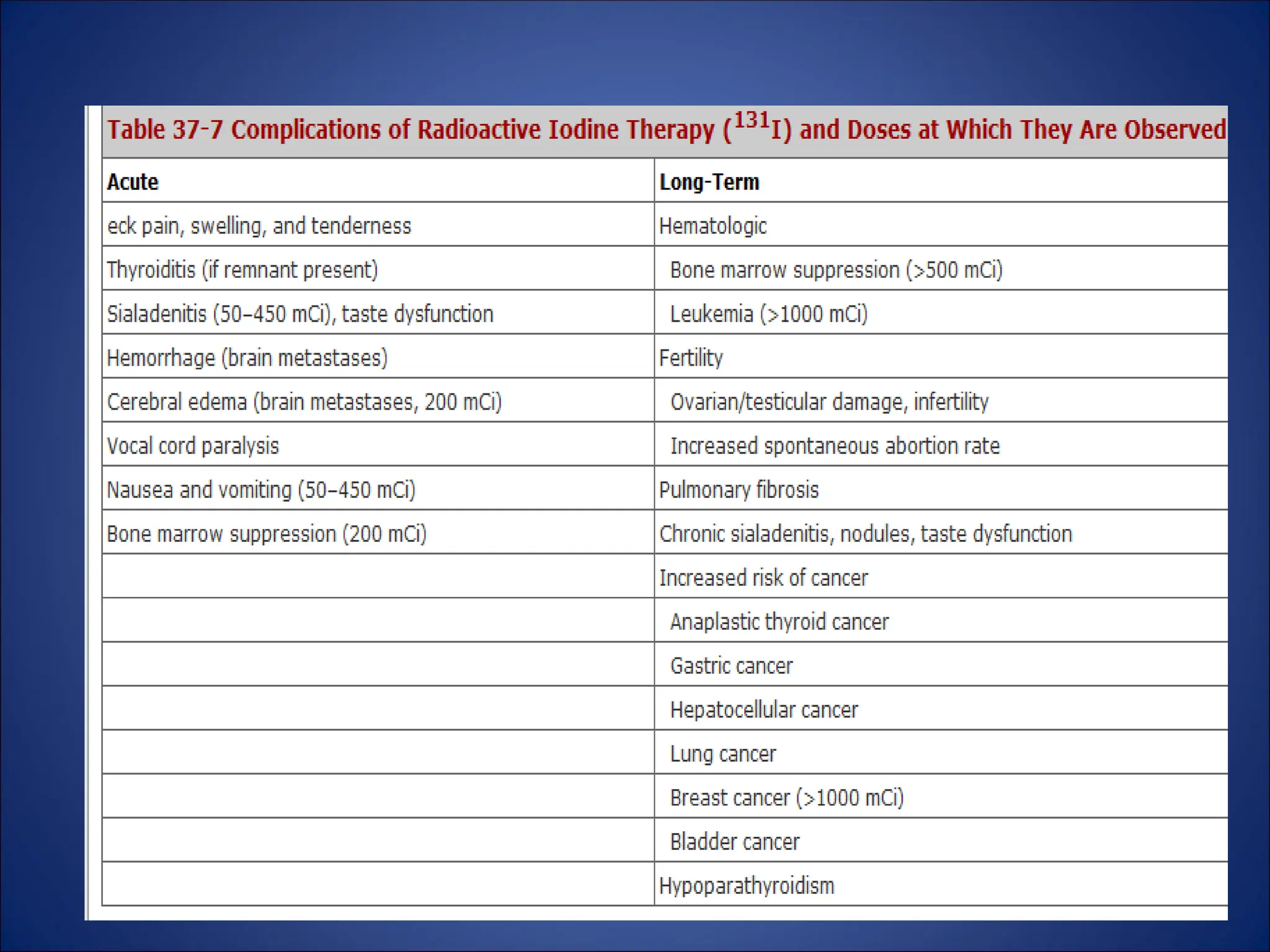
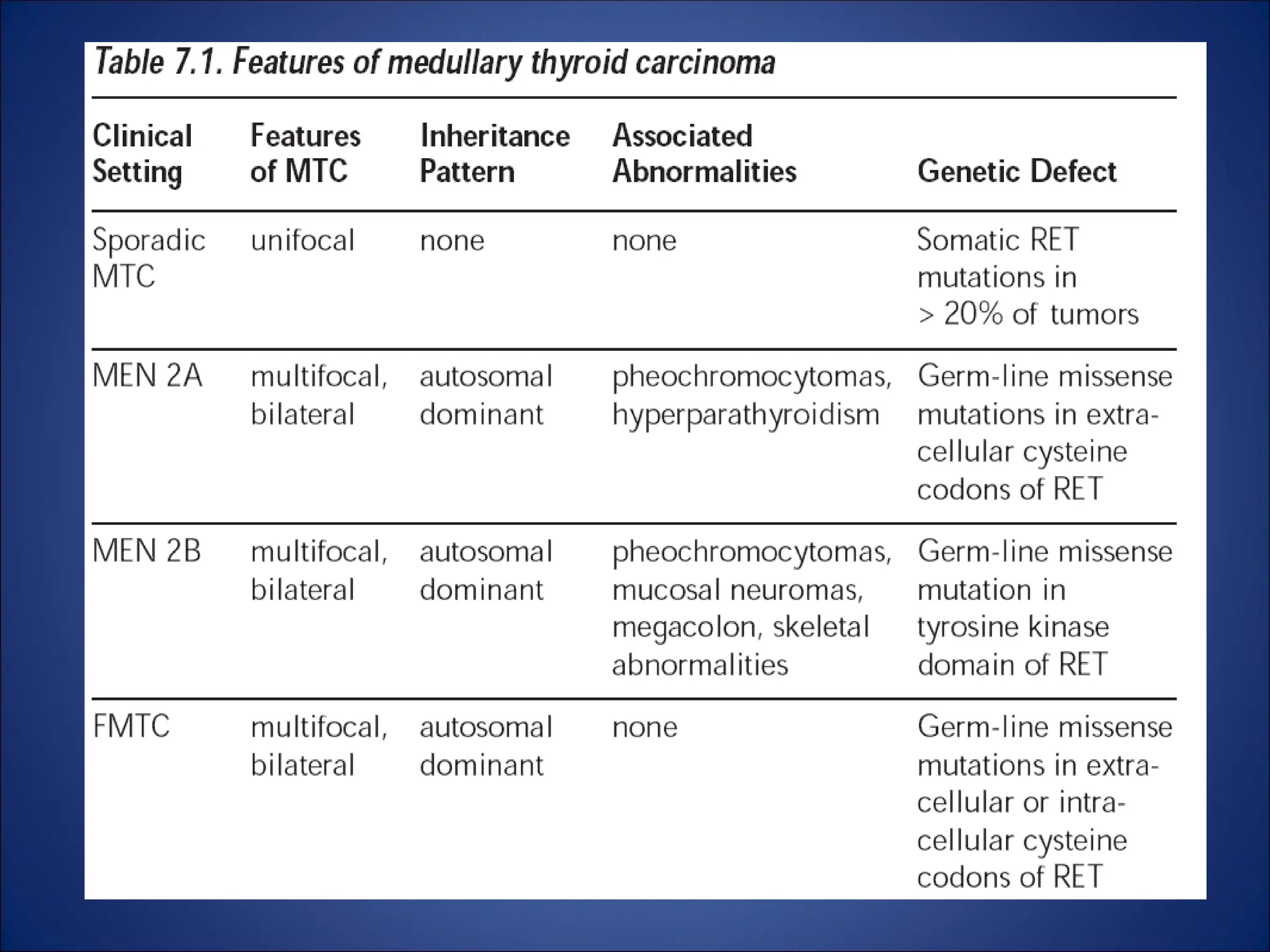
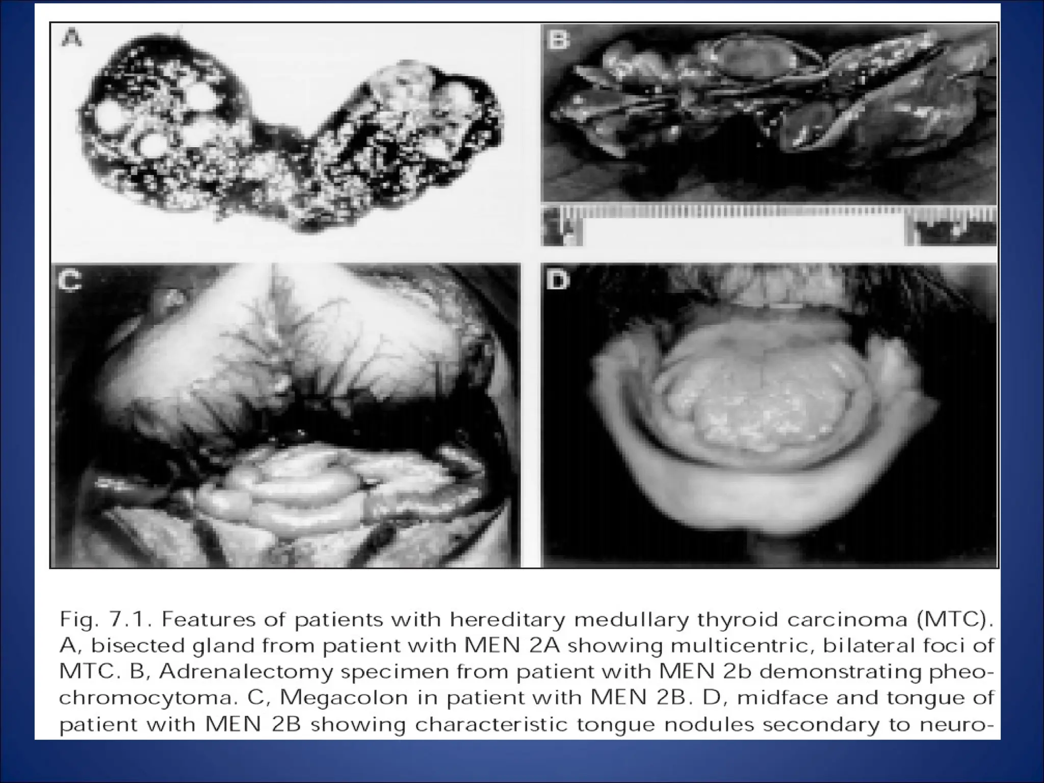
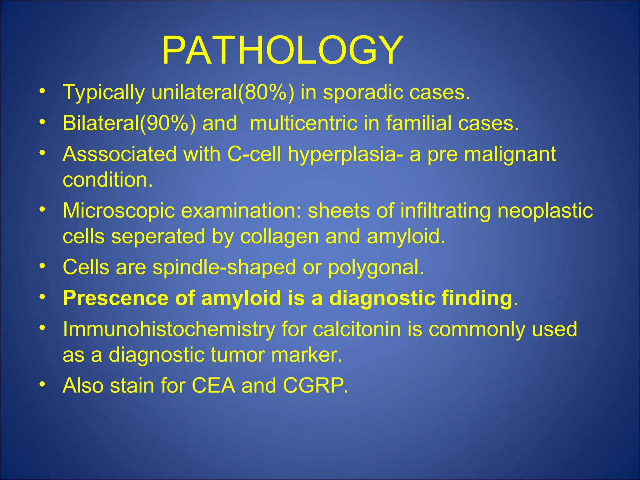
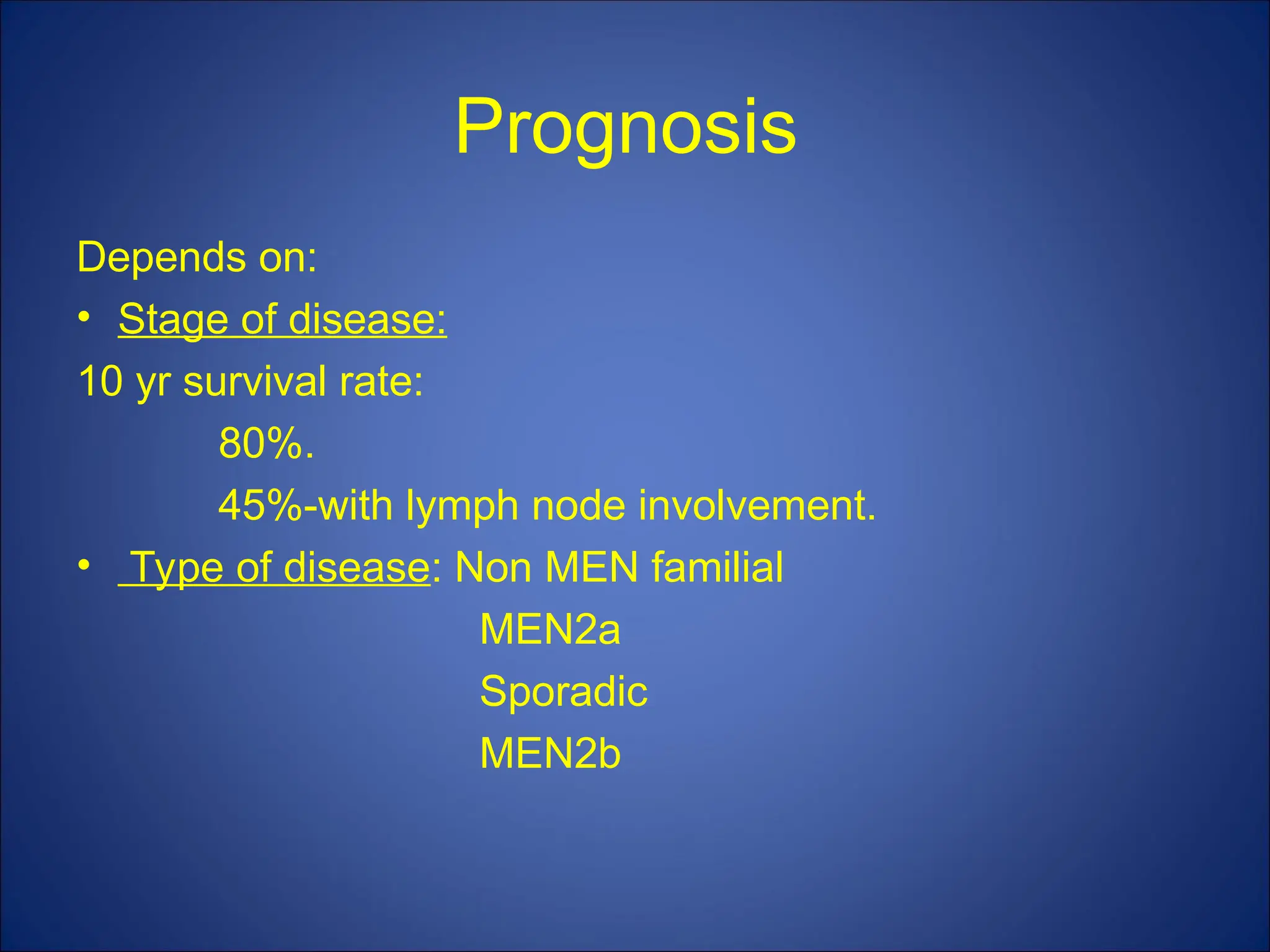
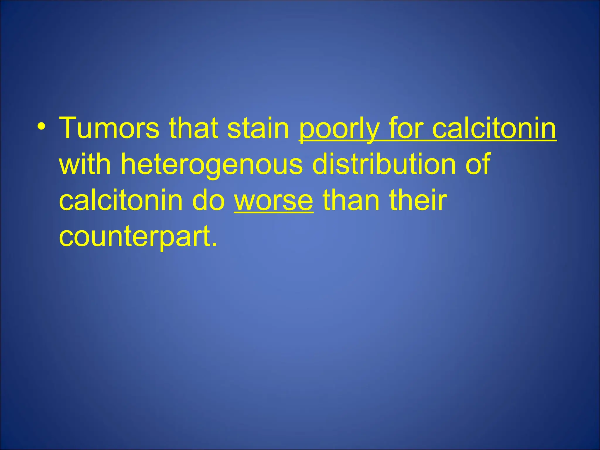
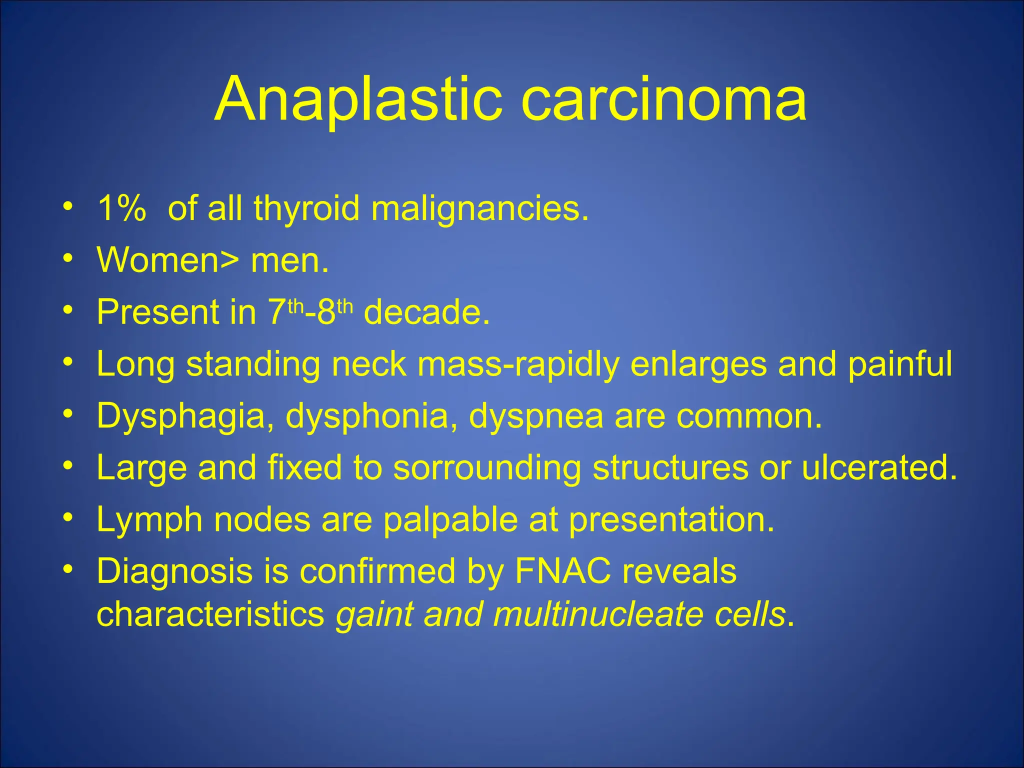
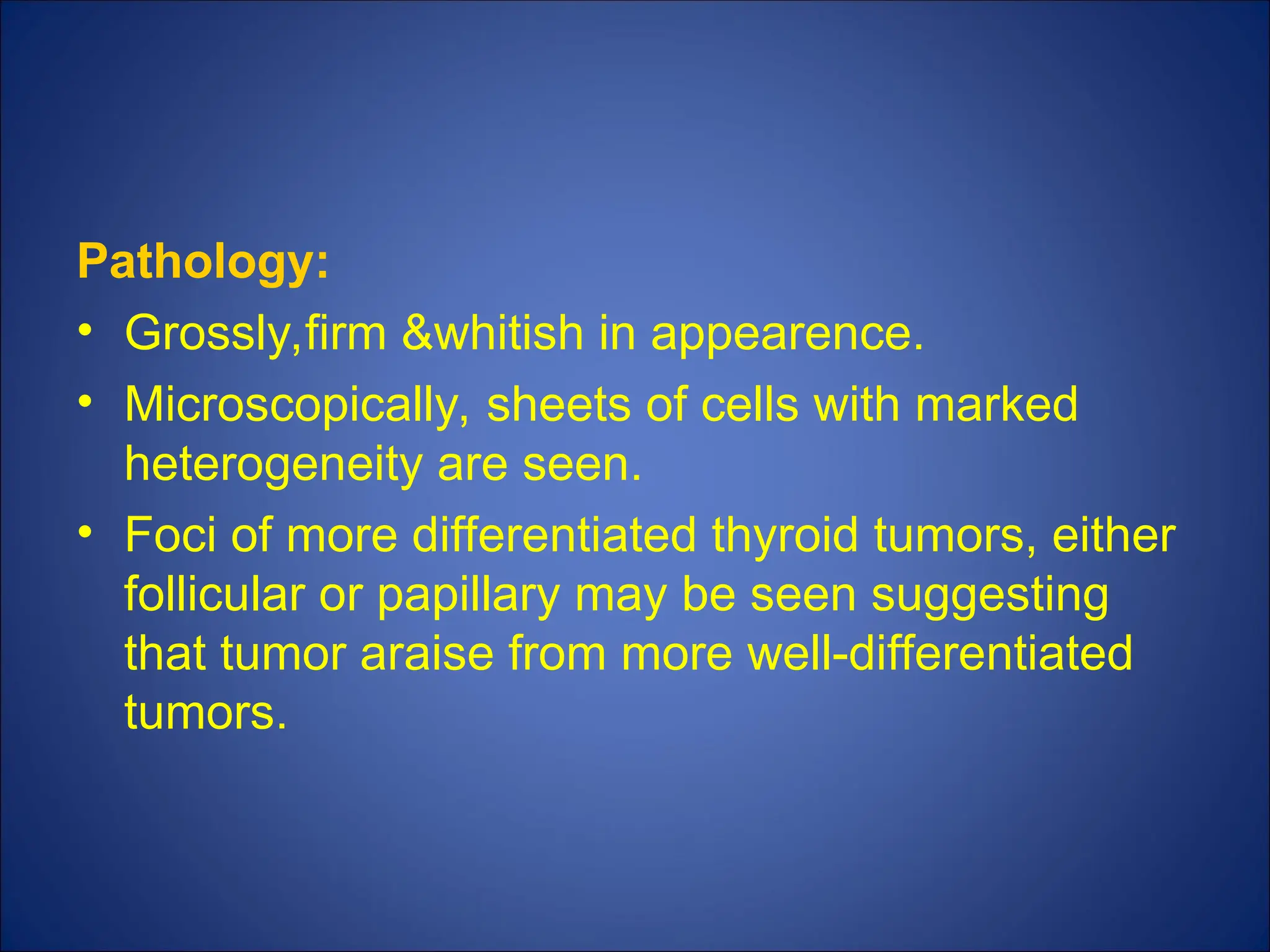
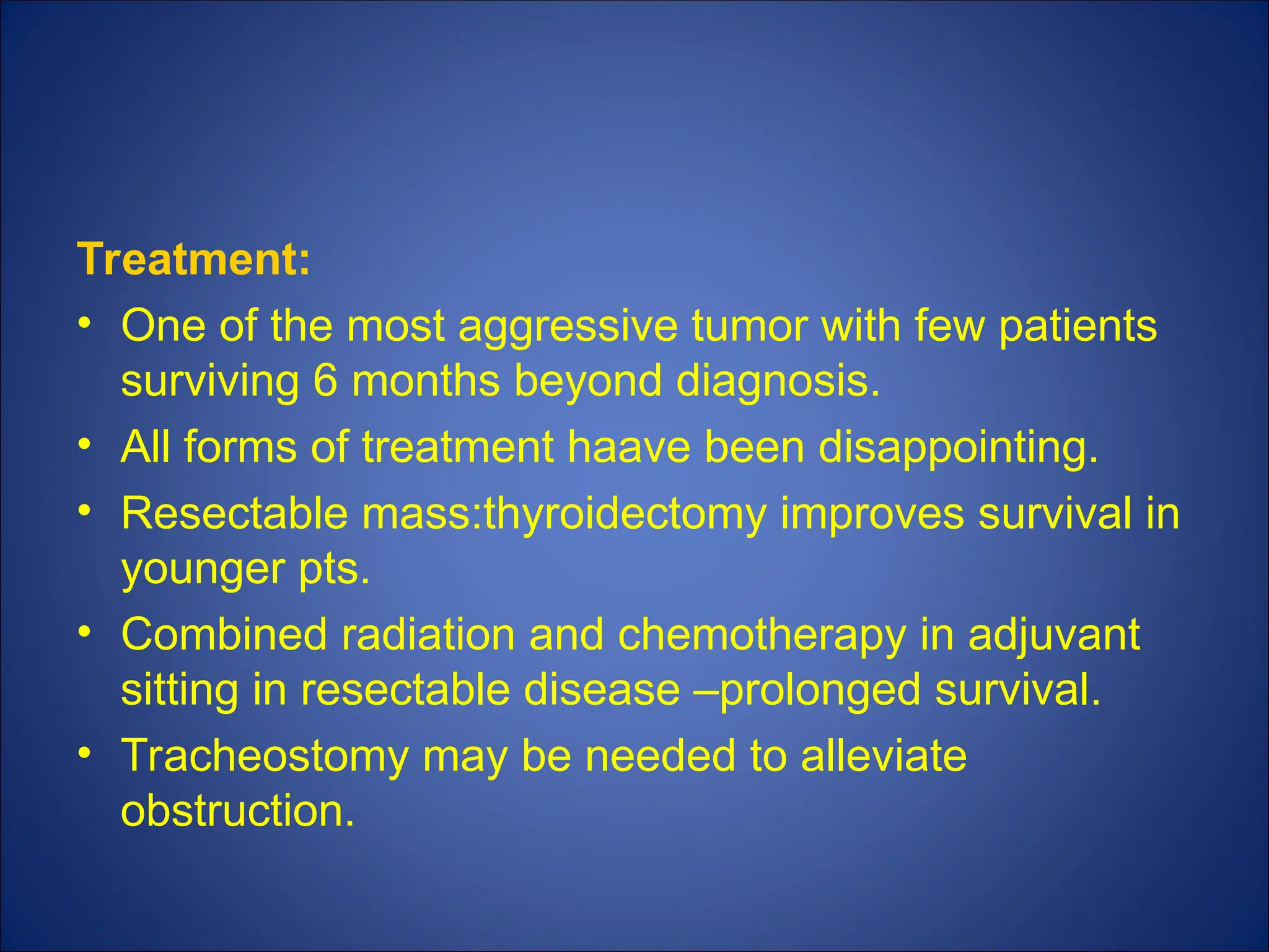
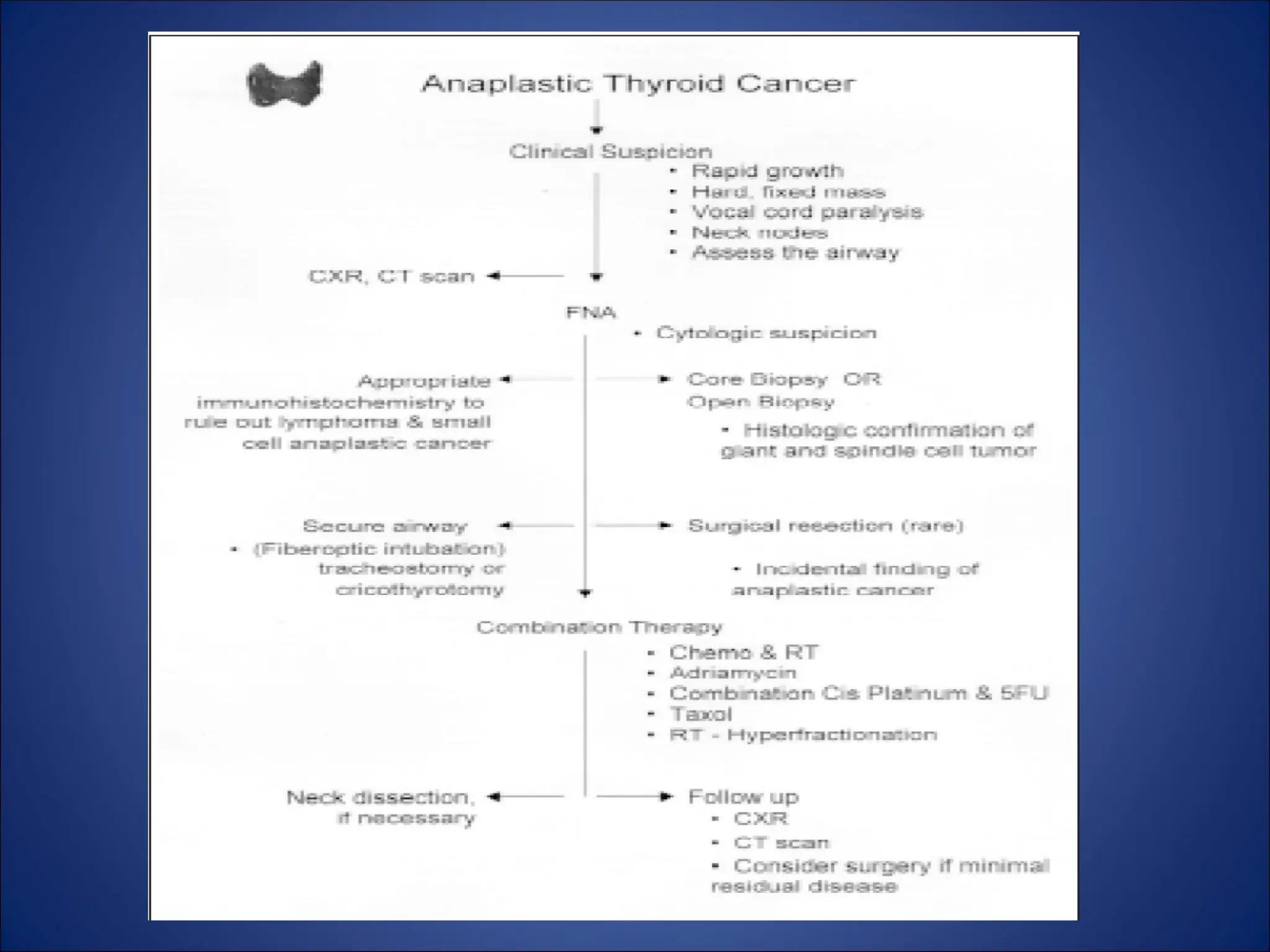
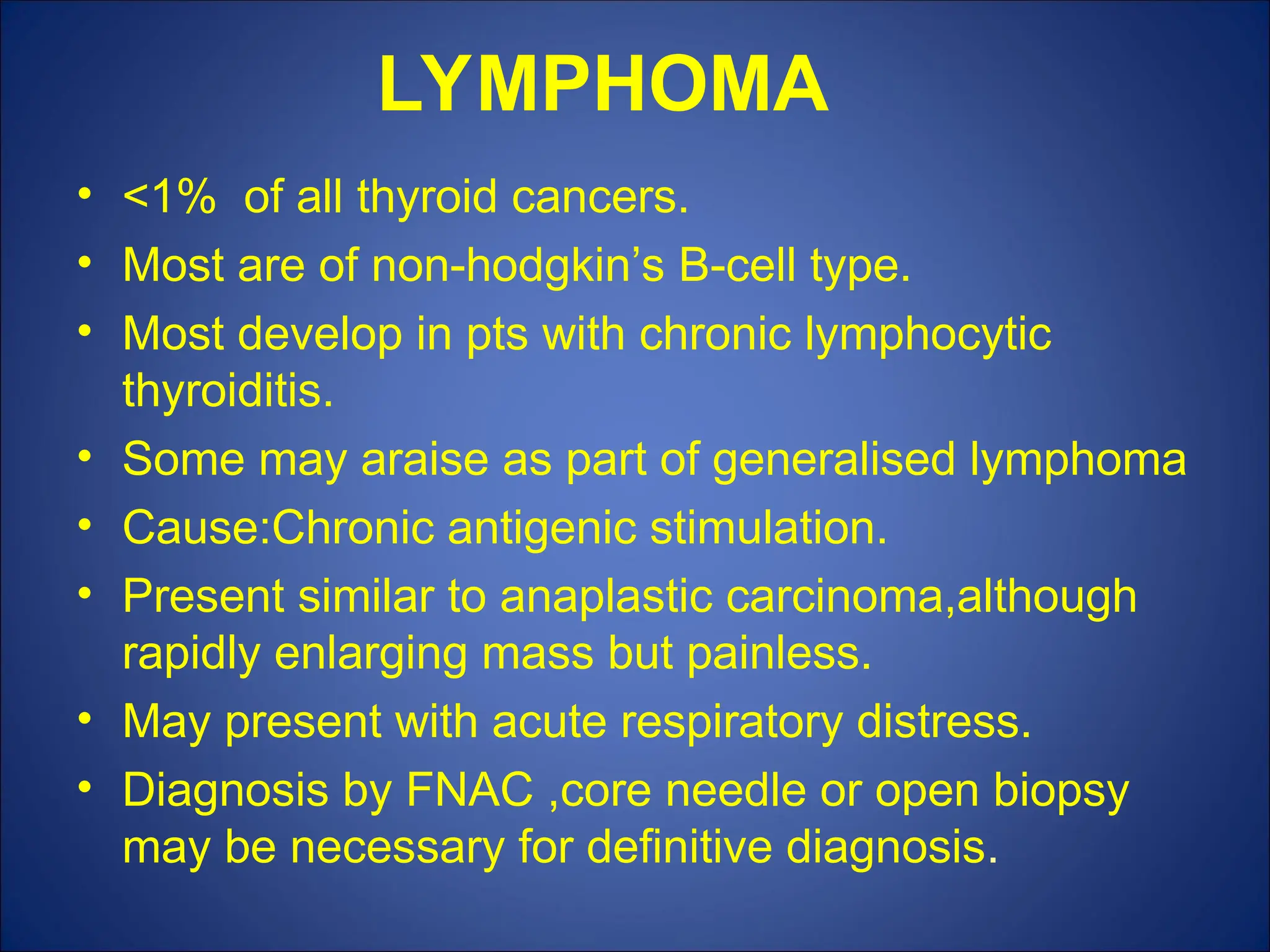
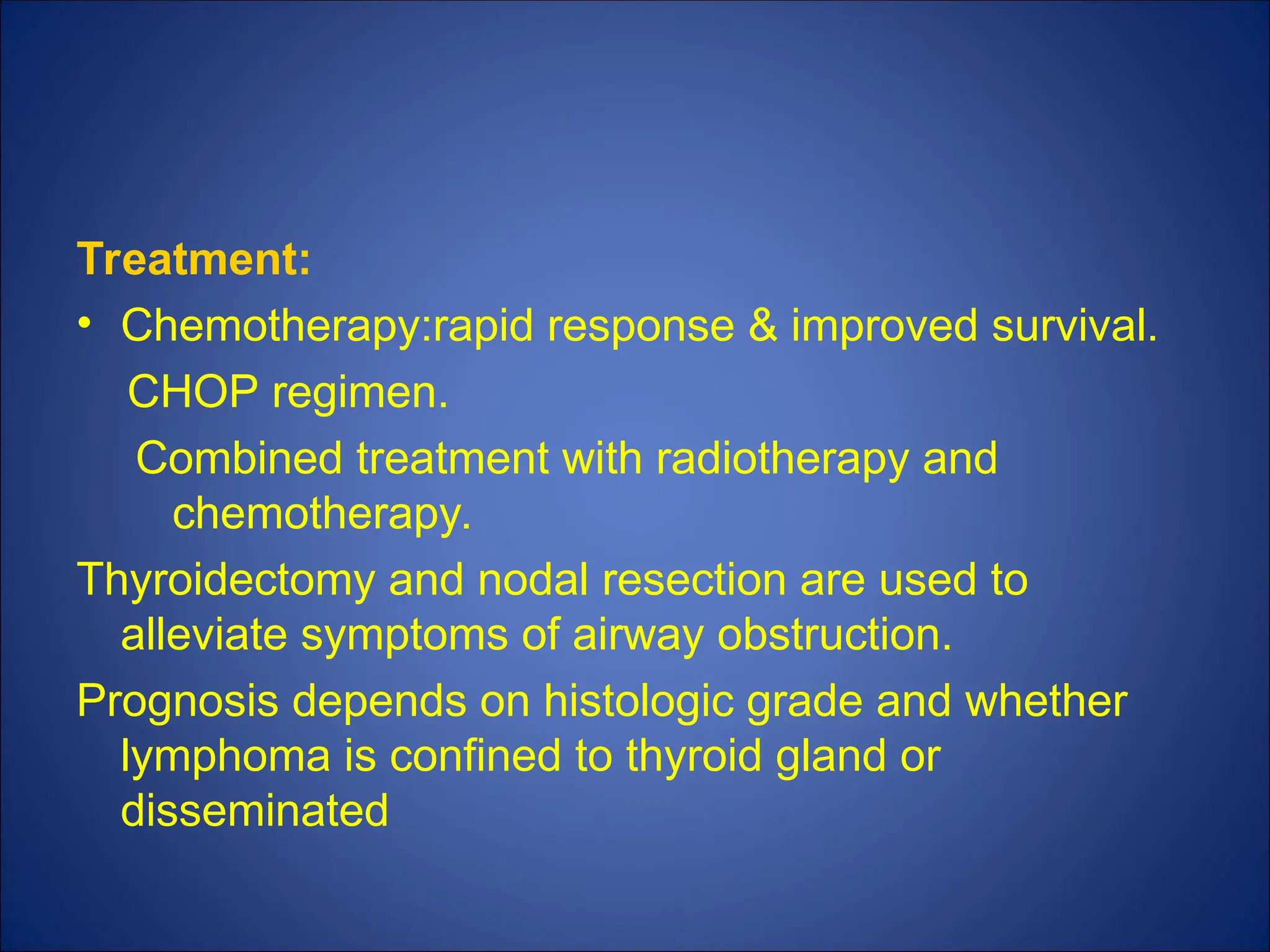
THYROID NEOPLASMS
DR.DHEERAJ
INTRODUCTION
• Possibility of neoplastic disease is of major
concern in patients with thyroid nodules.
• Fortunately,majority are non-neoplastic or
benign.
• Benign tumors outnumber
carcinomas(10:1)
• Carcinomas are uncommon (1%).
HISTOLOGY OF THE THYROID
GLAND
Follicular cells line
the colloid follicles,
concentrate iodine, and
produce thyroid hormone.
These cells give rise to
both well-differentiated
cancers and anaplastic
thyroid cancer.
The C or
parafollicular cell,
produces the hormone
calcitonin and is the cell of
origin for medullary
thyroid carcinoma.
Immune cells and
stromal cells of the thyroid
are responsible for
lymphoma and sarcoma,
respectively.
CLASSIFICATION OF THYROID
NEOPLASMS
BENIGN:
• Follicular adenoma
MALIGNANT:
Primary:
1)Follicular epithelium-differentiated
–Follicular
–Papillary
2)Follicular epithelium-undifferentiated
–Anaplastic
3)Parafollicular
–Medullary
4)Lymphoid cells
–Lymphoma
Secondaries:
–Metastatic
CLASSIFICATION OF
THYROID NEOPLASMS
RELATIVE INCIDENCE
• PAPILLARY - 80%
• FOLLICULAR - 17%
• ANAPLASTIC - 1.5%
• MEDULLARY - 0.5%
• OTHERS- 1%
Benign tumors
Follicular adenoma:
• Present as clinically solitary nodules.
• Distinction between adenoma and carcinoma can
only be made by histological examination.
• No invasion of capsule or of pericapsular blood
vessels-adenoma.
• Treatment: wide excision(lobectomy)
Papillary adenoma: as such an entity is doubtful
and all papillary tumors should be considered as
malignant even if encapsulated.
MALIGNANT TUMORS
Dunhill classified them histologically as differentiated
and undifferentiated.
ETIOLOGY:
• Irradiation –
– single most important factor in differentiated thyroid
carcinoma,particularly papillary carcinoma.
• Iodine deficiency-
– follicular carcinoma(due to TSH stimulation)
MALIGNANT TUMORS
• Autoimmune thyroiditis-
–malignant lymphomas
• Oncogenes-
–ret/PT3(aggressive papillary ca)
–ret/PTC1(less aggressive papillary
PAPILLARY CARCINOMA
• 80% of all thyroid malignancies.
• 90% of all radiation induced thyroid
malignancies.
• M:F = 1:3
• Mean age at presentation is 30 to 40 years.
CLINICAL FEATURES
• Slow growing painless mass in the neck
• Dysphagia,
• Dyspnea, and
• Dysphonia are usually associated with
locally advanced invasive disease.
• Lymph node metastases are common
especially in young adults and children and
may be the presenting complaint.
CLINICAL FEATURES
• LATERAL ABERRANT THYROID almost
always denotes a cervical lymph node that
has been invaded by metastatic cancer.
• Distant metastases are uncommon at
presentation,but may develop in 20% of
patients.
• Most common sites:
lungs>bone>liver>brain
Microscopic examination:
• Exhibit papillary projections,
• mixed pattern of papillary and follicular
structures, or
• pure follicular pattern(follicular variant).
ORPHAN ANNIE NUCLEI
• Cells are cuboidal with
pale abundant cytoplasm,
– grooving crowded
nuclei, and
– intranuclear
cytoplasmic
inclusions,leading to
the designation of
ORPHAN ANNIE
NUCLEI.
• Psammoma bodies :
–microscopic calcified deposits representing
clumps of sloughed cells.
• Multifocality: common(85%)and associated
with increased risk of cervical metastases.
• Rarely invade adjacent structures
(trachea,esophagus and recurrent laryngeal
nerve).
HISTOLOGY
Psammomma body
Psammoma bodies
Psammoma bodies are commonly seen in certain tumors such as:
• Papillary thyroid carcinoma
• Papillary renal cell carcinoma
• Micropapillary subtype of lung adenocarcinoma
• Ovarian papillary serous cystadenoma and cystadenocarcinoma
• Endometrial adenocarcinomas (Papillary serous carcinoma ~3%-
4%)
• Meningiomas, in the central nervous system
• Peritoneal and Pleural Mesothelioma
• Somatostatinoma (pancreas)
• Prolactinoma of the pituitary
• Mesothelioma
Other variants(1%):tall cell
insular
columnar
diffuse sclerosing
clear cell
trabecular
poorly differentiated
• All are associated with worse prognosis.
• Macroscopically, there are three forms based
on size and extent of primary disease.
1)MINIMAL/OCCULT/
MICROCARCINOMA :defined as tumors of 1cm
or less in size with
• no evidence of local invasiveness through
thyroid capsule
• or angioinvasion, and
• are not associated with lymph node
metastases.
• Nonpalpable
• Incidental findings at operative, histologic,or
autopsy examination.
• Present in 2-36% of thyroid glands removed at
autopsy.
• Recurrence rate :5%
• Mortality rate:0.5%
2)Intrathyroidal tumors: good prognosis.
3)Extrathyroidal tumors: invade through
capsule/adjacent structures.
PROGNOSTIC CLASSIFICATION
SYSTEMS IN WELL-DIFFERENTIATED
THYROID CARCINOMA
• AGES Age, grade of tumor, extent, size
• AMES Age, metastases, extent, size
• MACIS Metastases, age, completeness of
resection, invasion, size
AGES / AMES
LOW RISK HIGH RISK
Age <40 years >40 years
Grade Well differentiated Poorly differentiated
Metastasis None Regional or distant
Extent No local extension extra thyroidal
intrathyroidal extention
no capsular invasion Capsular invasion
Size <2 cm >4 cm
Sex Female Male
TNM CLASSIFICATION
A simplified system by degroot and
associates uses four groups:
• Class1(intrathyroidal)
• Class2 (cervical nodal metastses)
• Class3 (extrathyroidal invasion)
• Class4 (distant metastases).
Molecular and genetic markers:
• DNA aneuploidy,decreased cAMP
response to TSH, increased epidermal
growth factor binding,prescence of p53
mutations are associated with a worse
prognosis.
SURGICAL TREATMENT
High risk tumors or bilateral tumors:
• Total or near-total thyroidectomy.
Low risk(small,unilateral) tumors:
• Ipsilateral total and contralateral subtotal
thyroidectomy.
Minimal PTC: Complete ipsilateral lobectomy &
contralateral subtotal thyroid resection.
Lymph node metastases in lateral neck:
• Modified radical neck dissection.
• Dissection of posterior triangle and suprahyoid
dissection are usually not necessary unless there
is extensive metastatic disease in levels 2,3,and
4.
• Prophylactic neck node dissection is not
necessary in patients with PTC (do not
metastasize systemically from node and micro
mets are ablated with RAI
ADVANTAGES OF TOTAL THYROIDECTOMY:
1)facilitates the use of RAI to effectively detect and treat
residual thyroid tissue or metastatic disease.
2)makes serum Tg level a more sensitive marker.
3)eliminates the contralateral occult cancers as sites of
recurrence and improves survival
4)reduces the risk of recurrence
5)decreases the 1% risk of progression to undifferentiated
carcinoma.
6)reduces the need for reoperative surgery.
POSITIVE FACTORS FOR LOBECTOMY:
• Total thyroidectomy is associated with a
higher complication rate than lobectomy.
• Recurrence in the remaining thyroid tissue
is unusal(5%).
• Multicentricity seems to have little
prognostic signifiance.
FOLLICULAR CARCINOMA
• 10% of all thyroid cancers.
• More common in iodine deficient
areas.
• M:F = 1:3.
• Mean age at presentation-50 yrs.
PATHOLOGY
Gross examination:
• Solitary nodules with capsule.
Microscopic examination:
• Follicles are present, but devoid of colloid.
• Malignancy is defined as prescence of capsular and vascular invasion.
2 types:
Minimally invasive tumors:
• Grossly encapsulated with microscopic invasion through capsule and/or
invasion into small to medium sized vessels in or immediately outside
the capsule, but not within the tumor.
Widely invasive tumors:
• Large vessel invasion and/or broad invasion through the capsule.
• May be unencapsulated.
CLINICAL FEATURES
• Present as solitary nodules ,ocassionaly with
history of sudden increase in size or long
standing goitre.
• Pain is uncommon , unless hemorrhage has
occured into the nodule.
• Cervical lymphadenopathy is uncommon at
presentation but distant metastases may be
present.
• In 1% of cases, they may be hyperfunctionig-
thyrotoxicosis.
SURGICAL TREATMENT AND
PROGNOSIS
FNAC showing follicular lesion:
thyroid lobectomy(80% are benign).
Follicular lesion >4cm in older patients:
total thyroidectomy(50% are carcinomas).
Invasive follicular carcinoma:
total thyroidectomy.
Prophylactic nodal dissection is unwarranted because nodal
involvement is infrequent.
Unusal nodal metastases:
Therapeutic neck dissection.
Cumulative mortality:15% at 10yrs
30% at 20yrs.
Poor prognosis:
>50yrs
>4cm
high grade
marked vascular invasion
extrathyroidal invasion
distant metastases at diagnosis
HURTHLE CELL CARCINOMA
• 3% of all thyroid malingnancies.
• Subtype of follicular cancer.
• Characterised by capsular invasion and vascular
invasion.
• Contain esinophillic cells packed with mitochondria,
derived from oxyphilic cells of thyroid.
• Multifocal and bilateral
• Don’t take up RAI.
• More likely to metastasize to local nodes and distant
sites and are associated with high mortality.
MANAGEMENT
Unilateral hurthle cell adenoma:
Lobectomy and isthmusectomy
Invasive hurthle neoplasm:
• Total thyroidectomy.
• Routine central neck dissection is done
Palpable Lateral neck nodes:
• Modified radical neck dissection.
• RAI scanning and ablation are ineffective.
POSTOPERATIVE MANAGEMENT OF
DIFFERENCIATED THYROID CANCER
Thyroid harmones:
• Necessary not only as replacement therapy but also
for suppressing TSH and reducing the growth stimulus.
• Reduces tumor recurrence rates.
• Thyroxine should be administered to ensure that
patient remains euthyroid, with circulating TSH levels
at about 0.1mu/l in low-risk pt or <0.1mu/ml in high-risk
pt.
• Side effects: osteopenia,cardiac problems particularly
in older patients.
Thyroglobulin measurement:
• In pt who have undergone total thyroidectomy Tg
should be <2ng/ml when pt is taking T4 and
<5ng/ml when pt is hypothyroid.
• >2ng/ml – metastases or persistent normal thyroid
tissue(95%).
• Tg & anti Tg antibody levels: measured initially at
6-month intervals and then annually if the pt is
clinically free.
• High-risk pt: U/S of neck, CT or MRI scan for early
detection of any persistent or recurrent disease.
RADIO IODINE THERAPY:
• reduces recurrence and provides a small
improvement in survival even in low risk pts.
• Although screening with radioactive iodine for
detecting metstases sensitive than chest x-
ray,CT scanning but less sensitive than Tg
measurements(except hurthle cell tumors-only 5-
10% of the latter take up RAI)
• Screeening and treatment are facilitated by the
removal of all normal thyroid tissue.
• Metastatic differentiated thyroid carcinoma can
be detected and treated by I 131 in 75% of pts.
• Treats-70% of lung micrometastases whereas
only <10% of macrometastases.
• T4 should be discontinued for 6 wks prior to scanning with I-131.
• Should receive T3 during this period to decrease the period of
hypothyroidism.
• T3 has shorter half-life than T4(1day vs 1 wk)
• T3 needs to be discontinued for 2 wks to allow TSH levels to rise
prior to tretment.
• A low iodine diet is also recommended during this 2 wk period.
Usual protocol: administer screening dose 3mCi of I-131 and
measuring uptake after 24 hrs.
• A hot spot in the neck after initial screening usually represents
residual normal thyroid tissue.
• If there is significant uptake a therapeutic dose of I-
131 should be administered to patients.
low-risk pts:30-100 mCi
high-risk pts:100-200 mCi.
If pt has elevated Tg level, but negative RAI scan-treat
with 100 mCi of I-131 and repeat the scan 1-2 wks
later.
Alternate method: omit the screening dose to avoid
stunning of thyrocyte and subsequent requirement
of high treatment doses.
• Pt with previous positive scans and patients with
serum Tg levels >2 ng/ml- need another I-131
treatment after 6-12 months until one or two
negative scans are obtained.
• Follow-up scan be done after hormone withdrawl or
after recombinant TSH.
• Max dose at one time:200 mCi with cumulative dose
of 1000 to 1500 mCi.
If RAI scans are negative, but Tg levels remain elevated,
other
imaging studies-neck ultrasound, MRI scan are considered.
EXTERNAL BEAM RADIOTHERAPY:
• occasionally required to control unresectable,locally
invasive or recurrent disease and to treat metastases in
support bones to decrease the risk of fractures.
• To control pain in bony metastases.
CHEMOTHERAPY:
Adriamycin and taxol are most frequently used agents.
MEDULLARY CARCINOMA
• 5% of all thyroid malignancies.
• M:F = 1:1.5.
• Present between 50-60 years of age.
• Araise from the parafollicular cells or C-cells of thyroid
• These cells are concentrated superolaterally.
• C-cells secrete calcitonin.
• Occur in two forms:
sporadic
inherited(familial medullary thyroid
cancer,MEN2a,MEN2b)
all these variants are secondary to germline mutation in
RET proto-oncogenes.
PATHOLOGY
• Typically unilateral(80%) in sporadic cases.
• Bilateral(90%) and multicentric in familial cases.
• Asssociated with C-cell hyperplasia- a pre malignant
condition.
• Microscopic examination: sheets of infiltrating neoplastic
cells seperated by collagen and amyloid.
• Cells are spindle-shaped or polygonal.
• Prescence of amyloid is a diagnostic finding.
• Immunohistochemistry for calcitonin is commonly used
as a diagnostic tumor marker.
• Also stain for CEA and CGRP.
CLINICAL FEATURES
• Present with neck mass associated with palpable
cervical lymphadenopathy.
• Local pain or aching is more common.
• Local invasion-dysphagia,dysphonia,dyspnea.
• Distant metastases:liver, bone(osteoblastic),lung.
• Secrete:calcitonin
carcinoembryonic antigen
calcitonin gene related peptide
histaminadases
prostaglandins E2 & F2
serotonin.
• Patients with extensive metastastic
disease frequently develop diarrhea, which
may result from increased intestinal
motility and impaired intestinal water and
electrolyte flux.
• Develop cushings syndrome(2%)-ectopic
ACTH hormone .
Diagnosis
• Established by history, physical examination,
raised serum calcitonin or CEA levels, FNAC.
• All familial cases are screened for RET point
mutation,pheochromocytoma,hyperparathyroidism.
• It is important to rule out a co-existing
pheochromocytoma to avoid hypertensive crisis
and death.
• Screening for RET mutation has been replaced
with provocation testing using pentagastrin or
calcium stimulated calcitonin levels.
• Calcitonin and CEA are used to identify
persistent or recurrent MTC.
• Calcitonin –more sensitive marker
• CEA- better predictor of prognosis
TREATMENT
• Total thyroidectomy +B/L central neck node
dissection is the treatment of choice.
• Palpable lymph nodes or involved central nodes:
ipsilateral / bilateral modified radical neck dissection.
• Tumors > 1.5cm: ipsilateral prophylactic modified
radical neck dissection.
• Locally recurrent or metastatic disease:debulking of
the tumor.(to ameliorate flushing and diarrhea and to
decrease risk of death from central neck nodes)
• External beam radiotherapy for unresectable
residual or recurrent tumor.
• Liver metastases (>1.5 cm):laprascopic radio
frequency ablation.
• Tumors that express c-kit may also respond to
tyrosine kinase inhibitors(gleevec).
• In RET mutation carriers once mutation is confirmed
total thyroidectomy is indicated.
<6yrs in MEN2a
<1yr in MEN2b .
• Central neck dissection is avoided if RET
positive and calcitonin negative with
normal ultrasound examination.
• Calcitonin increased or ultrasound
suggests a thyroid cancer-prophylactic
central neck dissection.
Prognosis
Depends on:
• Stage of disease:
10 yr survival rate:
80%.
45%-with lymph node involvement.
• Type of disease: Non MEN familial
MEN2a
Sporadic
MEN2b
• Tumors that stain poorly for calcitonin
with heterogenous distribution of
calcitonin do worse than their
counterpart.
Anaplastic carcinoma
• 1% of all thyroid malignancies.
• Women> men.
• Present in 7th
-8th
decade.
• Long standing neck mass-rapidly enlarges and painful
• Dysphagia, dysphonia, dyspnea are common.
• Large and fixed to sorrounding structures or ulcerated.
• Lymph nodes are palpable at presentation.
• Diagnosis is confirmed by FNAC reveals
characteristics gaint and multinucleate cells.
Pathology:
• Grossly,firm &whitish in appearence.
• Microscopically, sheets of cells with marked
heterogeneity are seen.
• Foci of more differentiated thyroid tumors, either
follicular or papillary may be seen suggesting
that tumor araise from more well-differentiated
tumors.
Treatment:
• One of the most aggressive tumor with few patients
surviving 6 months beyond diagnosis.
• All forms of treatment haave been disappointing.
• Resectable mass:thyroidectomy improves survival in
younger pts.
• Combined radiation and chemotherapy in adjuvant
sitting in resectable disease –prolonged survival.
• Tracheostomy may be needed to alleviate
obstruction.
LYMPHOMA
• <1% of all thyroid cancers.
• Most are of non-hodgkin’s B-cell type.
• Most develop in pts with chronic lymphocytic
thyroiditis.
• Some may araise as part of generalised lymphoma
• Cause:Chronic antigenic stimulation.
• Present similar to anaplastic carcinoma,although
rapidly enlarging mass but painless.
• May present with acute respiratory distress.
• Diagnosis by FNAC ,core needle or open biopsy
may be necessary for definitive diagnosis.
Treatment:
• Chemotherapy:rapid response & improved survival.
CHOP regimen.
Combined treatment with radiotherapy and
chemotherapy.
Thyroidectomy and nodal resection are used to
alleviate symptoms of airway obstruction.
Prognosis depends on histologic grade and whether
lymphoma is confined to thyroid gland or
disseminated
• 5 yrs survival rate :50%; pts with extrathyroidal
disease –low survival rates.
METASTATIC CARCINOMA:
• Rare site.
• From kidney, breast, lung and melanoma.
• History and clinical examination suggest the source.
• Diagnosis is by FNAC.
• RESECTION (lobectomy) may be helpful depending
on the primary tumor.
•
THANK YOU

THYROID NEOPLASMS-1.ppt of the various thyroid malignancies

  • 1.
  • 2.
    INTRODUCTION • Possibility ofneoplastic disease is of major concern in patients with thyroid nodules. • Fortunately,majority are non-neoplastic or benign. • Benign tumors outnumber carcinomas(10:1) • Carcinomas are uncommon (1%).
  • 3.
    HISTOLOGY OF THETHYROID GLAND Follicular cells line the colloid follicles, concentrate iodine, and produce thyroid hormone. These cells give rise to both well-differentiated cancers and anaplastic thyroid cancer. The C or parafollicular cell, produces the hormone calcitonin and is the cell of origin for medullary thyroid carcinoma. Immune cells and stromal cells of the thyroid are responsible for lymphoma and sarcoma, respectively.
  • 4.
    CLASSIFICATION OF THYROID NEOPLASMS BENIGN: •Follicular adenoma MALIGNANT: Primary: 1)Follicular epithelium-differentiated –Follicular –Papillary 2)Follicular epithelium-undifferentiated –Anaplastic
  • 5.
  • 6.
    RELATIVE INCIDENCE • PAPILLARY- 80% • FOLLICULAR - 17% • ANAPLASTIC - 1.5% • MEDULLARY - 0.5% • OTHERS- 1%
  • 7.
    Benign tumors Follicular adenoma: •Present as clinically solitary nodules. • Distinction between adenoma and carcinoma can only be made by histological examination. • No invasion of capsule or of pericapsular blood vessels-adenoma. • Treatment: wide excision(lobectomy) Papillary adenoma: as such an entity is doubtful and all papillary tumors should be considered as malignant even if encapsulated.
  • 8.
    MALIGNANT TUMORS Dunhill classifiedthem histologically as differentiated and undifferentiated. ETIOLOGY: • Irradiation – – single most important factor in differentiated thyroid carcinoma,particularly papillary carcinoma. • Iodine deficiency- – follicular carcinoma(due to TSH stimulation)
  • 9.
    MALIGNANT TUMORS • Autoimmunethyroiditis- –malignant lymphomas • Oncogenes- –ret/PT3(aggressive papillary ca) –ret/PTC1(less aggressive papillary
  • 10.
    PAPILLARY CARCINOMA • 80%of all thyroid malignancies. • 90% of all radiation induced thyroid malignancies. • M:F = 1:3 • Mean age at presentation is 30 to 40 years.
  • 11.
    CLINICAL FEATURES • Slowgrowing painless mass in the neck • Dysphagia, • Dyspnea, and • Dysphonia are usually associated with locally advanced invasive disease. • Lymph node metastases are common especially in young adults and children and may be the presenting complaint.
  • 12.
    CLINICAL FEATURES • LATERALABERRANT THYROID almost always denotes a cervical lymph node that has been invaded by metastatic cancer. • Distant metastases are uncommon at presentation,but may develop in 20% of patients. • Most common sites: lungs>bone>liver>brain
  • 13.
    Microscopic examination: • Exhibitpapillary projections, • mixed pattern of papillary and follicular structures, or • pure follicular pattern(follicular variant).
  • 14.
    ORPHAN ANNIE NUCLEI •Cells are cuboidal with pale abundant cytoplasm, – grooving crowded nuclei, and – intranuclear cytoplasmic inclusions,leading to the designation of ORPHAN ANNIE NUCLEI.
  • 15.
    • Psammoma bodies: –microscopic calcified deposits representing clumps of sloughed cells. • Multifocality: common(85%)and associated with increased risk of cervical metastases. • Rarely invade adjacent structures (trachea,esophagus and recurrent laryngeal nerve).
  • 16.
  • 17.
    Psammoma bodies Psammoma bodiesare commonly seen in certain tumors such as: • Papillary thyroid carcinoma • Papillary renal cell carcinoma • Micropapillary subtype of lung adenocarcinoma • Ovarian papillary serous cystadenoma and cystadenocarcinoma • Endometrial adenocarcinomas (Papillary serous carcinoma ~3%- 4%) • Meningiomas, in the central nervous system • Peritoneal and Pleural Mesothelioma • Somatostatinoma (pancreas) • Prolactinoma of the pituitary • Mesothelioma
  • 18.
    Other variants(1%):tall cell insular columnar diffusesclerosing clear cell trabecular poorly differentiated • All are associated with worse prognosis.
  • 19.
    • Macroscopically, thereare three forms based on size and extent of primary disease. 1)MINIMAL/OCCULT/ MICROCARCINOMA :defined as tumors of 1cm or less in size with • no evidence of local invasiveness through thyroid capsule • or angioinvasion, and • are not associated with lymph node metastases. • Nonpalpable • Incidental findings at operative, histologic,or autopsy examination.
  • 20.
    • Present in2-36% of thyroid glands removed at autopsy. • Recurrence rate :5% • Mortality rate:0.5% 2)Intrathyroidal tumors: good prognosis. 3)Extrathyroidal tumors: invade through capsule/adjacent structures.
  • 21.
    PROGNOSTIC CLASSIFICATION SYSTEMS INWELL-DIFFERENTIATED THYROID CARCINOMA • AGES Age, grade of tumor, extent, size • AMES Age, metastases, extent, size • MACIS Metastases, age, completeness of resection, invasion, size
  • 22.
    AGES / AMES LOWRISK HIGH RISK Age <40 years >40 years Grade Well differentiated Poorly differentiated Metastasis None Regional or distant Extent No local extension extra thyroidal intrathyroidal extention no capsular invasion Capsular invasion Size <2 cm >4 cm Sex Female Male
  • 23.
  • 24.
    A simplified systemby degroot and associates uses four groups: • Class1(intrathyroidal) • Class2 (cervical nodal metastses) • Class3 (extrathyroidal invasion) • Class4 (distant metastases).
  • 25.
    Molecular and geneticmarkers: • DNA aneuploidy,decreased cAMP response to TSH, increased epidermal growth factor binding,prescence of p53 mutations are associated with a worse prognosis.
  • 26.
    SURGICAL TREATMENT High risktumors or bilateral tumors: • Total or near-total thyroidectomy. Low risk(small,unilateral) tumors: • Ipsilateral total and contralateral subtotal thyroidectomy. Minimal PTC: Complete ipsilateral lobectomy & contralateral subtotal thyroid resection.
  • 27.
    Lymph node metastasesin lateral neck: • Modified radical neck dissection. • Dissection of posterior triangle and suprahyoid dissection are usually not necessary unless there is extensive metastatic disease in levels 2,3,and 4. • Prophylactic neck node dissection is not necessary in patients with PTC (do not metastasize systemically from node and micro mets are ablated with RAI
  • 28.
    ADVANTAGES OF TOTALTHYROIDECTOMY: 1)facilitates the use of RAI to effectively detect and treat residual thyroid tissue or metastatic disease. 2)makes serum Tg level a more sensitive marker. 3)eliminates the contralateral occult cancers as sites of recurrence and improves survival 4)reduces the risk of recurrence 5)decreases the 1% risk of progression to undifferentiated carcinoma. 6)reduces the need for reoperative surgery.
  • 29.
    POSITIVE FACTORS FORLOBECTOMY: • Total thyroidectomy is associated with a higher complication rate than lobectomy. • Recurrence in the remaining thyroid tissue is unusal(5%). • Multicentricity seems to have little prognostic signifiance.
  • 30.
    FOLLICULAR CARCINOMA • 10%of all thyroid cancers. • More common in iodine deficient areas. • M:F = 1:3. • Mean age at presentation-50 yrs.
  • 31.
    PATHOLOGY Gross examination: • Solitarynodules with capsule. Microscopic examination: • Follicles are present, but devoid of colloid. • Malignancy is defined as prescence of capsular and vascular invasion. 2 types: Minimally invasive tumors: • Grossly encapsulated with microscopic invasion through capsule and/or invasion into small to medium sized vessels in or immediately outside the capsule, but not within the tumor. Widely invasive tumors: • Large vessel invasion and/or broad invasion through the capsule. • May be unencapsulated.
  • 33.
    CLINICAL FEATURES • Presentas solitary nodules ,ocassionaly with history of sudden increase in size or long standing goitre. • Pain is uncommon , unless hemorrhage has occured into the nodule. • Cervical lymphadenopathy is uncommon at presentation but distant metastases may be present. • In 1% of cases, they may be hyperfunctionig- thyrotoxicosis.
  • 35.
    SURGICAL TREATMENT AND PROGNOSIS FNACshowing follicular lesion: thyroid lobectomy(80% are benign). Follicular lesion >4cm in older patients: total thyroidectomy(50% are carcinomas). Invasive follicular carcinoma: total thyroidectomy. Prophylactic nodal dissection is unwarranted because nodal involvement is infrequent. Unusal nodal metastases: Therapeutic neck dissection. Cumulative mortality:15% at 10yrs 30% at 20yrs.
  • 37.
    Poor prognosis: >50yrs >4cm high grade markedvascular invasion extrathyroidal invasion distant metastases at diagnosis
  • 38.
    HURTHLE CELL CARCINOMA •3% of all thyroid malingnancies. • Subtype of follicular cancer. • Characterised by capsular invasion and vascular invasion. • Contain esinophillic cells packed with mitochondria, derived from oxyphilic cells of thyroid. • Multifocal and bilateral • Don’t take up RAI. • More likely to metastasize to local nodes and distant sites and are associated with high mortality.
  • 39.
    MANAGEMENT Unilateral hurthle celladenoma: Lobectomy and isthmusectomy Invasive hurthle neoplasm: • Total thyroidectomy. • Routine central neck dissection is done Palpable Lateral neck nodes: • Modified radical neck dissection. • RAI scanning and ablation are ineffective.
  • 40.
    POSTOPERATIVE MANAGEMENT OF DIFFERENCIATEDTHYROID CANCER Thyroid harmones: • Necessary not only as replacement therapy but also for suppressing TSH and reducing the growth stimulus. • Reduces tumor recurrence rates. • Thyroxine should be administered to ensure that patient remains euthyroid, with circulating TSH levels at about 0.1mu/l in low-risk pt or <0.1mu/ml in high-risk pt. • Side effects: osteopenia,cardiac problems particularly in older patients.
  • 41.
    Thyroglobulin measurement: • Inpt who have undergone total thyroidectomy Tg should be <2ng/ml when pt is taking T4 and <5ng/ml when pt is hypothyroid. • >2ng/ml – metastases or persistent normal thyroid tissue(95%). • Tg & anti Tg antibody levels: measured initially at 6-month intervals and then annually if the pt is clinically free. • High-risk pt: U/S of neck, CT or MRI scan for early detection of any persistent or recurrent disease.
  • 42.
    RADIO IODINE THERAPY: •reduces recurrence and provides a small improvement in survival even in low risk pts. • Although screening with radioactive iodine for detecting metstases sensitive than chest x- ray,CT scanning but less sensitive than Tg measurements(except hurthle cell tumors-only 5- 10% of the latter take up RAI)
  • 43.
    • Screeening andtreatment are facilitated by the removal of all normal thyroid tissue. • Metastatic differentiated thyroid carcinoma can be detected and treated by I 131 in 75% of pts. • Treats-70% of lung micrometastases whereas only <10% of macrometastases.
  • 45.
    • T4 shouldbe discontinued for 6 wks prior to scanning with I-131. • Should receive T3 during this period to decrease the period of hypothyroidism. • T3 has shorter half-life than T4(1day vs 1 wk) • T3 needs to be discontinued for 2 wks to allow TSH levels to rise prior to tretment. • A low iodine diet is also recommended during this 2 wk period. Usual protocol: administer screening dose 3mCi of I-131 and measuring uptake after 24 hrs. • A hot spot in the neck after initial screening usually represents residual normal thyroid tissue.
  • 46.
    • If thereis significant uptake a therapeutic dose of I- 131 should be administered to patients. low-risk pts:30-100 mCi high-risk pts:100-200 mCi. If pt has elevated Tg level, but negative RAI scan-treat with 100 mCi of I-131 and repeat the scan 1-2 wks later. Alternate method: omit the screening dose to avoid stunning of thyrocyte and subsequent requirement of high treatment doses.
  • 47.
    • Pt withprevious positive scans and patients with serum Tg levels >2 ng/ml- need another I-131 treatment after 6-12 months until one or two negative scans are obtained. • Follow-up scan be done after hormone withdrawl or after recombinant TSH. • Max dose at one time:200 mCi with cumulative dose of 1000 to 1500 mCi.
  • 48.
    If RAI scansare negative, but Tg levels remain elevated, other imaging studies-neck ultrasound, MRI scan are considered. EXTERNAL BEAM RADIOTHERAPY: • occasionally required to control unresectable,locally invasive or recurrent disease and to treat metastases in support bones to decrease the risk of fractures. • To control pain in bony metastases. CHEMOTHERAPY: Adriamycin and taxol are most frequently used agents.
  • 50.
    MEDULLARY CARCINOMA • 5%of all thyroid malignancies. • M:F = 1:1.5. • Present between 50-60 years of age. • Araise from the parafollicular cells or C-cells of thyroid • These cells are concentrated superolaterally. • C-cells secrete calcitonin. • Occur in two forms: sporadic inherited(familial medullary thyroid cancer,MEN2a,MEN2b) all these variants are secondary to germline mutation in RET proto-oncogenes.
  • 53.
    PATHOLOGY • Typically unilateral(80%)in sporadic cases. • Bilateral(90%) and multicentric in familial cases. • Asssociated with C-cell hyperplasia- a pre malignant condition. • Microscopic examination: sheets of infiltrating neoplastic cells seperated by collagen and amyloid. • Cells are spindle-shaped or polygonal. • Prescence of amyloid is a diagnostic finding. • Immunohistochemistry for calcitonin is commonly used as a diagnostic tumor marker. • Also stain for CEA and CGRP.
  • 55.
    CLINICAL FEATURES • Presentwith neck mass associated with palpable cervical lymphadenopathy. • Local pain or aching is more common. • Local invasion-dysphagia,dysphonia,dyspnea. • Distant metastases:liver, bone(osteoblastic),lung. • Secrete:calcitonin carcinoembryonic antigen calcitonin gene related peptide histaminadases prostaglandins E2 & F2 serotonin.
  • 56.
    • Patients withextensive metastastic disease frequently develop diarrhea, which may result from increased intestinal motility and impaired intestinal water and electrolyte flux. • Develop cushings syndrome(2%)-ectopic ACTH hormone .
  • 57.
    Diagnosis • Established byhistory, physical examination, raised serum calcitonin or CEA levels, FNAC. • All familial cases are screened for RET point mutation,pheochromocytoma,hyperparathyroidism. • It is important to rule out a co-existing pheochromocytoma to avoid hypertensive crisis and death. • Screening for RET mutation has been replaced with provocation testing using pentagastrin or calcium stimulated calcitonin levels.
  • 58.
    • Calcitonin andCEA are used to identify persistent or recurrent MTC. • Calcitonin –more sensitive marker • CEA- better predictor of prognosis
  • 59.
    TREATMENT • Total thyroidectomy+B/L central neck node dissection is the treatment of choice. • Palpable lymph nodes or involved central nodes: ipsilateral / bilateral modified radical neck dissection. • Tumors > 1.5cm: ipsilateral prophylactic modified radical neck dissection. • Locally recurrent or metastatic disease:debulking of the tumor.(to ameliorate flushing and diarrhea and to decrease risk of death from central neck nodes)
  • 60.
    • External beamradiotherapy for unresectable residual or recurrent tumor. • Liver metastases (>1.5 cm):laprascopic radio frequency ablation. • Tumors that express c-kit may also respond to tyrosine kinase inhibitors(gleevec). • In RET mutation carriers once mutation is confirmed total thyroidectomy is indicated. <6yrs in MEN2a <1yr in MEN2b .
  • 62.
    • Central neckdissection is avoided if RET positive and calcitonin negative with normal ultrasound examination. • Calcitonin increased or ultrasound suggests a thyroid cancer-prophylactic central neck dissection.
  • 63.
    Prognosis Depends on: • Stageof disease: 10 yr survival rate: 80%. 45%-with lymph node involvement. • Type of disease: Non MEN familial MEN2a Sporadic MEN2b
  • 64.
    • Tumors thatstain poorly for calcitonin with heterogenous distribution of calcitonin do worse than their counterpart.
  • 65.
    Anaplastic carcinoma • 1%of all thyroid malignancies. • Women> men. • Present in 7th -8th decade. • Long standing neck mass-rapidly enlarges and painful • Dysphagia, dysphonia, dyspnea are common. • Large and fixed to sorrounding structures or ulcerated. • Lymph nodes are palpable at presentation. • Diagnosis is confirmed by FNAC reveals characteristics gaint and multinucleate cells.
  • 66.
    Pathology: • Grossly,firm &whitishin appearence. • Microscopically, sheets of cells with marked heterogeneity are seen. • Foci of more differentiated thyroid tumors, either follicular or papillary may be seen suggesting that tumor araise from more well-differentiated tumors.
  • 67.
    Treatment: • One ofthe most aggressive tumor with few patients surviving 6 months beyond diagnosis. • All forms of treatment haave been disappointing. • Resectable mass:thyroidectomy improves survival in younger pts. • Combined radiation and chemotherapy in adjuvant sitting in resectable disease –prolonged survival. • Tracheostomy may be needed to alleviate obstruction.
  • 69.
    LYMPHOMA • <1% ofall thyroid cancers. • Most are of non-hodgkin’s B-cell type. • Most develop in pts with chronic lymphocytic thyroiditis. • Some may araise as part of generalised lymphoma • Cause:Chronic antigenic stimulation. • Present similar to anaplastic carcinoma,although rapidly enlarging mass but painless. • May present with acute respiratory distress. • Diagnosis by FNAC ,core needle or open biopsy may be necessary for definitive diagnosis.
  • 70.
    Treatment: • Chemotherapy:rapid response& improved survival. CHOP regimen. Combined treatment with radiotherapy and chemotherapy. Thyroidectomy and nodal resection are used to alleviate symptoms of airway obstruction. Prognosis depends on histologic grade and whether lymphoma is confined to thyroid gland or disseminated
  • 71.
    • 5 yrssurvival rate :50%; pts with extrathyroidal disease –low survival rates. METASTATIC CARCINOMA: • Rare site. • From kidney, breast, lung and melanoma. • History and clinical examination suggest the source. • Diagnosis is by FNAC. • RESECTION (lobectomy) may be helpful depending on the primary tumor.
  • 72.