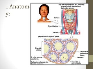
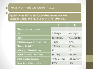
By:Aymen hasseb
1
Introduction:
2
The thyroid gland was described by Galen and was named "glandulae
thyroidaeae" by Wharton in 1656.
The thyroid gland is the source of two
fundamentally different types of hormones.
•The iodothyronine hormones include
Thyroxine (T4
3,5,3’-triiodothyronine (T3)
•They are essential for normal growth and development and play an important role in energy
metabolism.
Anatom
y:
3
The ratio of T4 and T3 secreted :- 10:1
Approximate Values for Thyroid Hormone Plasma
Concentrations and Various Kinetic Parameters.
4
T4 T3
Plasma concentration
Total 7.77 mg/dL 0.14 mg/ dL
Free 1.554 ng/dL 0.389 ng/dL
Total hormone in free form 0.02% 0.3%
Plasma half-life 6.7 days 0.75 days
Volume of distribution 10 L 40 L
Metabolic clearancerate 1.1L/day 24 L/day
Total production rate 85.47 mg/day 33.6 mg/day
From thyroid secretion 100% 20%
 Ac t i o n s of ThyroidHormones :
5
Evaluation of Thyroid
Diseases
:Thyroid Function test :
• commonly in use for estimating the iodine, TBG, free
T4 and free T3 concentrations are as follow:
• Protein-bound iodine:
• It is a measure of organically bound iodine in blood
(normal 4-8 ug/dl: myxoedema 2ug/dl;
thyrotoxicosis 10-12 ug/dl).
• Serum T4:
• It is measured by radio-immuno assay (normal 5-12
ug/dl). Serum T3 assay can also be done (normal
80-180 ng/dl).
6
Serum TBG:
• It is measured by radio-immuno assay or immuno electrophoresis, and the ratio
of T4 to TBG is used as an index of free hormone activity.
Free thyroxine index and effective thyroxine ratio: These are
calculated from the serum T4 and
• serum TBG level. There is a good correlation between these values and free T4
levels. Direct free T4 measurement are tedious and difficult.
Scintillography:
• The radio iodine uptake by the gland is recorded photographically and scanned.
7
T3 suppression test:
• In the standard test, the pretreatment radioactive iodine
uptake (RAIU) is determined. Then T3 (as Cytomel) 25 ug
tid, is given for 7-10 days. On the last day a repeat RAIU
is performed. In a normal person the RAIU will be
suppressed by 50% or less of the original value.
• Whereas in Graves’ disease there is no suppresion as the
thyroid is functioning autonomously.
Clinical history and physical examination :
8
 T hy ro i d Imaging
: Radioactive imaging
Ultrasound waves
CT/MRI scan
9
Diseases of the Thyroid :
• Two significant functional disorders
characterized by distinct clinical syndromes are :
10
Hyperthyroidism :
• Associated with excessive release of Thyroid
hormones.
Hypothyroidism :
• Associated with Thyroid hormone deficiency.
1) Hyperthyroidism :
The term hyperthyroidism is restricted to those conditions in which thyroid
hormone production and release are increased due to gland hyper
function.
The condition is more frequent in females and is associated with rise in
both T3and T4 levels in blood, though the increase in T3 is generally
greater than that of T4.
Etiopathogenesis :
P r i m a r y : Graves’ disease
• Toxic multi-nodular goiter Toxic
adenoma
Secondary : TSH hyper secretion by a
•pituitary tumor
14
Mainly referable to
• Hypermetabolic state
• Over activity of the
sympathetic nervous
system
12
 Diagnosis
:
1
3
Autonomous thyroid function
• Low TSH
• Elevated T3 / T4
Thyroid scan
• diffuse elevated iodine uptake Thyroid
ultrasound
 Treatment
:
14
Choices:
• Antithyroid drugs
• Radioactive iodine therapy
• Surgery
• Choice depends on:
• Age
• Severity of the disease
• Size of the gland
• Coexistent pathology (Ophthalmoplegia)
• Other factors:
• Patient’s preference
• Pregnancy
1. Antithyroid drugs:
15
Propyl thiouracil (PTU) = 100-300 mg TID
Methimazole (Tapazole) = 10-20 mg TID then OD
Carbimazole = 40 mg OD
MOA :
• Inhibits the organic binding of iodine and coupling of iodotyrosine
• PTU can also lower conversion of T4 to T3; it can also decrease
thyroid autoantibody levels.
Disadvantages of thesedrugs :
16
Crosses the placenta --> inhibits fetal thyroid
•function
Excreted in breast milk Side effects:
Skin rashes
Fever
Peripheral neuritis
Polyarthritis
Granulocytopenia (reversible)
2. Radioactive Iodine Therapy:
17
MOA :
• 131
I is taken up and trapped
• Emission of α-particle Destroy thyroid tissue
Advantages :
Avoidance of surgery (no injury to nerve / parathyroid
gland)
Reduce cost & ease of treatment
 Disadvantages :
18
Lifelong thyroxin replacement therapy
Slower correction of hyperthyroidism
Higher relapse rate
Adverse effect of ophthalmopathy
Development of Hypothyroidism after thyroid ablation
Suitable for :
• Small or moderate size goiter
• Relapse after medical and surgical therapy
• Antithyroid drug and surgery are contraindicated
Contraindicated :
• Pregnant / breast feeding
• Ophthalmopathy (progression of eye signs)
• Young age (children/adolescence)
• ----> Infertility / carcinoma
3. Thyroid Surgery:
19
Mainly Suitable for :
• Young patient
• With Graves’ ophthalmopathy
• Pregnant
Advantages :
• Immediate cure of the disease
• Low incidence of hypothyroidism
• Potential removal of coexisting thyroid
• carcinoma
Disadvantages :
• Complication ---> nerve injury (1%) and hypoparathyroidism (13%
transient/ 1% permanent).
• Hematoma
• Hypertrophic scar formation
2) Hypothyroidism :
20
Hypothyroidism is a hypometabolic clinical state resulting from
either
• Inadequate production of thyroid hormones
• for prolonged periods,
• From resistance of the peripheral tissues to the effects of thyroid hormones (rarely).
Depending on whether the hypothyroidism arises from an intrinsic
abnormality in the thyroid or results from hypothalamic or
pituitary disease, divided into primary and secondary categories.
 Causes ofhypothyroidism :
21
P r i m a r y
Postablative (after s u rg er y or radioiodine th er a p y)
P r i m a r y idiopathic h y p o t h y ro i d i s m
Ha s h imo to thyroiditis*
Iodine deficiency*
Congenital biosynthetic defect (dyshormonogenetic goiter)*
Secondary
P i t u i t a r y or h yp o th a la mic failure (u n co mmo n)
Mainly referable to
•Cretinism
•Myxoedema
22
Cretinism :
• A cretinism is a child with severe hypothyroidism present at
birth or developing within first two years of postnatal life.
Clinical features :
• Impaired development of skeletal system & CNS
• Severe mental retardation
• Coarse facial features
• A protruding tongue
• Umbilical hernia
23
 Treatment of Cretinism :
24
Iodine only if iodine deficiency is the cause.
Levothyroxine (T4
• Average dose 1.6 ug/kg
• Age > 50-60 or cardiac disease:
• àmust start at a low dose (25 ug/d)
• Recheck thyroid hormone levels every 4-6 weeks
after a dose change
• Aim for a normal TSH level
Liothyronine (T3):
• Tablet (Cytomel®) : 5-10 ug/d (starting)
• : 25 ug/d (maintenance)
• Injection (Triostat®) : 50-100 ug
Myxoedema :
• Myxoedema coma is a rare syndrome that represents the
extreme expression of severe, long- standing hypothyroidism.
• Common precipitating factors include :
• Pulmonary infections,
• Cerebrovascular accidents,
• Congestive heart failure
Ä Clinical features :
• Hypothermia, which may be profound;
• Respiratory depression
• Unconsciousness
• Dry & rough skin 25
 Treatment of Myxoedema :
26
Levothyroxine
• à500 mg/day
Livothyronine
• à75 mg/day
Other
Rewarming with blankets
Correction of hyponatremia
Treatment of the precipitating incident
27
Introduction:
28
The parathyroid glands are usually 4 in number:
• The superior pair derived from the 3rd branchial pouch
• Inferior pair from the 4th branchial pouch of
• primitive foregut
Anatomy
:
— composed of
solid sheets and cords of
parenchymal cells
29
 Ac t i o n s of ParathyroidHormones :
30
Diseases of the Parathyroid
:
31
The major parathyroid disorders
are its functional disorders:
• Hypoparathyroidism
• Hyperparathyroidism
1) Hypoparathyroidism :
32
When the parathyroid glands do not secrete sufficient PTH, the
osteocytic reabsorption of exchangeable calcium decreases and
the osteoclasts become almost totally inactive.
As a result, calcium reabsorption from the bones is so depressed
that the level of calcium in the body fluids decreases.
When the parathyroid glands are suddenly removed, the calcium
level in the blood falls from the normal of 9.4 mg/dl to 6 to 7
mg/dl within 2 to 3 days.
 
Aetiology:
Most common cause : Surgery for thyroid diseases
• Neck exploration Adenoma
Clinical manifestation:
• Most of due to Hypocalcaemia
• àIncreased neuromuscular excitablity Major symptoms
• Numbness around mouth
• Muscle spasm
• Irritability
• Cataract
• Positive chvostek’s sign
• Positive trousseau’s sign 37
 
Treatment:
34
For sever, acute treatment:
• 10% calcium gluconate IV injection
For chronic treatment:
• PTH therapy (not currently practised)
• Maintenance treatment: Vitamin D
preparations
Drug Preparations Activity
Ergocalciferol Calciferol injection 7.5 mg Requires renal and
(3000000 units/ml) hepatic activation
( calciferol, vitamine D2)
Calciferol tablets 250µg(10000
units) and 1.25mg (50 000 units)
Calcium and ergocalciferol tablet
(2.4 mmolof calcium + 400 units
of ergocalciferol)
Colecalciferol • Arange of preparation Requires renal and
containing calcium (500-600mg) hepatic activation
( vitamin D3)
and colecalciferol (200-440units)
35
Alfacalcidiol
(1α-hydroxycolecalciferol)
Alfacalcidiol capsule 250ng,
500ng and 1µg
Alfacalcidiol injection
2µg/ml
Requires hepatic
activation
Calcitriol capsule250ng
and 500 ng
Calcitriol injection 1µg/ml
36
ActiveCalcitriol (1, 25 –
dihydroxycolecalciferol)
Dihydrotachysterol •Dihydrotachysterol oral
injection 250mg/ml
Requires hepatic
activation
2) Hyperparathyroidism :
Hyperparathyroidism is the clinical state that results
from increased production of PTH by the parathyroid
gland.
Hyperparathyroidism is further categorized as
follow:
• Primary Hyperparathyroidism
• Secondary Hyperparathyroidism
• Tertiary Hyperparathyroidism
37
I. Primary Hyperparathyroidism:
 Aetiology:
Cause of primary hyperparathyroidism is a tumor of one of the parathyroid gland.
Much more frequently in women.
Common causes:
• Parathyroid adenomas 80% Carcinoma of parathyroid 2-3% Primary hyperplasia 15%
Ä Clinical features:
Elevated levels of parathyroid hormone
Hypercalcaemia
Hypercalciuria
Kidney stones
42
II.Secondary Hyperparathyroidism:
39
— In secondary hyperparathyroidism, high levels of PTH occur
as a compensation for hypocalcemia rather than as a primary
abnormality of the parathyroid glands.
Ä Etiology:
• Chronic renal insufficiency Vitamin D deficiency
• Intestinal malabsorption syndromes
Ä Clinical features:
• Mild hypocalcaemia
• Renal osteodystrophy
• Soft tissue calcification
III.Tertiary Hyperparathyroidism:
40
Tertiary hyperparathyroidism is a complication of
secondary hyperparathyroidism in which hyper function
in spit of removal of the cause of secondary hyperplasia.
Possibly, hyperplastic nodule in the parathyroid gland
develops which becomes partially autonomous and
continue to secrete large quantities of parathyroid
hormone without regard to the needs of the body.
 Treatment of Hyperparathyroidism:
41
Surgical removal of the diseased gland.
•Intravenous saline infusion to correct
dehydration.
•Intravenous infusion of 0.1 M solution of basic
•sodium phosphate to promote calcium excretion.
•Isotonic sodium sulphate and sodium chloride
administered intravenously to induce calciuresis.
Disodium edetate (EDTA). It chelates
calcium, but is too toxic for routine
use. In an emergency 50 mg/kg in 500
ml saline may be given intravenously.
•Mithramycin. It is a cytotoxic agent and reduces
serum calcium levels. In an emergency 25
mcg/kg/day may be given IV for 3 to 4 days.
(vii)
42
Calcitonin 5 to 25 mcg/kg may be of therapeutic value, but experience with
this agent is limited.
(viii) Glucocorticosteroids may be tried. They are claimed
to be effective in hypercalcaemia of vitamin D therapy.
(ix) Haemodialysis may be of value when all other
measures have failed.
— Thus, effective pharmacotherapy for hyperparathyroidism is
not available. Mainly the treatment is operative.
Reference
s:
43
44

Thyroid and parathyroid

  • 1.
  • 2.
    Introduction: 2 The thyroid glandwas described by Galen and was named "glandulae thyroidaeae" by Wharton in 1656. The thyroid gland is the source of two fundamentally different types of hormones. •The iodothyronine hormones include Thyroxine (T4 3,5,3’-triiodothyronine (T3) •They are essential for normal growth and development and play an important role in energy metabolism.
  • 3.
  • 4.
    The ratio ofT4 and T3 secreted :- 10:1 Approximate Values for Thyroid Hormone Plasma Concentrations and Various Kinetic Parameters. 4 T4 T3 Plasma concentration Total 7.77 mg/dL 0.14 mg/ dL Free 1.554 ng/dL 0.389 ng/dL Total hormone in free form 0.02% 0.3% Plasma half-life 6.7 days 0.75 days Volume of distribution 10 L 40 L Metabolic clearancerate 1.1L/day 24 L/day Total production rate 85.47 mg/day 33.6 mg/day From thyroid secretion 100% 20%
  • 5.
     Ac ti o n s of ThyroidHormones : 5
  • 6.
    Evaluation of Thyroid Diseases :ThyroidFunction test : • commonly in use for estimating the iodine, TBG, free T4 and free T3 concentrations are as follow: • Protein-bound iodine: • It is a measure of organically bound iodine in blood (normal 4-8 ug/dl: myxoedema 2ug/dl; thyrotoxicosis 10-12 ug/dl). • Serum T4: • It is measured by radio-immuno assay (normal 5-12 ug/dl). Serum T3 assay can also be done (normal 80-180 ng/dl). 6
  • 7.
    Serum TBG: • Itis measured by radio-immuno assay or immuno electrophoresis, and the ratio of T4 to TBG is used as an index of free hormone activity. Free thyroxine index and effective thyroxine ratio: These are calculated from the serum T4 and • serum TBG level. There is a good correlation between these values and free T4 levels. Direct free T4 measurement are tedious and difficult. Scintillography: • The radio iodine uptake by the gland is recorded photographically and scanned. 7
  • 8.
    T3 suppression test: •In the standard test, the pretreatment radioactive iodine uptake (RAIU) is determined. Then T3 (as Cytomel) 25 ug tid, is given for 7-10 days. On the last day a repeat RAIU is performed. In a normal person the RAIU will be suppressed by 50% or less of the original value. • Whereas in Graves’ disease there is no suppresion as the thyroid is functioning autonomously. Clinical history and physical examination : 8
  • 9.
     T hyro i d Imaging : Radioactive imaging Ultrasound waves CT/MRI scan 9
  • 10.
    Diseases of theThyroid : • Two significant functional disorders characterized by distinct clinical syndromes are : 10 Hyperthyroidism : • Associated with excessive release of Thyroid hormones. Hypothyroidism : • Associated with Thyroid hormone deficiency.
  • 11.
    1) Hyperthyroidism : Theterm hyperthyroidism is restricted to those conditions in which thyroid hormone production and release are increased due to gland hyper function. The condition is more frequent in females and is associated with rise in both T3and T4 levels in blood, though the increase in T3 is generally greater than that of T4. Etiopathogenesis : P r i m a r y : Graves’ disease • Toxic multi-nodular goiter Toxic adenoma Secondary : TSH hyper secretion by a •pituitary tumor 14
  • 12.
    Mainly referable to •Hypermetabolic state • Over activity of the sympathetic nervous system 12
  • 13.
     Diagnosis : 1 3 Autonomous thyroidfunction • Low TSH • Elevated T3 / T4 Thyroid scan • diffuse elevated iodine uptake Thyroid ultrasound
  • 14.
     Treatment : 14 Choices: • Antithyroiddrugs • Radioactive iodine therapy • Surgery • Choice depends on: • Age • Severity of the disease • Size of the gland • Coexistent pathology (Ophthalmoplegia) • Other factors: • Patient’s preference • Pregnancy
  • 15.
    1. Antithyroid drugs: 15 Propylthiouracil (PTU) = 100-300 mg TID Methimazole (Tapazole) = 10-20 mg TID then OD Carbimazole = 40 mg OD MOA : • Inhibits the organic binding of iodine and coupling of iodotyrosine • PTU can also lower conversion of T4 to T3; it can also decrease thyroid autoantibody levels.
  • 16.
    Disadvantages of thesedrugs: 16 Crosses the placenta --> inhibits fetal thyroid •function Excreted in breast milk Side effects: Skin rashes Fever Peripheral neuritis Polyarthritis Granulocytopenia (reversible)
  • 17.
    2. Radioactive IodineTherapy: 17 MOA : • 131 I is taken up and trapped • Emission of α-particle Destroy thyroid tissue Advantages : Avoidance of surgery (no injury to nerve / parathyroid gland) Reduce cost & ease of treatment
  • 18.
     Disadvantages : 18 Lifelongthyroxin replacement therapy Slower correction of hyperthyroidism Higher relapse rate Adverse effect of ophthalmopathy Development of Hypothyroidism after thyroid ablation Suitable for : • Small or moderate size goiter • Relapse after medical and surgical therapy • Antithyroid drug and surgery are contraindicated Contraindicated : • Pregnant / breast feeding • Ophthalmopathy (progression of eye signs) • Young age (children/adolescence) • ----> Infertility / carcinoma
  • 19.
    3. Thyroid Surgery: 19 MainlySuitable for : • Young patient • With Graves’ ophthalmopathy • Pregnant Advantages : • Immediate cure of the disease • Low incidence of hypothyroidism • Potential removal of coexisting thyroid • carcinoma Disadvantages : • Complication ---> nerve injury (1%) and hypoparathyroidism (13% transient/ 1% permanent). • Hematoma • Hypertrophic scar formation
  • 20.
    2) Hypothyroidism : 20 Hypothyroidismis a hypometabolic clinical state resulting from either • Inadequate production of thyroid hormones • for prolonged periods, • From resistance of the peripheral tissues to the effects of thyroid hormones (rarely). Depending on whether the hypothyroidism arises from an intrinsic abnormality in the thyroid or results from hypothalamic or pituitary disease, divided into primary and secondary categories.
  • 21.
     Causes ofhypothyroidism: 21 P r i m a r y Postablative (after s u rg er y or radioiodine th er a p y) P r i m a r y idiopathic h y p o t h y ro i d i s m Ha s h imo to thyroiditis* Iodine deficiency* Congenital biosynthetic defect (dyshormonogenetic goiter)* Secondary P i t u i t a r y or h yp o th a la mic failure (u n co mmo n)
  • 22.
  • 23.
    Cretinism : • Acretinism is a child with severe hypothyroidism present at birth or developing within first two years of postnatal life. Clinical features : • Impaired development of skeletal system & CNS • Severe mental retardation • Coarse facial features • A protruding tongue • Umbilical hernia 23
  • 24.
     Treatment ofCretinism : 24 Iodine only if iodine deficiency is the cause. Levothyroxine (T4 • Average dose 1.6 ug/kg • Age > 50-60 or cardiac disease: • àmust start at a low dose (25 ug/d) • Recheck thyroid hormone levels every 4-6 weeks after a dose change • Aim for a normal TSH level Liothyronine (T3): • Tablet (Cytomel®) : 5-10 ug/d (starting) • : 25 ug/d (maintenance) • Injection (Triostat®) : 50-100 ug
  • 25.
    Myxoedema : • Myxoedemacoma is a rare syndrome that represents the extreme expression of severe, long- standing hypothyroidism. • Common precipitating factors include : • Pulmonary infections, • Cerebrovascular accidents, • Congestive heart failure Ä Clinical features : • Hypothermia, which may be profound; • Respiratory depression • Unconsciousness • Dry & rough skin 25
  • 26.
     Treatment ofMyxoedema : 26 Levothyroxine • à500 mg/day Livothyronine • à75 mg/day Other Rewarming with blankets Correction of hyponatremia Treatment of the precipitating incident
  • 27.
  • 28.
    Introduction: 28 The parathyroid glandsare usually 4 in number: • The superior pair derived from the 3rd branchial pouch • Inferior pair from the 4th branchial pouch of • primitive foregut
  • 29.
    Anatomy : — composed of solidsheets and cords of parenchymal cells 29
  • 30.
     Ac ti o n s of ParathyroidHormones : 30
  • 31.
    Diseases of theParathyroid : 31 The major parathyroid disorders are its functional disorders: • Hypoparathyroidism • Hyperparathyroidism
  • 32.
    1) Hypoparathyroidism : 32 Whenthe parathyroid glands do not secrete sufficient PTH, the osteocytic reabsorption of exchangeable calcium decreases and the osteoclasts become almost totally inactive. As a result, calcium reabsorption from the bones is so depressed that the level of calcium in the body fluids decreases. When the parathyroid glands are suddenly removed, the calcium level in the blood falls from the normal of 9.4 mg/dl to 6 to 7 mg/dl within 2 to 3 days.
  • 33.
      Aetiology: Most commoncause : Surgery for thyroid diseases • Neck exploration Adenoma Clinical manifestation: • Most of due to Hypocalcaemia • àIncreased neuromuscular excitablity Major symptoms • Numbness around mouth • Muscle spasm • Irritability • Cataract • Positive chvostek’s sign • Positive trousseau’s sign 37
  • 34.
      Treatment: 34 For sever,acute treatment: • 10% calcium gluconate IV injection For chronic treatment: • PTH therapy (not currently practised) • Maintenance treatment: Vitamin D preparations
  • 35.
    Drug Preparations Activity ErgocalciferolCalciferol injection 7.5 mg Requires renal and (3000000 units/ml) hepatic activation ( calciferol, vitamine D2) Calciferol tablets 250µg(10000 units) and 1.25mg (50 000 units) Calcium and ergocalciferol tablet (2.4 mmolof calcium + 400 units of ergocalciferol) Colecalciferol • Arange of preparation Requires renal and containing calcium (500-600mg) hepatic activation ( vitamin D3) and colecalciferol (200-440units) 35
  • 36.
    Alfacalcidiol (1α-hydroxycolecalciferol) Alfacalcidiol capsule 250ng, 500ngand 1µg Alfacalcidiol injection 2µg/ml Requires hepatic activation Calcitriol capsule250ng and 500 ng Calcitriol injection 1µg/ml 36 ActiveCalcitriol (1, 25 – dihydroxycolecalciferol) Dihydrotachysterol •Dihydrotachysterol oral injection 250mg/ml Requires hepatic activation
  • 37.
    2) Hyperparathyroidism : Hyperparathyroidismis the clinical state that results from increased production of PTH by the parathyroid gland. Hyperparathyroidism is further categorized as follow: • Primary Hyperparathyroidism • Secondary Hyperparathyroidism • Tertiary Hyperparathyroidism 37
  • 38.
    I. Primary Hyperparathyroidism: Aetiology: Cause of primary hyperparathyroidism is a tumor of one of the parathyroid gland. Much more frequently in women. Common causes: • Parathyroid adenomas 80% Carcinoma of parathyroid 2-3% Primary hyperplasia 15% Ä Clinical features: Elevated levels of parathyroid hormone Hypercalcaemia Hypercalciuria Kidney stones 42
  • 39.
    II.Secondary Hyperparathyroidism: 39 — Insecondary hyperparathyroidism, high levels of PTH occur as a compensation for hypocalcemia rather than as a primary abnormality of the parathyroid glands. Ä Etiology: • Chronic renal insufficiency Vitamin D deficiency • Intestinal malabsorption syndromes Ä Clinical features: • Mild hypocalcaemia • Renal osteodystrophy • Soft tissue calcification
  • 40.
    III.Tertiary Hyperparathyroidism: 40 Tertiary hyperparathyroidismis a complication of secondary hyperparathyroidism in which hyper function in spit of removal of the cause of secondary hyperplasia. Possibly, hyperplastic nodule in the parathyroid gland develops which becomes partially autonomous and continue to secrete large quantities of parathyroid hormone without regard to the needs of the body.
  • 41.
     Treatment ofHyperparathyroidism: 41 Surgical removal of the diseased gland. •Intravenous saline infusion to correct dehydration. •Intravenous infusion of 0.1 M solution of basic •sodium phosphate to promote calcium excretion. •Isotonic sodium sulphate and sodium chloride administered intravenously to induce calciuresis. Disodium edetate (EDTA). It chelates calcium, but is too toxic for routine use. In an emergency 50 mg/kg in 500 ml saline may be given intravenously. •Mithramycin. It is a cytotoxic agent and reduces serum calcium levels. In an emergency 25 mcg/kg/day may be given IV for 3 to 4 days.
  • 42.
    (vii) 42 Calcitonin 5 to25 mcg/kg may be of therapeutic value, but experience with this agent is limited. (viii) Glucocorticosteroids may be tried. They are claimed to be effective in hypercalcaemia of vitamin D therapy. (ix) Haemodialysis may be of value when all other measures have failed. — Thus, effective pharmacotherapy for hyperparathyroidism is not available. Mainly the treatment is operative.
  • 43.
  • 44.