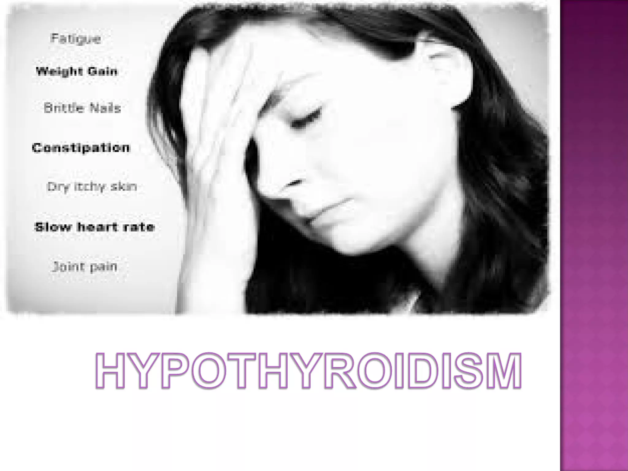
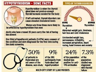
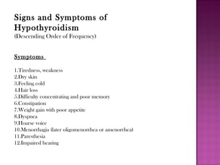
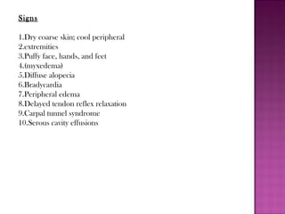
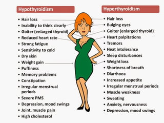
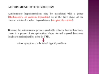
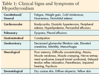
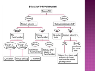
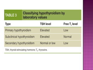
Causes of Hypothyroidism
Primary
Autoimmune hypothyroidism: Hashimoto’s thyroiditis, atrophic
thyroiditis
Iatrogenic: I 131 treatment, subtotal or total thyroidectomy, external
irradiation of neck for lymphoma or cancer
Drugs: iodine excess (including iodine-containing contrast media and
amiodarone),lithium, antithyroid drugs, p-aminosalicylic acid, interferon
α and other cytokines, aminoglutethimide, tyrosine kinase inhibitors
(e.g., sunitinib)
Congenital hypothyroidism: absent or ectopic thyroid gland,
dyshormonogenesis, TSH-R mutation
Iodine deficiency
Infiltrative disorders: amyloidosis, sarcoidosis, hemochromatosis,
scleroderma, cystinosis, Riedel’s thyroiditis
Overexpression of type 3 deiodinase in infantile hemangioma and
other tumors
Transient
Silent thyroiditis, including postpartum thyroiditis
 Subacute thyroiditis
Withdrawal of supraphysiologic thyroxine treatment in individuals
with an intact thyroid
After 131I treatment or subtotal thyroidectomy for Graves’ disease
Secondary
Hypopituitarism: tumors, pituitary surgery or irradiation, infiltrative
disorders, Sheehan’s syndrome, trauma, genetic forms of combined
pituitary hormone deficiencies
Isolated TSH deficiency or inactivity
Bexarotene treatment
Hypothalamic disease: tumors, trauma, infiltrative disorders,
idiopathic
Signs and Symptoms of
Hypothyroidism
(Descending Order of Frequency)
Symptoms
1.Tiredness, weakness
2.Dry skin
3.Feeling cold
4.Hair loss
5.Difficulty concentrating and poor memory
6.Constipation
7.Weight gain with poor appetite
8.Dyspnea
9.Hoarse voice
10.Menorrhagia (later oligomenorrhea or amenorrhea)
11.Paresthesia
12.Impaired hearing
Signs
1.Dry coarse skin; cool peripheral
2.extremities
3.Puffy face, hands, and feet
4.(myxedema)
5.Diffuse alopecia
6.Bradycardia
7.Peripheral edema
8.Delayed tendon reflex relaxation
9.Carpal tunnel syndrome
10.Serous cavity effusions
Congenital hypothyroidism
Hypothyroidism occurs in about 1 in 4000 newborns.
It may be transient, especially if the mother has TSH-R blocking
antibodies
or has received antithyroid drugs, but permanent hypothyroidism
occurs in the majority.
Neonatal hypothyroidism is due to:
 thyroid gland dysgenesis in 80–85%
 inborn errors of thyroid hormone synthesis in 10–15%
 TSH-R antibody-mediated in 5%
The developmental abnormalities are twice as common in girls.
Mutations that cause congenital hypothyroidism are being
increasingly identified, but most remain idiopathic
The majority of infants appear normal at birth and <10% are
diagnosed based on clinical features,
-prolonged jaundice, feeding problems, hypotonia, enlarged tongue,
delayed bone maturation, and umbilical hernia.
Importantly, permanent neurologic damage results if treatment is
delayed.
Typical features of adult hypothyroidism may also be present
Other congenital malformations, especially cardiac, are four times
more common in congenital hypothyroidism.
Diagnosis and Treatment
Because of the severe neurologic consequences of untreated congenital
hypothyroidism, neonatal screening programs have been established.
These are generally based on measurement of TSH or T4 levels in
heel-prick blood specimens.
When the diagnosis is confirmed, T4 is instituted at a dose of 10–15
g/kg per dayμ , and the dose is adjusted by close monitoring of TSH
levels.
T4 requirements are relatively great during the first year of life, and a
high circulating T4 level is usually needed to normalize TSH.
Early treatment with T4 results in normal IQ levels, but subtle
neurodevelopmental abnormalities may occur in those with the most
severe hypothyroidism at diagnosis or when treatment is delayed or
suboptimal.
AUTOIMMUNE HYPOTHYROIDISM
Autoimmune hypothyroidism may be associated with a goiter
(Hashimoto’s, or goitrous thyroiditis) or, at the later stages of the
disease, minimal residual thyroid tissue (atrophic thyroiditis).
Because the autoimmune process gradually reduces thyroid function,
there is a phase of compensation when normal thyroid hormone
levels are maintained by a rise in TSH.
minor symptoms, subclinical hypothyroidism.
Later,
unbound T4 levels fall and TSH levels rise further
symptoms become more readily apparent at this stage
(usually TSH >10 mIU/L)
-clinical hypothyroidism or overt hypothyroidism.
In Hashimoto’s thyroiditis:
there is a marked lymphocytic infiltration of the thyroid with
germinal center formation
atrophy of the thyroid follicles accompanied by oxyphil metaplasia
absence of colloid
 mild to moderate fibrosis.
In atrophic thyroiditis:
the fibrosis is much more extensive
 lymphocyte infiltration is less pronounced
thyroid follicles are almost completely absent
Atrophic thyroiditis likely represents the end stage of Hashimoto’s
thyroiditis rather than a distinct disorder.
susceptibility to autoimmune
hypothyroidism is determined by a combination of genetic and environmental
factors, and the risk of either autoimmune hypothyroidism or
Graves’ disease is increased among siblings. HLA-DR polymorphisms
are the best documented genetic risk factors for autoimmune hypothyroidism,
especially HLA-DR3, -DR4, and -DR5 in Caucasians. A
weak association also exists between polymorphisms in CTLA-4, a T
cell–regulatory gene, and autoimmune hypothyroidism. Both of these
genetic associations are shared by other autoimmune diseases, which
may explain the relationship between autoimmune hypothyroidism and
other autoimmune diseases, especially type 1 diabetes mellitus, Addison’s
disease, pernicious anemia, and vitiligo. HLA-DR and CTLA-4 polymorphisms
account for approximately half of the genetic susceptibility to
autoimmune hypothyroidism. Other contributory loci remain to be
identified. A gene on chromosome 21 may be responsible for the association
between autoimmune hypothyroidism and Down’s syndrome. The
female preponderance of thyroid autoimmunity is most likely due to sex
steroid effects on the immune response, but an X chromosome–related
genetic factor is also possible and may account for the high frequency
of autoimmune hypothyroidism in Turner’s syndrome. Environmental
susceptibility factors are poorly defined at present. A high iodine intake
and decreased exposure to microorganisms in childhood increase the
risk of autoimmune hypothyroidism. These factors may account for the
increase in prevalence over the last two to three decades.
The thyroid lymphocytic infiltrate in autoimmune hypothyroidism
is composed of activated CD4+ and CD8+ T cells as well as B cells.
Thyroid cell destruction is primarily mediated by the CD8+ cytotoxic
T cells, which destroy their targets by either perforin-induced cell
necrosis or granzyme B–induced apoptosis. In addition, local T cell
production of cytokines, such as tumor necrosis factor (TNF), IL-1,
and interferon γ (IFN-γ), may render thyroid cells more susceptible
to apoptosis mediated by death receptors, such as Fas, which are
activated by their respective ligands on T cells. These cytokines also
impair thyroid cell function directly and induce the expression of
other proinflammatory molecules by the thyroid cells themselves, such
as cytokines, HLA class I and class II molecules, adhesion molecules,
CD40, and nitric oxide. Administration of high concentrations of cytokines
for therapeutic purposes (especially IFN-α) is associated with
increased autoimmune thyroid disease, possibly through mechanisms
similar to those in sporadic disease.
Antibodies to TPO and Tg are clinically useful markers of thyroid
autoimmunity, but any pathogenic effect is restricted to a secondary
role in amplifying an ongoing autoimmune response. TPO antibodies
fix complement, and complement membrane-attack complexes
are present in the thyroid in autoimmune hypothyroidism. However,
transplacental passage of Tg or TPO antibodies has no effect on the
fetal thyroid, which suggests that T cell–mediated injury is required to
initiate autoimmune damage to the thyroid.
Up to 20% of patients with autoimmune hypothyroidism have antibodies
against the TSH-R, which, in contrast to TSI, do not stimulate
the receptor but prevent the binding of TSH. These TSH-R-blocking
antibodies, therefore, cause hypothyroidism and, especially in Asian
patients, thyroid atrophy. Their transplacental passage may induce
transient neonatal hypothyroidism.
Rarely, patients have a mixture of
TSI and TSH-R-blocking antibodies, and thyroid function can oscillate
between hyperthyroidism and hypothyroidism as one or the other
antibody becomes dominant. Predicting the course of disease in such
individuals is difficult, and they require close monitoring of thyroid
function. Bioassays can be used to document that TSH-R-blocking
antibodies reduce the cyclic AMP–inducing effect of TSH on cultured
TSH-R-expressing cells, but these assays are difficult to perform.
Thyrotropin-binding inhibitory immunoglobulin (TBII) assays that
measure the binding of antibodies to the receptor by competition
with radiolabeled TSH do not distinguish between TSI- and TSH-Rblocking
antibodies, but a positive result in a patient with spontaneous
hypothyroidism is strong evidence for the presence of blocking
antibodies. The use of these assays does not generally alter clinicalmanagement,
although it may be useful to confirm the cause of tran- 2291
sient neonatal hypothyroidism.
CLINICAL FEATURES
The onset is usually insidious, and the patient may become
aware of symptoms only when euthyroidism is restored.
Patients with Hashimoto’s thyroiditis may present because of
goiter rather than symptoms of hypothyroidism.
The goiter may not be large, but it is usually irregular and firm
in consistency.
It is often possible to palpate a pyramidal lobe, normally a
vestigial remnant of the thyroglossal duct.
Rarely is uncomplicated Hashimoto’s thyroiditis associated
with pain.
Patients with atrophic thyroiditis or the late stage of Hashimoto’s
thyroiditis present with symptoms and signs of hypothyroidism.
 The skin is dry, and there is decreased sweating, thinning of the
epidermis, and hyperkeratosis of the stratum corneum.
Increased dermal glycosaminoglycan content traps water, giving rise
to skin thickening without pitting (myxedema).
Typical features include a puffy face with edematous eyelids and
nonpitting pretibial edema
There is pallor, often with a yellow tinge to the skin due to carotene
accumulation.
Nail growth is retarded, and hair is dry, brittle, difficult to manage,
and falls out easily.
In addition to diffuse alopecia, there is thinning of the outer third of
the eyebrows, although this is not a specific sign of hypothyroidism.
Other common features include constipation and weight gain
(despite a poor appetite). In contrast to popular perception, the
weight gain is usually modest and due mainly to fluid retention in the
myxedematous tissues.
Libido is decreased in both sexes, and there may be oligomenorrhea
or amenorrhea in long-standing disease, but menorrhagia is also
common. Fertility is reduced, and the incidence of miscarriage is
increased. Prolactin levels are often modestly increased and may
contribute to alterations in libido and fertility and cause galactorrhea.
Pulmonary function is generally normal, but dyspnea may be caused
by pleural effusion, impaired respiratory muscle function, diminished
ventilatory drive, or sleep apnea
Myocardial contractility and pulse rate are reduced, leading to a
reduced stroke volume and bradycardia. Increased peripheral
resistance may be accompanied by hypertension, particularly
diastolic. Blood flow is diverted from the skin, producing cool
extremities. Pericardial effusions occur in up to 30% of patients
but rarely compromise cardiac function. Although alterations in
myosin heavy chain isoform expression have been documented,
cardiomyopathy is unusual.
Fluid may also accumulate in other serous cavities and in the
middle ear, giving rise to conductive deafness.
Carpal tunnel and other entrapment syndromes are common, as
is impairment of muscle function with stiffness, cramps, and
pain. On examination, there may be slow relaxation of tendon
reflexes and pseudomyotonia.
Memory and concentration are impaired.
Experimentally, positron emission tomography (PET) scans
examining glucose metabolism in hypothyroid subjects show lower
regional activity in the amygdala, hippocampus, and perigenual
anterior cingulated cortex, among other regions, and this activity
corrects after thyroxine replacement.
Rare neurologic problems include reversible cerebellar ataxia,
dementia, psychosis, and myxedema coma.
Hashimoto’s encephalopathy has been defined as a steroid-responsive
syndrome associated with TPO antibodies, myoclonus, and slow-wave
activity on electroencephalography, but the relationship with thyroid
autoimmunity or hypothyroidism is not established.
The hoarse voice and occasionally clumsy speech of hypothyroidism
reflect fluid accumulation in the vocal cords and tongue.
Autoimmune hypothyroidism may be associated with signs or symptoms
of other autoimmune diseases, particularly vitiligo, pernicious anemia,
Addison’s disease, alopecia areata, and type 1 diabetes mellitus. Less
common associations include celiac disease, dermatitis herpetiformis,
chronic active hepatitis, rheumatoid arthritis, systemic lupus
erythematosus (SLE), myasthenia gravis, and Sjogren’s syndrome.
Thyroid-associated ophthalmopathy, which usually occurs in Graves’
disease , occurs in about 5% of patients with autoimmune
hypothyroidism.
Autoimmune hypothyroidism is uncommon in children and usually
presents :
1.Slow growth
2.Delayed facial maturation.
3.The appearance of permanent teeth is also delayed.
4.Myopathy, with muscle swelling, is more common in children than in
adults.
5.In most cases, Puberty is delayed, but precocious puberty sometimes
occurs.
6.There may be intellectual impairment if the onset is before 3 years
and the hormone deficiency is severe.
If the TSH is elevated, an unbound T4 level is needed to confirm
the presence of clinical hypothyroidism, but T4 is inferior to
TSH when used as a screening test, because it will not detect
subclinical hypothyroidism.
Circulating unbound T3 levels are normal in about 25% of
patients, reflecting adaptive deiodinase responses to
hypothyroidism.
T3 measurements are, therefore, not indicated.
Once clinical or subclinical hypothyroidism is confirmed, the etiology
is usually easily established by demonstrating the presence of TPO
antibodies, which are present in >90% of patients with autoimmune
hypothyroidism.
TBII can be found in 10–20% of patients, but measurement is not
needed routinely.
If there is any doubt about the cause of a goiter associated with
hypothyroidism, FNA biopsy can be used to confirm the presence of
autoimmune thyroiditis.
Other abnormal laboratory findings in hypothyroidism may include:
increased creatine phosphokinase,
elevated cholesterol and triglycerides
anemia (usually normocytic or macrocytic).
Except when accompanied by iron deficiency, the anemia and other
abnormalities gradually resolve with thyroxine replacement.
Differential Diagnosis
An asymmetric goiter in Hashimoto’s thyroiditis may be confused
with a MNG or thyroid carcinoma, in which thyroid antibodies may
also be present.
Ultrasound can be used to show the presence of a solitary lesion or an
MNG rather than the heterogeneous thyroid enlargement typical of
Hashimoto’s thyroiditis.
FNA biopsy is useful in the investigation of focal nodules.
Iatrogenic hypothyroidism is a common cause of hypothyroidism and
can often be detected by screening before symptoms develop. In the
first 3–4 months after radioiodine treatment, transient hypothyroidism
may occur due to reversible radiation damage.
Low-dose thyroxine treatment can be withdrawn if recovery occurs.
Because TSH levels are suppressed by hyperthyroidism, unbound T4
levels are a better measure of thyroid function than TSH in the
months following radioiodine treatment.
Mild hypothyroidism after subtotal thyroidectomy may also resolve
after several months, as the gland remnant is stimulated by increased
TSH levels.
Iodine deficiency is responsible for endemic goiter and cretinism but is
an uncommon cause of adult hypothyroidism unless the iodine intake
is very low or there are complicating factors, such as the consumption
of thiocyanates in cassava or selenium deficiency.
Although hypothyroidism due to iodine deficiency can be treated with
thyroxine, public health measures to improve iodine intake should be
advocated to eliminate this problem.
Iodized salt or bread or a single bolus of oral or intramuscular iodized
oil have all been used successfully.
Paradoxically, chronic iodine excess can also induce goiter and
hypothyroidism. The intracellular events that account for this effect
are unclear, but individuals with autoimmune thyroiditis are
especially susceptible. Iodine excess is responsible for the
hypothyroidism that occurs in up to 13% of patients treated with
amiodarone. Other drugs, particularly lithium, may also cause
hypothyroidism. Transient hypothyroidism caused by thyroiditis is
discussed below.
Secondary hypothyroidism is usually diagnosed in the context of
other anterior pituitary hormone deficiencies; isolated TSH
deficiency is very rare .TSH levels may be low, normal, or even
slightly increased in secondary hypothyroidism; the latter is due to
secretion of immunoactive but bioinactive forms of TSH.
The diagnosis is confirmed by detecting a low unbound T4 level.
The goal of treatment is to maintain T4 levels in the upper half of
the reference range, because TSH levels cannot be used to monitor
therapy.
CLINICAL HYPOTHYROIDISM
If there is no residual thyroid function, the daily replacement dose of
levothyroxine is usually 1.6 g/kg body weight (typically 100–150 g),μ μ
ideally taken at least 30 min before breakfast.
In many patients, however, lower doses suffice until residual thyroid
tissue is destroyed.
In patients who develop hypothyroidism after the treatment of Graves’
disease, there is often underlying autonomous function, necessitating
lower replacement doses (typically 75–125 μg/d).
Adult patients under 60 years old without evidence of heart disease
may be started on 50–100 g levothyroxineμ (T4) daily.
The dose is adjusted on the basis of TSH levels, with the goal of
treatment being a normal TSH, ideally in the lower half of the
reference range.
TSH responses are gradual and should be measured about 2 months
after instituting treatment or after any subsequent change in
levothyroxine dosage.
The clinical effects of levothyroxine replacement are slow to appear.
Patients may not experience full relief from symptoms until 3–6
months after normal TSH levels are restored. Adjustment of
levothyroxine dosage is made in 12.5- or 25- g increments if the TSHμ
is high; decrements of the same magnitude should be made if the
TSH is suppressed.
Patients with a suppressed TSH of any cause, including T4
overtreatment, have an increased risk of atrial fibrillation and reduced
bone density.
Although desiccated animal thyroid preparations (thyroid extract
USP) are available, they are not recommended because the ratio of
T3 to T4 is nonphysiologic.
The use of levothyroxine combined with liothyronine
(triiodothyronine, T3) has been investigated, but benefit has not
been confirmed in prospective studies.
There is no place for liothyronine alone as long-term replacement,
because the short half-life necessitates three or four daily doses and
is associated with fluctuating T3 levels.
Once full replacement is achieved and TSH levels are stable,
follow-up measurement of TSH is recommended at annual
intervals and may be extended to every 2–3 years if a normal TSH
is maintained over several years. It is important to ensure ongoing
adherence, however, as patients do not feel any symptomatic
difference after missing a few doses of levothyroxine, and this
sometimes leads to self-discontinuation.
In patients of normal body weight who are taking ≥200
g of levothyroxine per day, an elevated TSH level isμ
often a sign of poor adherence to treatment.
This is also the likely explanation for fluctuating TSH levels,
despite a constant levothyroxine dosage.
Such patients often have normal or high unbound T4 levels,
despite an elevated TSH, because they remember to take
medication for a few days before testing; this is sufficient to
normalize T4, but not TSH levels.
It is important to consider variable adherence, because this pattern
of thyroid function tests is otherwise suggestive of disorders
associated with inappropriate TSH secretion .
Because T4 has a long half-life (7 days), patients who miss a dose
can be advised to take two doses of the skipped tablets at once.
Other causes of increased levothyroxine requirements must be
excluded, particularly malabsorption (e.g., celiac disease, small-
bowel surgery), estrogen or selective estrogen receptor modulator
therapy, ingestion with a meal, and drugs that interfere with T4
absorption or metabolism such as cholestyramine, ferrous sulfate,
calcium supplements, proton pump inhibitors, lovastatin, aluminum
hydroxide, rifampicin, amiodarone, carbamazepine, phenytoin, and
tyrosine kinase inhibitors.
SUBCLINICAL HYPOTHYROIDISM
By definition, subclinical hypothyroidism refers to biochemical
evidence of thyroid hormone deficiency in patients who have few or
no apparent clinical features of hypothyroidism.
There are no universally accepted recommendations for the
management of subclinical hypothyroidism, but levothyroxine is
recommended if the patient is a woman who wishes to conceive or is
pregnant, or when TSH levels are above 10 mIU/L. When TSH
levels are below 10 mIU/L, treatment should be considered when
patients have suggestive symptoms of hypothyroidism, positive TPO
antibodies, or any evidence of heart disease.
It is important to confirm that any elevation of TSH is
sustained over a 3-month period before treatment is given.
As long as excessive treatment is avoided, there is no risk in
correcting a slightly increased TSH.
Treatment is administered by starting with a low dose of
levothyroxine (25–50 g/d) with the goal of normalizingμ
TSH.
If levothyroxine is not given, thyroid function should be
evaluated annually
SPECIAL TREATMENT CONSIDERATIONS
Rarely, levothyroxine replacement is associated with pseudotumor
cerebri in children. Presentation appears to be idiosyncratic and
occurs months after treatment has begun.
Women with a history or high risk of hypothyroidism should ensure
that they are euthyroid prior to conception and during early
pregnancy because maternal hypothyroidism may adversely affect fetal
neural development and cause preterm delivery.
The presence of thyroid autoantibodies alone, in a euthyroid patient,
is also associated with miscarriage and preterm delivery; it is unclear if
levothyroxine therapy improves outcomes.
Thyroid function should be evaluated immediately after pregnancy is
confirmed and every 4 weeks during the first half of the pregnancy, with
less frequent testing after 20 weeks’ gestation (every 6–8 weeks
depending on whether levothyroxine dose adjustment is ongoing).
The levothyroxine dose may need to be increased by up to 50% during
pregnancy, with a goal TSH of less than 2.5 mIU/L during the first
trimester and less than 3.0 mIU/L during the second and third
trimesters.
After delivery, thyroxine doses typically return to prepregnancy levels.
Pregnant women should be counseled to separate ingestion of prenatal
vitamins and iron supplements from levothyroxine by at least 4 h.
Elderly patients may require 20% less thyroxine than younger
patients. In the elderly, especially patients with known coronary
artery disease, the starting dose of levothyroxine is 12.5–25 g/dμ
with similar increments every 2–3 months until TSH is normalized.
In some patients, it may be impossible to achieve full replacement
despite optimal antianginal treatment.
Emergency surgery is generally safe in patients with untreated
hypothyroidism, although routine surgery in a hypothyroid patient
should be deferred until euthyroidism is achieved.
Myxedema coma still has a 20–40% mortality rate, despite intensive
treatment, and outcomes are independent of the T4 and TSH
levels.
Clinical manifestations include :
reduced level of consciousness
sometimes associated with seizures
other features of hypothyroidism.
Hypothermia can reach 23°C (74°F).
There may be a history of treated hypothyroidism with poor
compliance, or the patient may be previously undiagnosed.
Myxedema coma almost always occurs in the elderly and is usually
precipitated :
By factors that impair respiration, such as drugs (especially sedatives,
anesthetics, and antidepressants), pneumonia, congestive heart failure,
myocardial infarction, gastrointestinal bleeding, or cerebrovascular
accidents.
Sepsis should also be suspected.
Exposure to cold may also be a risk factor.
Hypoventilation, leading to hypoxia and hypercapnia, plays a major role
in pathogenesis; hypoglycemia and dilutional hyponatremia also
contribute to the development of myxedema coma.
Levothyroxine can initially be administered as a single IV bolus of 500
g, which serves as a loading dose. Although further levothyroxine is notμ
strictly necessary for several days, it is usually continued at a dose of 50–
100 g/d.μ
If suitable IV preparation is not available, the same initial dose of
levothyroxine can be given by nasogastric tube (although absorption may
be impaired in myxedema).
An alternative is to give liothyronine (T3) intravenously or via
nasogastric tube, in doses ranging from 10 to 25 g every 8–12 h.μ
This treatment has been advocated because T4 T3 conversion is→
impaired in myxedemacoma. However, excess liothyronine has the
potential to provoke arrhythmias.
Another option is to combine levothyroxine (200 g) and liothyronineμ
(25 g) as a single, initial IV bolus followed by daily treatment withμ
levothyroxine (50–100 g/d) and liothyronine (10 g every 8 h).μ μ
Supportive therapy should be provided to correct any associated
metabolic disturbances.
External warming is indicated only if the temperature is <30°C, as
it can result in cardiovascular collapse .Space blankets should be
used to prevent further heat loss.
Parenteral hydrocortisone (50 mg every 6 h) should be
administered, because there is impaired adrenal reserve in
profound hypothyroidism.
Any precipitating factors should be treated, including the early
use of broad-spectrum antibiotics, pending the exclusion of
infection.
Ventilatory support with regular blood gas analysis is usually needed
during the first 48 h.
Hypertonic saline or IV glucose may be needed if there is severe
hyponatremia or hypoglycemia; hypotonic IV fluids should be
avoided because they may exacerbate water retention secondary to
reduced renal perfusion and inappropriate vasopressin secretion.
The metabolism of most medications is impaired, and sedatives
should be avoided if possible or used in reduced doses. Medication
blood levels should be monitored, when available, to guide dosage
DIETARY ADVICE
Goitrogens are chemicals that are naturally occurring and are ingested
in foods or drugs. These chemicals can affect thyroid function,
rendering it unable to produce some of the hormones necessary for
regulating metabolism.
It is important to note that the Goitrogenic properties can be lessened
by cooking. The process of steaming or cooking alters the molecular
structure of the Isothiocyanates – eliminating the goitrogenic effect. A
person does not have to completely eliminate certain foods. Let
moderation, preparation  and how you feel be a guide.
Foods that are known goitrogens include:
- soy products, and from the chemical group called
Isothiocyanates. Isothiocyanates are found in Cabbage,
Kale, Broccoli, Turnips, Peaches, Mustard greens, and
Pears. Due to small levels of compounds, other foods
that are potentially goitrogenic are peanuts, peaches,
spinach, millet, pine nuts, and strawberries.
Make sure that you include the following in your daily diet:
#1. Seafood and other iodine-rich foods
It’s no secret that iodine is needed to produce thyroid hormone in
your body. If you have hypothyroidism, diet should always include
foods that are high in iodine content such as seafood like sushi,
seaweed, and saltwater fish. Iodine can also be found in bananas and
parsley. Also, you should make sure to use iodized salt instead of plain
rock salt in cooking.
#2. Water
You need to drink lots of water, at least 64 ounces or 8 glasses a day.
Remember when you are dehydrated, your body stores more fat and
making you gain more weight.
#3. Fiber
Fiber is beneficial to you if you have hypothyroidism. Diets high in
fiber will not only help you lose weight but also promote regularity
in your bowel movement and can help lower your cholesterol.
How does it make you lose weight? Fiber makes you feel full so it
lessens your hunger levels, and will simply make you burn more
calories. Most fruits and vegetables are high in fiber content.
#4. Coconut Oil
It has been identified that consumption of coconut oil stimulates
the production of thyroid hormone. It increases your metabolism
and is considered as an energy booster but this should be taken in
moderation, appropriate to your age and body structure.
#5. Green Tea
Green tea helps you lose weight by raising your metabolism and also
through fat oxidation. You can start drinking a cup of green tea
everyday.
#6. Tyrosine-rich Foods
Tyrosine is needed for your thyroid hormone production. Increase
your intake of lean meats, fish, lentils, and low-fat milk as these are
rich in tyrosine.
#7. Selenium-rich Foods
Just like tyrosine, this is also important in helping to produce your
thyroid hormone. Foods such as tuna, brown rice, Brazil nuts, and
salmon can provide the selenium that your body needs
Common psychological problems:
• Mental lethargy, general dulling of the personality and
slowing of all cognitive functions.
• Complains of thickness in the head or of ‘feeling of fog’
• Memory affected from early stages
• Marked inability to sustain mental exertion, and increasing
slowness of uptake and grasp delay to seek medical help
• Irritability
Psychosis and dementia with myxoedema
No form of psychosis specific to myxodema, but rather a variety
of ‘reaction types’ .
The only unifying feature is the frequency of a paranoid
colouring whatever form the psychosis may take.
The commonest is an organic psychosis ,sometimes developing
acutely or sometimes running a subacute course over several
weeks or months
In other cases a picture of dementia develops insidiously and
may progress over several years to an advanced degree.
More rarely a severe depressive psychosis may emerge or a
typical schizophrenia may be precipitated.
In Asher’s (1949) classical paper on ‘myxoedematous madness’ :
5-organic reaction with hallucinations and persecutory ideas
5 -schizophrenia with marked paranoid colouring
2 -advanced dementia
2 -depressive features
Common organic psychosis:
Features of delirium
Florid delusions of persecutions
 Auditory hallucinations
 Mental confusion and impairment of cognitive functions and
particularly of recent memory.
Impairment of consciousness
Dementia develops as an extension of the mental impairment.
Olivarus and Roder considered myxoedema to be the most
important and the most frequently overlooked of the metabolic
causes of reversible organic intellectual impairment
Neurological abnormalities in myxoedema
Slowing of tendon reflexes
Jellinek (1962) has drawn attention to the occurrence of fits, faints and
cerebrovascular accidents in myxoedematous patients.
4 patients were reported with grandmal attacks which responded to
thyroid replacement ,others with attacks of syncope and others with
unusual confusional episodes which suggested temporal lobe
dysfunction.
Cerebrovascular accidents were found to have occurred in several
patients and evidence of attacks of TIA s in several more.
Jellinek and Kelly(1960) have described other cases of myxoedema
presenting with cerebellar disturbance in the form of ataxia, tremor,
dysarthria and nystagmus and remitting promptly with replacement
therapy.
Differential diagnosis
Patients may be labelled as
neurotic, hypochondriacal or
personality disordered.
Aetiology of mental disturbances
Changes in cerebral metabolism.
Decreased cerebral blood flow(decreased cardiac output) with unaltered
metabolic needs of glucose and oxygen
cerebral hypoxia
coexisting anaemia
hypoxia worsens
symptoms
The acute organic psychosis are likely to be due to some additional
aspect of the intracerebral metabolic disturbance
Other possible etiologies: electrolyte changes and catecholamine
disturbances
Non myxoedematous hypometabolism
Kurland et al (1955) reported a syndrome of metabolic insufficiecy
in which the basal metabolic rate is low in spite of a normal serum
protein bound iodine and radioactive iodine uptake.
Treatment with thyroxine was ineffective but triiodothyronine raised
BMR and produced clinical improvement
Levin(1960)- found no benefit with either.
The patients complaints were more typical of neurotic disorder.
Outcome of mental disturbances in myxoedema
Prognosis is good with good response to thyroxine therapy.
Additional measures in the form of phenothiazines, antidepressant
medication or electroconvulsive therapy may be necessary in severe
psychotic disorders .
Phenothiazines carry some risk of precipitating hypothermic coma in
hypothyroid patients as in the case reported by Mitchell et al.(1959)
Hypothyroidism management and psychiatric aspects

Hypothyroidism management and psychiatric aspects

  • 2.
    Causes of Hypothyroidism Primary Autoimmunehypothyroidism: Hashimoto’s thyroiditis, atrophic thyroiditis Iatrogenic: I 131 treatment, subtotal or total thyroidectomy, external irradiation of neck for lymphoma or cancer Drugs: iodine excess (including iodine-containing contrast media and amiodarone),lithium, antithyroid drugs, p-aminosalicylic acid, interferon α and other cytokines, aminoglutethimide, tyrosine kinase inhibitors (e.g., sunitinib)
  • 3.
    Congenital hypothyroidism: absentor ectopic thyroid gland, dyshormonogenesis, TSH-R mutation Iodine deficiency Infiltrative disorders: amyloidosis, sarcoidosis, hemochromatosis, scleroderma, cystinosis, Riedel’s thyroiditis Overexpression of type 3 deiodinase in infantile hemangioma and other tumors
  • 4.
    Transient Silent thyroiditis, includingpostpartum thyroiditis  Subacute thyroiditis Withdrawal of supraphysiologic thyroxine treatment in individuals with an intact thyroid After 131I treatment or subtotal thyroidectomy for Graves’ disease
  • 5.
    Secondary Hypopituitarism: tumors, pituitarysurgery or irradiation, infiltrative disorders, Sheehan’s syndrome, trauma, genetic forms of combined pituitary hormone deficiencies Isolated TSH deficiency or inactivity Bexarotene treatment Hypothalamic disease: tumors, trauma, infiltrative disorders, idiopathic
  • 7.
    Signs and Symptomsof Hypothyroidism (Descending Order of Frequency) Symptoms 1.Tiredness, weakness 2.Dry skin 3.Feeling cold 4.Hair loss 5.Difficulty concentrating and poor memory 6.Constipation 7.Weight gain with poor appetite 8.Dyspnea 9.Hoarse voice 10.Menorrhagia (later oligomenorrhea or amenorrhea) 11.Paresthesia 12.Impaired hearing
  • 8.
    Signs 1.Dry coarse skin;cool peripheral 2.extremities 3.Puffy face, hands, and feet 4.(myxedema) 5.Diffuse alopecia 6.Bradycardia 7.Peripheral edema 8.Delayed tendon reflex relaxation 9.Carpal tunnel syndrome 10.Serous cavity effusions
  • 10.
    Congenital hypothyroidism Hypothyroidism occursin about 1 in 4000 newborns. It may be transient, especially if the mother has TSH-R blocking antibodies or has received antithyroid drugs, but permanent hypothyroidism occurs in the majority. Neonatal hypothyroidism is due to:  thyroid gland dysgenesis in 80–85%  inborn errors of thyroid hormone synthesis in 10–15%  TSH-R antibody-mediated in 5% The developmental abnormalities are twice as common in girls. Mutations that cause congenital hypothyroidism are being increasingly identified, but most remain idiopathic
  • 11.
    The majority ofinfants appear normal at birth and <10% are diagnosed based on clinical features, -prolonged jaundice, feeding problems, hypotonia, enlarged tongue, delayed bone maturation, and umbilical hernia. Importantly, permanent neurologic damage results if treatment is delayed. Typical features of adult hypothyroidism may also be present Other congenital malformations, especially cardiac, are four times more common in congenital hypothyroidism.
  • 12.
    Diagnosis and Treatment Becauseof the severe neurologic consequences of untreated congenital hypothyroidism, neonatal screening programs have been established. These are generally based on measurement of TSH or T4 levels in heel-prick blood specimens. When the diagnosis is confirmed, T4 is instituted at a dose of 10–15 g/kg per dayμ , and the dose is adjusted by close monitoring of TSH levels. T4 requirements are relatively great during the first year of life, and a high circulating T4 level is usually needed to normalize TSH. Early treatment with T4 results in normal IQ levels, but subtle neurodevelopmental abnormalities may occur in those with the most severe hypothyroidism at diagnosis or when treatment is delayed or suboptimal.
  • 13.
    AUTOIMMUNE HYPOTHYROIDISM Autoimmune hypothyroidismmay be associated with a goiter (Hashimoto’s, or goitrous thyroiditis) or, at the later stages of the disease, minimal residual thyroid tissue (atrophic thyroiditis). Because the autoimmune process gradually reduces thyroid function, there is a phase of compensation when normal thyroid hormone levels are maintained by a rise in TSH. minor symptoms, subclinical hypothyroidism.
  • 14.
    Later, unbound T4 levelsfall and TSH levels rise further symptoms become more readily apparent at this stage (usually TSH >10 mIU/L) -clinical hypothyroidism or overt hypothyroidism.
  • 15.
    In Hashimoto’s thyroiditis: thereis a marked lymphocytic infiltration of the thyroid with germinal center formation atrophy of the thyroid follicles accompanied by oxyphil metaplasia absence of colloid  mild to moderate fibrosis. In atrophic thyroiditis: the fibrosis is much more extensive  lymphocyte infiltration is less pronounced thyroid follicles are almost completely absent Atrophic thyroiditis likely represents the end stage of Hashimoto’s thyroiditis rather than a distinct disorder.
  • 16.
    susceptibility to autoimmune hypothyroidismis determined by a combination of genetic and environmental factors, and the risk of either autoimmune hypothyroidism or Graves’ disease is increased among siblings. HLA-DR polymorphisms are the best documented genetic risk factors for autoimmune hypothyroidism, especially HLA-DR3, -DR4, and -DR5 in Caucasians. A weak association also exists between polymorphisms in CTLA-4, a T cell–regulatory gene, and autoimmune hypothyroidism. Both of these genetic associations are shared by other autoimmune diseases, which may explain the relationship between autoimmune hypothyroidism and other autoimmune diseases, especially type 1 diabetes mellitus, Addison’s disease, pernicious anemia, and vitiligo. HLA-DR and CTLA-4 polymorphisms account for approximately half of the genetic susceptibility to autoimmune hypothyroidism. Other contributory loci remain to be identified. A gene on chromosome 21 may be responsible for the association between autoimmune hypothyroidism and Down’s syndrome. The female preponderance of thyroid autoimmunity is most likely due to sex steroid effects on the immune response, but an X chromosome–related genetic factor is also possible and may account for the high frequency of autoimmune hypothyroidism in Turner’s syndrome. Environmental susceptibility factors are poorly defined at present. A high iodine intake and decreased exposure to microorganisms in childhood increase the risk of autoimmune hypothyroidism. These factors may account for the increase in prevalence over the last two to three decades.
  • 17.
    The thyroid lymphocyticinfiltrate in autoimmune hypothyroidism is composed of activated CD4+ and CD8+ T cells as well as B cells. Thyroid cell destruction is primarily mediated by the CD8+ cytotoxic T cells, which destroy their targets by either perforin-induced cell necrosis or granzyme B–induced apoptosis. In addition, local T cell production of cytokines, such as tumor necrosis factor (TNF), IL-1, and interferon γ (IFN-γ), may render thyroid cells more susceptible to apoptosis mediated by death receptors, such as Fas, which are activated by their respective ligands on T cells. These cytokines also impair thyroid cell function directly and induce the expression of other proinflammatory molecules by the thyroid cells themselves, such as cytokines, HLA class I and class II molecules, adhesion molecules, CD40, and nitric oxide. Administration of high concentrations of cytokines for therapeutic purposes (especially IFN-α) is associated with increased autoimmune thyroid disease, possibly through mechanisms similar to those in sporadic disease.
  • 18.
    Antibodies to TPOand Tg are clinically useful markers of thyroid autoimmunity, but any pathogenic effect is restricted to a secondary role in amplifying an ongoing autoimmune response. TPO antibodies fix complement, and complement membrane-attack complexes are present in the thyroid in autoimmune hypothyroidism. However, transplacental passage of Tg or TPO antibodies has no effect on the fetal thyroid, which suggests that T cell–mediated injury is required to initiate autoimmune damage to the thyroid. Up to 20% of patients with autoimmune hypothyroidism have antibodies against the TSH-R, which, in contrast to TSI, do not stimulate the receptor but prevent the binding of TSH. These TSH-R-blocking antibodies, therefore, cause hypothyroidism and, especially in Asian patients, thyroid atrophy. Their transplacental passage may induce transient neonatal hypothyroidism.
  • 19.
    Antibodies to TPOand Tg are clinically useful markers of thyroid autoimmunity, but any pathogenic effect is restricted to a secondary role in amplifying an ongoing autoimmune response. TPO antibodies fix complement, and complement membrane-attack complexes are present in the thyroid in autoimmune hypothyroidism. However, transplacental passage of Tg or TPO antibodies has no effect on the fetal thyroid, which suggests that T cell–mediated injury is required to initiate autoimmune damage to the thyroid. Up to 20% of patients with autoimmune hypothyroidism have antibodies against the TSH-R, which, in contrast to TSI, do not stimulate the receptor but prevent the binding of TSH. These TSH-R-blocking antibodies, therefore, cause hypothyroidism and, especially in Asian patients, thyroid atrophy. Their transplacental passage may induce transient neonatal hypothyroidism.
  • 20.
    Rarely, patients havea mixture of TSI and TSH-R-blocking antibodies, and thyroid function can oscillate between hyperthyroidism and hypothyroidism as one or the other antibody becomes dominant. Predicting the course of disease in such individuals is difficult, and they require close monitoring of thyroid function. Bioassays can be used to document that TSH-R-blocking antibodies reduce the cyclic AMP–inducing effect of TSH on cultured TSH-R-expressing cells, but these assays are difficult to perform. Thyrotropin-binding inhibitory immunoglobulin (TBII) assays that measure the binding of antibodies to the receptor by competition with radiolabeled TSH do not distinguish between TSI- and TSH-Rblocking antibodies, but a positive result in a patient with spontaneous hypothyroidism is strong evidence for the presence of blocking antibodies. The use of these assays does not generally alter clinicalmanagement, although it may be useful to confirm the cause of tran- 2291 sient neonatal hypothyroidism.
  • 21.
    CLINICAL FEATURES The onsetis usually insidious, and the patient may become aware of symptoms only when euthyroidism is restored. Patients with Hashimoto’s thyroiditis may present because of goiter rather than symptoms of hypothyroidism. The goiter may not be large, but it is usually irregular and firm in consistency. It is often possible to palpate a pyramidal lobe, normally a vestigial remnant of the thyroglossal duct. Rarely is uncomplicated Hashimoto’s thyroiditis associated with pain.
  • 22.
    Patients with atrophicthyroiditis or the late stage of Hashimoto’s thyroiditis present with symptoms and signs of hypothyroidism.  The skin is dry, and there is decreased sweating, thinning of the epidermis, and hyperkeratosis of the stratum corneum. Increased dermal glycosaminoglycan content traps water, giving rise to skin thickening without pitting (myxedema). Typical features include a puffy face with edematous eyelids and nonpitting pretibial edema There is pallor, often with a yellow tinge to the skin due to carotene accumulation. Nail growth is retarded, and hair is dry, brittle, difficult to manage, and falls out easily. In addition to diffuse alopecia, there is thinning of the outer third of the eyebrows, although this is not a specific sign of hypothyroidism.
  • 23.
    Other common featuresinclude constipation and weight gain (despite a poor appetite). In contrast to popular perception, the weight gain is usually modest and due mainly to fluid retention in the myxedematous tissues. Libido is decreased in both sexes, and there may be oligomenorrhea or amenorrhea in long-standing disease, but menorrhagia is also common. Fertility is reduced, and the incidence of miscarriage is increased. Prolactin levels are often modestly increased and may contribute to alterations in libido and fertility and cause galactorrhea. Pulmonary function is generally normal, but dyspnea may be caused by pleural effusion, impaired respiratory muscle function, diminished ventilatory drive, or sleep apnea
  • 24.
    Myocardial contractility andpulse rate are reduced, leading to a reduced stroke volume and bradycardia. Increased peripheral resistance may be accompanied by hypertension, particularly diastolic. Blood flow is diverted from the skin, producing cool extremities. Pericardial effusions occur in up to 30% of patients but rarely compromise cardiac function. Although alterations in myosin heavy chain isoform expression have been documented, cardiomyopathy is unusual. Fluid may also accumulate in other serous cavities and in the middle ear, giving rise to conductive deafness. Carpal tunnel and other entrapment syndromes are common, as is impairment of muscle function with stiffness, cramps, and pain. On examination, there may be slow relaxation of tendon reflexes and pseudomyotonia. Memory and concentration are impaired.
  • 25.
    Experimentally, positron emissiontomography (PET) scans examining glucose metabolism in hypothyroid subjects show lower regional activity in the amygdala, hippocampus, and perigenual anterior cingulated cortex, among other regions, and this activity corrects after thyroxine replacement. Rare neurologic problems include reversible cerebellar ataxia, dementia, psychosis, and myxedema coma. Hashimoto’s encephalopathy has been defined as a steroid-responsive syndrome associated with TPO antibodies, myoclonus, and slow-wave activity on electroencephalography, but the relationship with thyroid autoimmunity or hypothyroidism is not established. The hoarse voice and occasionally clumsy speech of hypothyroidism reflect fluid accumulation in the vocal cords and tongue.
  • 26.
    Autoimmune hypothyroidism maybe associated with signs or symptoms of other autoimmune diseases, particularly vitiligo, pernicious anemia, Addison’s disease, alopecia areata, and type 1 diabetes mellitus. Less common associations include celiac disease, dermatitis herpetiformis, chronic active hepatitis, rheumatoid arthritis, systemic lupus erythematosus (SLE), myasthenia gravis, and Sjogren’s syndrome. Thyroid-associated ophthalmopathy, which usually occurs in Graves’ disease , occurs in about 5% of patients with autoimmune hypothyroidism.
  • 28.
    Autoimmune hypothyroidism isuncommon in children and usually presents : 1.Slow growth 2.Delayed facial maturation. 3.The appearance of permanent teeth is also delayed. 4.Myopathy, with muscle swelling, is more common in children than in adults. 5.In most cases, Puberty is delayed, but precocious puberty sometimes occurs. 6.There may be intellectual impairment if the onset is before 3 years and the hormone deficiency is severe.
  • 31.
    If the TSHis elevated, an unbound T4 level is needed to confirm the presence of clinical hypothyroidism, but T4 is inferior to TSH when used as a screening test, because it will not detect subclinical hypothyroidism. Circulating unbound T3 levels are normal in about 25% of patients, reflecting adaptive deiodinase responses to hypothyroidism. T3 measurements are, therefore, not indicated.
  • 33.
    Once clinical orsubclinical hypothyroidism is confirmed, the etiology is usually easily established by demonstrating the presence of TPO antibodies, which are present in >90% of patients with autoimmune hypothyroidism. TBII can be found in 10–20% of patients, but measurement is not needed routinely. If there is any doubt about the cause of a goiter associated with hypothyroidism, FNA biopsy can be used to confirm the presence of autoimmune thyroiditis. Other abnormal laboratory findings in hypothyroidism may include: increased creatine phosphokinase, elevated cholesterol and triglycerides anemia (usually normocytic or macrocytic). Except when accompanied by iron deficiency, the anemia and other abnormalities gradually resolve with thyroxine replacement.
  • 34.
    Differential Diagnosis An asymmetricgoiter in Hashimoto’s thyroiditis may be confused with a MNG or thyroid carcinoma, in which thyroid antibodies may also be present. Ultrasound can be used to show the presence of a solitary lesion or an MNG rather than the heterogeneous thyroid enlargement typical of Hashimoto’s thyroiditis. FNA biopsy is useful in the investigation of focal nodules.
  • 35.
    Iatrogenic hypothyroidism isa common cause of hypothyroidism and can often be detected by screening before symptoms develop. In the first 3–4 months after radioiodine treatment, transient hypothyroidism may occur due to reversible radiation damage. Low-dose thyroxine treatment can be withdrawn if recovery occurs. Because TSH levels are suppressed by hyperthyroidism, unbound T4 levels are a better measure of thyroid function than TSH in the months following radioiodine treatment. Mild hypothyroidism after subtotal thyroidectomy may also resolve after several months, as the gland remnant is stimulated by increased TSH levels.
  • 36.
    Iodine deficiency isresponsible for endemic goiter and cretinism but is an uncommon cause of adult hypothyroidism unless the iodine intake is very low or there are complicating factors, such as the consumption of thiocyanates in cassava or selenium deficiency. Although hypothyroidism due to iodine deficiency can be treated with thyroxine, public health measures to improve iodine intake should be advocated to eliminate this problem. Iodized salt or bread or a single bolus of oral or intramuscular iodized oil have all been used successfully.
  • 37.
    Paradoxically, chronic iodineexcess can also induce goiter and hypothyroidism. The intracellular events that account for this effect are unclear, but individuals with autoimmune thyroiditis are especially susceptible. Iodine excess is responsible for the hypothyroidism that occurs in up to 13% of patients treated with amiodarone. Other drugs, particularly lithium, may also cause hypothyroidism. Transient hypothyroidism caused by thyroiditis is discussed below. Secondary hypothyroidism is usually diagnosed in the context of other anterior pituitary hormone deficiencies; isolated TSH deficiency is very rare .TSH levels may be low, normal, or even slightly increased in secondary hypothyroidism; the latter is due to secretion of immunoactive but bioinactive forms of TSH. The diagnosis is confirmed by detecting a low unbound T4 level. The goal of treatment is to maintain T4 levels in the upper half of the reference range, because TSH levels cannot be used to monitor therapy.
  • 39.
    CLINICAL HYPOTHYROIDISM If thereis no residual thyroid function, the daily replacement dose of levothyroxine is usually 1.6 g/kg body weight (typically 100–150 g),μ μ ideally taken at least 30 min before breakfast. In many patients, however, lower doses suffice until residual thyroid tissue is destroyed. In patients who develop hypothyroidism after the treatment of Graves’ disease, there is often underlying autonomous function, necessitating lower replacement doses (typically 75–125 μg/d).
  • 40.
    Adult patients under60 years old without evidence of heart disease may be started on 50–100 g levothyroxineμ (T4) daily. The dose is adjusted on the basis of TSH levels, with the goal of treatment being a normal TSH, ideally in the lower half of the reference range. TSH responses are gradual and should be measured about 2 months after instituting treatment or after any subsequent change in levothyroxine dosage. The clinical effects of levothyroxine replacement are slow to appear. Patients may not experience full relief from symptoms until 3–6 months after normal TSH levels are restored. Adjustment of levothyroxine dosage is made in 12.5- or 25- g increments if the TSHμ is high; decrements of the same magnitude should be made if the TSH is suppressed. Patients with a suppressed TSH of any cause, including T4 overtreatment, have an increased risk of atrial fibrillation and reduced bone density.
  • 41.
    Although desiccated animalthyroid preparations (thyroid extract USP) are available, they are not recommended because the ratio of T3 to T4 is nonphysiologic. The use of levothyroxine combined with liothyronine (triiodothyronine, T3) has been investigated, but benefit has not been confirmed in prospective studies. There is no place for liothyronine alone as long-term replacement, because the short half-life necessitates three or four daily doses and is associated with fluctuating T3 levels. Once full replacement is achieved and TSH levels are stable, follow-up measurement of TSH is recommended at annual intervals and may be extended to every 2–3 years if a normal TSH is maintained over several years. It is important to ensure ongoing adherence, however, as patients do not feel any symptomatic difference after missing a few doses of levothyroxine, and this sometimes leads to self-discontinuation.
  • 42.
    In patients ofnormal body weight who are taking ≥200 g of levothyroxine per day, an elevated TSH level isμ often a sign of poor adherence to treatment. This is also the likely explanation for fluctuating TSH levels, despite a constant levothyroxine dosage. Such patients often have normal or high unbound T4 levels, despite an elevated TSH, because they remember to take medication for a few days before testing; this is sufficient to normalize T4, but not TSH levels. It is important to consider variable adherence, because this pattern of thyroid function tests is otherwise suggestive of disorders associated with inappropriate TSH secretion .
  • 43.
    Because T4 hasa long half-life (7 days), patients who miss a dose can be advised to take two doses of the skipped tablets at once. Other causes of increased levothyroxine requirements must be excluded, particularly malabsorption (e.g., celiac disease, small- bowel surgery), estrogen or selective estrogen receptor modulator therapy, ingestion with a meal, and drugs that interfere with T4 absorption or metabolism such as cholestyramine, ferrous sulfate, calcium supplements, proton pump inhibitors, lovastatin, aluminum hydroxide, rifampicin, amiodarone, carbamazepine, phenytoin, and tyrosine kinase inhibitors.
  • 44.
    SUBCLINICAL HYPOTHYROIDISM By definition,subclinical hypothyroidism refers to biochemical evidence of thyroid hormone deficiency in patients who have few or no apparent clinical features of hypothyroidism. There are no universally accepted recommendations for the management of subclinical hypothyroidism, but levothyroxine is recommended if the patient is a woman who wishes to conceive or is pregnant, or when TSH levels are above 10 mIU/L. When TSH levels are below 10 mIU/L, treatment should be considered when patients have suggestive symptoms of hypothyroidism, positive TPO antibodies, or any evidence of heart disease.
  • 45.
    It is importantto confirm that any elevation of TSH is sustained over a 3-month period before treatment is given. As long as excessive treatment is avoided, there is no risk in correcting a slightly increased TSH. Treatment is administered by starting with a low dose of levothyroxine (25–50 g/d) with the goal of normalizingμ TSH. If levothyroxine is not given, thyroid function should be evaluated annually
  • 47.
    SPECIAL TREATMENT CONSIDERATIONS Rarely,levothyroxine replacement is associated with pseudotumor cerebri in children. Presentation appears to be idiosyncratic and occurs months after treatment has begun. Women with a history or high risk of hypothyroidism should ensure that they are euthyroid prior to conception and during early pregnancy because maternal hypothyroidism may adversely affect fetal neural development and cause preterm delivery. The presence of thyroid autoantibodies alone, in a euthyroid patient, is also associated with miscarriage and preterm delivery; it is unclear if levothyroxine therapy improves outcomes.
  • 48.
    Thyroid function shouldbe evaluated immediately after pregnancy is confirmed and every 4 weeks during the first half of the pregnancy, with less frequent testing after 20 weeks’ gestation (every 6–8 weeks depending on whether levothyroxine dose adjustment is ongoing). The levothyroxine dose may need to be increased by up to 50% during pregnancy, with a goal TSH of less than 2.5 mIU/L during the first trimester and less than 3.0 mIU/L during the second and third trimesters. After delivery, thyroxine doses typically return to prepregnancy levels. Pregnant women should be counseled to separate ingestion of prenatal vitamins and iron supplements from levothyroxine by at least 4 h.
  • 49.
    Elderly patients mayrequire 20% less thyroxine than younger patients. In the elderly, especially patients with known coronary artery disease, the starting dose of levothyroxine is 12.5–25 g/dμ with similar increments every 2–3 months until TSH is normalized. In some patients, it may be impossible to achieve full replacement despite optimal antianginal treatment. Emergency surgery is generally safe in patients with untreated hypothyroidism, although routine surgery in a hypothyroid patient should be deferred until euthyroidism is achieved.
  • 50.
    Myxedema coma stillhas a 20–40% mortality rate, despite intensive treatment, and outcomes are independent of the T4 and TSH levels. Clinical manifestations include : reduced level of consciousness sometimes associated with seizures other features of hypothyroidism. Hypothermia can reach 23°C (74°F). There may be a history of treated hypothyroidism with poor compliance, or the patient may be previously undiagnosed.
  • 51.
    Myxedema coma almostalways occurs in the elderly and is usually precipitated : By factors that impair respiration, such as drugs (especially sedatives, anesthetics, and antidepressants), pneumonia, congestive heart failure, myocardial infarction, gastrointestinal bleeding, or cerebrovascular accidents. Sepsis should also be suspected. Exposure to cold may also be a risk factor. Hypoventilation, leading to hypoxia and hypercapnia, plays a major role in pathogenesis; hypoglycemia and dilutional hyponatremia also contribute to the development of myxedema coma.
  • 52.
    Levothyroxine can initiallybe administered as a single IV bolus of 500 g, which serves as a loading dose. Although further levothyroxine is notμ strictly necessary for several days, it is usually continued at a dose of 50– 100 g/d.μ If suitable IV preparation is not available, the same initial dose of levothyroxine can be given by nasogastric tube (although absorption may be impaired in myxedema). An alternative is to give liothyronine (T3) intravenously or via nasogastric tube, in doses ranging from 10 to 25 g every 8–12 h.μ This treatment has been advocated because T4 T3 conversion is→ impaired in myxedemacoma. However, excess liothyronine has the potential to provoke arrhythmias. Another option is to combine levothyroxine (200 g) and liothyronineμ (25 g) as a single, initial IV bolus followed by daily treatment withμ levothyroxine (50–100 g/d) and liothyronine (10 g every 8 h).μ μ
  • 53.
    Supportive therapy shouldbe provided to correct any associated metabolic disturbances. External warming is indicated only if the temperature is <30°C, as it can result in cardiovascular collapse .Space blankets should be used to prevent further heat loss. Parenteral hydrocortisone (50 mg every 6 h) should be administered, because there is impaired adrenal reserve in profound hypothyroidism. Any precipitating factors should be treated, including the early use of broad-spectrum antibiotics, pending the exclusion of infection.
  • 54.
    Ventilatory support withregular blood gas analysis is usually needed during the first 48 h. Hypertonic saline or IV glucose may be needed if there is severe hyponatremia or hypoglycemia; hypotonic IV fluids should be avoided because they may exacerbate water retention secondary to reduced renal perfusion and inappropriate vasopressin secretion. The metabolism of most medications is impaired, and sedatives should be avoided if possible or used in reduced doses. Medication blood levels should be monitored, when available, to guide dosage
  • 55.
    DIETARY ADVICE Goitrogens arechemicals that are naturally occurring and are ingested in foods or drugs. These chemicals can affect thyroid function, rendering it unable to produce some of the hormones necessary for regulating metabolism. It is important to note that the Goitrogenic properties can be lessened by cooking. The process of steaming or cooking alters the molecular structure of the Isothiocyanates – eliminating the goitrogenic effect. A person does not have to completely eliminate certain foods. Let moderation, preparation  and how you feel be a guide.
  • 56.
    Foods that areknown goitrogens include: - soy products, and from the chemical group called Isothiocyanates. Isothiocyanates are found in Cabbage, Kale, Broccoli, Turnips, Peaches, Mustard greens, and Pears. Due to small levels of compounds, other foods that are potentially goitrogenic are peanuts, peaches, spinach, millet, pine nuts, and strawberries.
  • 57.
    Make sure thatyou include the following in your daily diet: #1. Seafood and other iodine-rich foods It’s no secret that iodine is needed to produce thyroid hormone in your body. If you have hypothyroidism, diet should always include foods that are high in iodine content such as seafood like sushi, seaweed, and saltwater fish. Iodine can also be found in bananas and parsley. Also, you should make sure to use iodized salt instead of plain rock salt in cooking. #2. Water You need to drink lots of water, at least 64 ounces or 8 glasses a day. Remember when you are dehydrated, your body stores more fat and making you gain more weight.
  • 58.
    #3. Fiber Fiber isbeneficial to you if you have hypothyroidism. Diets high in fiber will not only help you lose weight but also promote regularity in your bowel movement and can help lower your cholesterol. How does it make you lose weight? Fiber makes you feel full so it lessens your hunger levels, and will simply make you burn more calories. Most fruits and vegetables are high in fiber content. #4. Coconut Oil It has been identified that consumption of coconut oil stimulates the production of thyroid hormone. It increases your metabolism and is considered as an energy booster but this should be taken in moderation, appropriate to your age and body structure.
  • 59.
    #5. Green Tea Greentea helps you lose weight by raising your metabolism and also through fat oxidation. You can start drinking a cup of green tea everyday. #6. Tyrosine-rich Foods Tyrosine is needed for your thyroid hormone production. Increase your intake of lean meats, fish, lentils, and low-fat milk as these are rich in tyrosine. #7. Selenium-rich Foods Just like tyrosine, this is also important in helping to produce your thyroid hormone. Foods such as tuna, brown rice, Brazil nuts, and salmon can provide the selenium that your body needs
  • 61.
    Common psychological problems: •Mental lethargy, general dulling of the personality and slowing of all cognitive functions. • Complains of thickness in the head or of ‘feeling of fog’ • Memory affected from early stages • Marked inability to sustain mental exertion, and increasing slowness of uptake and grasp delay to seek medical help • Irritability
  • 62.
    Psychosis and dementiawith myxoedema No form of psychosis specific to myxodema, but rather a variety of ‘reaction types’ . The only unifying feature is the frequency of a paranoid colouring whatever form the psychosis may take. The commonest is an organic psychosis ,sometimes developing acutely or sometimes running a subacute course over several weeks or months In other cases a picture of dementia develops insidiously and may progress over several years to an advanced degree. More rarely a severe depressive psychosis may emerge or a typical schizophrenia may be precipitated.
  • 63.
    In Asher’s (1949)classical paper on ‘myxoedematous madness’ : 5-organic reaction with hallucinations and persecutory ideas 5 -schizophrenia with marked paranoid colouring 2 -advanced dementia 2 -depressive features
  • 64.
    Common organic psychosis: Featuresof delirium Florid delusions of persecutions  Auditory hallucinations  Mental confusion and impairment of cognitive functions and particularly of recent memory. Impairment of consciousness Dementia develops as an extension of the mental impairment. Olivarus and Roder considered myxoedema to be the most important and the most frequently overlooked of the metabolic causes of reversible organic intellectual impairment
  • 65.
    Neurological abnormalities inmyxoedema Slowing of tendon reflexes Jellinek (1962) has drawn attention to the occurrence of fits, faints and cerebrovascular accidents in myxoedematous patients. 4 patients were reported with grandmal attacks which responded to thyroid replacement ,others with attacks of syncope and others with unusual confusional episodes which suggested temporal lobe dysfunction. Cerebrovascular accidents were found to have occurred in several patients and evidence of attacks of TIA s in several more. Jellinek and Kelly(1960) have described other cases of myxoedema presenting with cerebellar disturbance in the form of ataxia, tremor, dysarthria and nystagmus and remitting promptly with replacement therapy.
  • 66.
    Differential diagnosis Patients maybe labelled as neurotic, hypochondriacal or personality disordered.
  • 67.
    Aetiology of mentaldisturbances Changes in cerebral metabolism. Decreased cerebral blood flow(decreased cardiac output) with unaltered metabolic needs of glucose and oxygen cerebral hypoxia coexisting anaemia hypoxia worsens symptoms The acute organic psychosis are likely to be due to some additional aspect of the intracerebral metabolic disturbance Other possible etiologies: electrolyte changes and catecholamine disturbances
  • 68.
    Non myxoedematous hypometabolism Kurlandet al (1955) reported a syndrome of metabolic insufficiecy in which the basal metabolic rate is low in spite of a normal serum protein bound iodine and radioactive iodine uptake. Treatment with thyroxine was ineffective but triiodothyronine raised BMR and produced clinical improvement Levin(1960)- found no benefit with either. The patients complaints were more typical of neurotic disorder.
  • 69.
    Outcome of mentaldisturbances in myxoedema Prognosis is good with good response to thyroxine therapy. Additional measures in the form of phenothiazines, antidepressant medication or electroconvulsive therapy may be necessary in severe psychotic disorders . Phenothiazines carry some risk of precipitating hypothermic coma in hypothyroid patients as in the case reported by Mitchell et al.(1959)