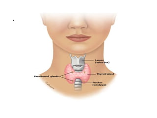
• The thyroidgland consists of two lobes and
is situated in the lower neck.
• The gland synthesizes, stores and releases
two major metabolically active hormones:
tetra-iodothyronine (thyroxine [T4]) and tri-
iodothyronine (T3).
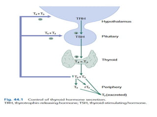
• Regulation of hormone synthesis is by
variable secretion of the glycoprotein
hormone thyroid stimulating hormone (TSH)
from the anterior pituitary.
4.
• In turnTSH is regulated by hypothalamic secretion
of the tripeptide thyrotrophin-releasing hormone
(TRH).
• Low circulating levels of thyroid hormones initiate
the release of TSH and probably also TRH.
• Rising levels of TSH promote increased iodide
trapping by the gland and a subsequent increase in
thyroid hormone synthesis.
• The increase in circulating hormone levels feeds
back on the pituitary and hypothalamus, shutting off
TRH, TSH and further hormone synthesis.
6.
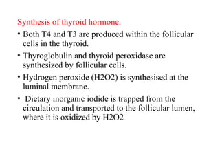
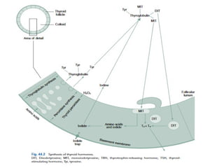
Synthesis of thyroidhormone.
• Both T4 and T3 are produced within the follicular
cells in the thyroid.
• Thyroglobulin and thyroid peroxidase are
synthesized by follicular cells.
• Hydrogen peroxide (H2O2) is synthesised at the
luminal membrane.
• Dietary inorganic iodide is trapped from the
circulation and transported to the follicular lumen,
where it is oxidized by H2O2
7.
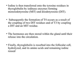
• Iodine isthen transferred onto the tyrosine residues in
thyroglobulin by iodinase enzymes forming
monoiodotyrosine (MIT) and diiodotyrosine (DIT).
• Subsequently the formation of T4 occurs as a result of
the coupling of two DIT residues and of T3 by coupling
a DIT and an MIT residue.
• The hormones are then stored within the gland until their
release into the circulation.
• Finally, thyroglobulin is resorbed into the follicular cell,
hydrolyzed, and its amino acids and remaining iodine
reused
9.
• The T4:T3ratio secreted by the thyroid gland is
approximately 10:1
• However, only 10% of circulating T3 is derived from direct
thyroidal secretion, the remaining 90% being produced by
peripheral conversion from T4.
• T4 can therefore be considered a prohormone that is
converted in the peripheral tissues (liver, kidney and brain)
either to the active hormone T3 or to the biologically
inactive reverse T3 (rT3)
10.
• T4 is99.98% bound, with only 0.02% circulating
free.
• T3 is slightly less protein bound (99.8%), resulting
in a considerably higher circulating free fraction
(0.2%).
• The hormones are metabolized in the periphery
(kidney, liver and heart) by deiodination.
• The half-life of T4 in plasma is about 6–7 days and
that of T3 is 24–36 hours in euthyroid adults.
• The apparent volume of distribution for T4 is about
10 L and for T3 about 40 L.
11.
Epidemiology
• Thyroid diseaseis common, affecting approximately
5% to 15%of the general population.
• Women are three to four times more likely than
men to experience any type of thyroid disease.
• The prevalence of hypothyroidism is 1.4% to 2% in
women and 0.1% to 0.2% in men.
• The incidence increases in persons older than 60
years, to 6% of women and 2.5% of men.
12.
• Hyperthyroidism affectsabout 2% of women and
about 0.1% of men.
• The prevalence of hyperthyroidism in older
patients varies between 0.5% and 2.3% but
accounts for 10% to 15% of all thyrotoxic patients,
13.
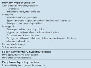
Hypothyroidism
• Hypothyroidism isthe clinical state resulting from
decreased production of thyroid hormones or rarely
from tissue resistance.
• Iodine deficiency is the commonest cause of
hypothyroidism globally
• Iodine replete regions autoimmune disease is the
common cause of primary hypo thyroidism and
accounts for more than 95% of adult cases.
14.
• It isusually due to a failure of the thyroid gland itself as
a result of autoimmune destruction, or the effects of
treatment of thyrotoxicosis.
• Hypothyroidism may be drug induced. Amiodarone and
lithium cause hypothyroidism in around 10% of patients
treated.
• Secondary disease is due to hypopituitarism, and
tertiary disease is due to failure of the hypothalamus
• Peripheral hypothyroidism is due to tissue insensitivity
to the action of thyroid hormones.
15.
• Iodides mayproduce hypothyroidism in patients who
are particularly sensitive to their ability to block the
active transport pump of the thyroid gland.
• Iodine absorption from topical iodine- containing
antiseptics has been shown to cause hypothyroidism in
neonates.
• This is potentially very dangerous at a critical time of
neurological development in the newborn infant.
• Transient hypothyroidism may be seen in 25% of iodine
exposed infants.
17.
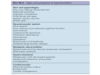
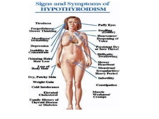
Clinical manifestations
• Hypothyroidismcan affect multiple body systems,
but symptoms are mainly nonspecific and gradual
in onset .
• The most useful clinical signs are myotonic (slow
relaxing) tendon relexes, bradycardia, hair loss and
cool, dry skin.
• Effusions may occur into pericardial, pleural,
peritoneal or joint spaces.
18.
• Mild anemiaof a macrocytic type is quite common
and responds to thyroxine replacement.
• Pernicious anemia is a frequent concomitant
finding in hypothyroidism.
• Other, organ speciic autoimmune diseases such as
Addison’s disease may be associated.
21.
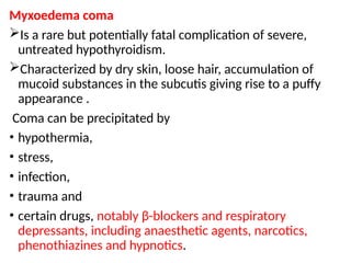
Myxoedema coma
Is arare but potentially fatal complication of severe,
untreated hypothyroidism.
Characterized by dry skin, loose hair, accumulation of
mucoid substances in the subcutis giving rise to a puffy
appearance .
Coma can be precipitated by
• hypothermia,
• stress,
• infection,
• trauma and
• certain drugs, notably β-blockers and respiratory
depressants, including anaesthetic agents, narcotics,
phenothiazines and hypnotics.
22.
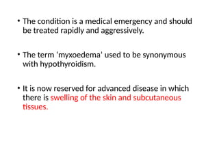
• The conditionis a medical emergency and should
be treated rapidly and aggressively.
• The term ‘myxoedema’ used to be synonymous
with hypothyroidism.
• It is now reserved for advanced disease in which
there is swelling of the skin and subcutaneous
tissues.
23.
Investigations of hypothyroidism
•Usually clinical assessment, combined with a single
estimation of thyroid hormones and TSH, is suficient to
make the diagnosis.
• In primary disease, the levels of free T4 and T3 are low,
and the TSH level rises markedly.
• A chest radiograph may detect the presence of
effusions, and an electrocardiogram is useful, especially
in patients with angina or coronary heart disease, in
whom replacement therapy needs to be introduced
gradually.
24.
Investigations
• Thyroid-stimulating hormone(TSH) test
• Positive results:
• High TSH levels: This is the primary indicator of an
underactive thyroid. Your pituitary gland produces TSH to
stimulate your thyroid to produce hormones. When your
thyroid isn't producing enough hormones, your pituitary
gland releases more TSH in an attempt to compensate.
• T4 (thyroxine) test
• Positive results:
• Low T4 levels: T4 is the main thyroid hormone
produced by your thyroid gland. Low levels
confirm the diagnosis of hypothyroidism,
especially when combined with high TSH levels.
25.
Cont……..
• Thyroid antibodiestest:
• Positive results:
• Presence of antibodies: These tests (like anti-
thyroid peroxidase antibodies (TPA)and anti-
thyroglobulin antibodies) can help determine if
your hypothyroidism is caused by an autoimmune
condition like Hashimoto's disease.
26.
Treatment
• Levothyroxine (l-thyroxine)is the preferred thyroid
replacement preparation.
• The signs and symptoms of hypothyroidism can be easily
corrected in most patients by the administration of l-
thyroxine on an empty stomach at an oral replacement
dosage
• Exceptions include older patients, patients with severe and
long standing hypothyroidism, and patients with cardiac
disease, in whom administration of full replacement doses
might cause cardiac toxicity
• In such patients, minute T4 doses should be started
initially, and the dosage titrated upward as tolerated;
27.
• complete reversalof hypothyroidism might not be
indicated or possible
• In myxedema coma, intravenous (IV) therapy with a
large initial loading dose of l-thyroxine is necessary
to reduce the high mortality rate. it is controversial
whether T4 replacement therapy is beneficial.
• There is no justification for treating patients with
hypothyroid symptoms and normal TSH findings
with T4.
28.
• The goalof therapy is to reverse the signs and symptoms
of hypothyroidism and normalize the TSH and FT4 levels.
• Some improvement of hypothyroid symptoms is often
evident within 2 to 3 weeks of starting T4 therapy.
NB
• Over replacement of l-thyroxine is associated with
osteoporosis and cardiac changes
• The optimal T4 replacement dosage must be administered
for approx imately 6 to 8 weeks before steady-state levels
are reached.
• Evaluation of thyroid function tests before this time is
misleading.
29.
Once a euthyroidstate is attained, laboratory tests
can be monitored every 3 to 6 months for the first
year and then yearly thereafter
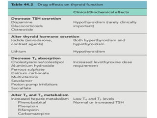
Medications that interfere with T4 absorption e.g.,
• iron,
• aluminum-containing products,
• some calcium preparations [e.g., carbonate],
• cholesterol resin and
• phosphate binders,
should be separated by at least 4 hours from
concomitant T4 administration.
32.
Hyperthyroidism/thyrotoxicosis
• Is thehyper metabolic syndrome that occurs when the production of
thyroid hormone is excessive.
• Thyrotoxicosis refers to the clinical syndrome associated with
prolonged exposure to elevated levels of thyroid hormone.
Aetiology
• Hyperthyroidism is a disorder of various aetiologies.
• In clinical terms, thyrotoxicosis is the result of persistently elevated
levels of thyroid hormones.
33.
1.Graves’ disease
• Graves’disease is the commonest cause of
thyrotoxicosis.
• It is an autoimmune condition and results from
production of an abnormal immunoglobulin G (IgG) that
is able to occupy the TSH receptor on the thyroid
follicular cell.
• Here it mimics the effect of TSH, causing cell division
and stimulating thyroid hormone secretion
34.
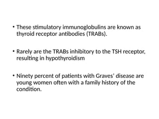
• These stimulatoryimmunoglobulins are known as
thyroid receptor antibodies (TRABs).
• Rarely are the TRABs inhibitory to the TSH receptor,
resulting in hypothyroidism
• Ninety percent of patients with Graves’ disease are
young women often with a family history of the
condition.
35.
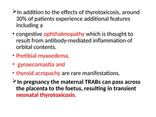
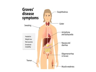
In addition tothe effects of thyrotoxicosis, around
30% of patients experience additional features
including a
• congestive ophthalmopathy which is thought to
result from antibody-mediated inflammation of
orbital contents.
• Pretibial myxoedema,
• gynaecomastia and
• thyroid acropachy are rare manifestations.
In pregnancy the maternal TRABs can pass across
the placenta to the foetus, resulting in transient
neonatal thyrotoxicosis.
38.
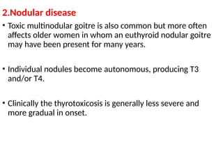
2.Nodular disease
• Toxicmultinodular goitre is also common but more often
affects older women in whom an euthyroid nodular goitre
may have been present for many years.
• Individual nodules become autonomous, producing T3
and/or T4.
• Clinically the thyrotoxicosis is generally less severe and
more gradual in onset.
39.
• Often onlyT3 levels are elevated, although the TSH
will be suppressed in all cases.
• Thyrotoxicosis may also be caused by single
autonomous thyroid adenomas.
• These are benign, well-differentiated tumours that
secrete excessive amounts of thyroid hormones.
41.
3.Thyroiditis
• If thethyroid is inflamed by viral or rapid
autoimmune attack, the resulting follicular cell
death will result in the release of pre-formed
thyroid hormones.
• This usually presents as a painful mildly enlarged
and tender thyroid.
• There is a brief period of hyperthyroidism before
thyroid hormone levels fall to subnormal.
• .
42.
• Most oftenthis period of hyperthyroidism does not
lead to clinically apparent thyrotoxicosis
• and in any event is brief, but it is common for these
patients to be prescribed thionamides which
compound the ensuing hypothyroidism.
43.
Clinical manifestation
• Thyrotoxicosisis characterised by increases in
metabolic rate and activity of many systems due to
excessive circulating quantities of thyroid
hormones.
• There is a wide spectrum of clinical disturbance.
The signs and symptoms reflect increased
adrenergic activity, especially in the cardiovascular
and neurological systems
NB
Not all manifestations will be seen in every patient.
Additional clinical features will depend on the
underlying cause of the thyrotoxicosis
44.
• The clinicalfeatures of thyrotoxicosis in the elderly
may not be so obvious.
• Signs and symptoms of cardiovascular disturbance
tend to predominate, atrial fibrillation is frequent
and the patient may experience congestive heart
failure.
• Unexplained heart failure after middle age should
always arouse suspicion of thyrotoxicosis.
45.
• The extrathyroidalmanifestations of Graves’
disease deserve separate mention.
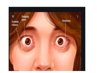
• Most frequent is ophthalmopathy due to
inflammation and expansion of the contents of the
orbit.
• The eye is pushed forward (proptosis) such that
white sclera appears between the iris and the lower
lid.
• Congestive changes develop including peri-orbital
oedema, conjunctival swelling and redness.
46.
• The extraocularmuscles are swollen and become
tethered,leading to failure of movement of the
globe of the eye and thus diplopia.
• Severe disease causes pressure in the orbit, which
cancompress the optic nerve leading to blindness.
• The cutaneous features of Graves’ disease include
thickening of the pretibial skin (myxoedema),
onycholysis (separation of the nail from the nail
bed) and acropachy (similar to inger clubbing) .
48.
• Investigation
• Plasmafree T4 (and/or T3) levels are elevated.
• The TSH level is suppressed to subnormal levels in all
causes of thyrotoxicosis, except the exceptionally rare
cases ofTSH-secreting pituitary adenomas.
• Radioactive iodine uptake scans will differentiate those
patients with thyroiditis.
• Measurement of TRABs will identify patients with Graves’
disease.
• If the diagnosis is still equivocal, the clinical findings
should be reassessed and particular attention paid to the
patient’s drug history.
49.
Treatment
• A numberof factors need to be considered when
choosing the most appropriate form of therapy for
an individual patient
• . Usually a number of therapeutic options are
available, and the patient should be involved in the
decision on what treatment to have.
• The decision may also be influenced by
physician preference,
which in turn can depend on the facilities available
50.
• Three formsof therapy are available, including
antithyroid drugs,
surgery and
radioactive iodine.
51.
There is nogeneral agreement as to the
specific indications for each form of therapy,
and none of them is ideal, because all are
associated with both short- and long-term
sequelae.
Neither surgery nor radioactive iodine should
be given until the patient has been rendered
euthyroid because of the risk of inducing a
thyroid crisis
52.
• In children,surgery may be dificult and the
complication rate is higher. Also radioiodine has
been avoided because of concern about the
potential development of thyroid malignancy.
• In pregnancy, radioiodine is not used because of
the likelihood of the neonate having
hypothyroidism.
• Thyroid surgery during pregnancy should be
deferred until the second trimester if possible, and
most patients’ symptoms can be controlled with
drugs
53.
• Thionamide dosesshould be kept as low as possible,
especially in the last 2 months of preg-nancy,
because excessive treatment may produce goitre in
the foetus.
•
• Aplasia cutis is said to occur after carbimazole
therapy, so propylthiouracil is usually preferred over
the former during the first trimester of pregnancy.
• Pregnant patients with thyrotoxicosis should be
under the care of a specialist endocrine unit
54.
Immediate treatment ofthyrotoxicosis
Patients need to have their symptoms addressed
and their thyrotoxicosis controlled.
• Non-selective β-blockers in standard
antihypertensive doses are effective within a matter
of hours and should be offered to all non-
asthmatics with severe thyro-toxicosis.
• These agents help to alleviate symptoms such as
tremors, palpitations and anxiety which are
generally associated with sympathetic over-activity.
55.
• Carbimazole (40mg once a day) or propylthiouracil
(150 mg twice a day) will render most patients
euthyroid within 6 weeks.
• Adjunctive treatment of cardiac disease and
anxiety/sleeplessness may be required.
56.
Graves’ disease
• Aproportion (40–50%) of patients with Graves’
disease will achieve a long-lasting remission after a
period of euthyroidism while receiving
thionamides.
• The optimal duration of antithyroid treatment is
unknown but in most units the length of the
treatment course has fallen to between 6 and 12
months.
57.
Remission of Graves’disease is much less likely in
those with
• very large goiters,
• those who require high-dose thionamide treatment
to maintain euthyroidism,
• those with high TRAB titres and
• patients who have relapsed once after a course of
drug treatment.
Such patients should therefore be rendered
euthyroid and then have a discussion about either
surgical or radioiodine thyroid ablation
58.
Nodular thyroid disease
•As the nodules function autonomously and
thyrotoxicosis will always recur when thionamides
are stopped,
• there is no value in attempting to achieve a
remission of nodular thyroid disease using
prolonged medical treatment.
• Patients should be rendered euthyroid with drugs
and then have a discussion about radioiodine
ablative (RIA) therapy
59.
Antithyroid drugs
The thionamides,
•propylthiouracil,
• thiamazole (methimazole) and its precursor
• carbimazole,
Are equally effective pharmacological therapies for
thyrotoxicosis.
These drugs prevent thyroid hormone synthesis by
inhibiting the oxidative binding of iodide and its coupling
to tyrosine residues.
Propylthiouracil, but not carbimazole, inhibits the
peripheral deiodination of T4 to T3.
Thionamides may also have an immunosuppressive
action
61.
• Adverse effects.
•The most common adverse effect of antithyroid
treatment is rash and arthropathy (5%) and
• less commonly agranulocytosis, hepatitis, aplastic
anaemia and lupus-like syndromes .
• These side effects usually occur during the first 6 weeks
of treatment, but this is not invariable.
• Cross-sensitivity between carbimazole and
propylthiouracil is around 10%, and the patient can
often be safely changed to the alternative agent if an
adverse event occurs
62.
At the timeof prescription,
• all patients should be counselled about the possible
implication of sore throat, mouth ulcers and
pyrexia, and instructed to seek an urgent (within
hours) full blood count.
• This verbal information should be backed up by
written advice which should specify where the
patient should go for the blood test.
• An abnormal white cell count should prompt urgent
admission under a specialist endocrine team.
63.
The regimen
• Carbimazoleis usually given initially at a dosage up
to 40–60 mg daily, depending on the severity of the
condition.
• It can be given as a single daily dose in multiples of
20 mg tablets to aid adherence.
• Although the plasma half-life is short (4–6 hours),
the biological effect lasts longer (up to 40 hours).
64.
• T4 concentrationsare checked at 6-week intervals
until the patient is clinically euthyroid and the T4
and T3 levels are normalised.
• (TSH remains suppressed for at least 4 weeks after
resolution of significant thyrotoxicosis, so TSH levels
are unhelpful in the early stages of treatment
65.
• Pregnancy isa specific situation, however, in which
tailored-dose propylthiouracil is preferred over
carbimazole during the first trimester.
• Both the immunoglobulins, which cause Graves’
disease, and thionamide drugs cross the placenta
and will affect the fetal thyroid, but maternal
levothyroxine is not able to reach the foetus.
• Thus, the lowest possible dose of propylthiouracil
or carbimazole in pregnancy should be used and
the foetus closely monitored for heart rate and
growth.
• Breastfeeding is considered to be safe when
mothers are tak-ing thionamide
66.
Thyroid ablative therapy
Thyroidablation is required for all patients with
• toxic multinodular goiters,
• those who have relapsed or are likely to relapse
• after drug therapy for Graves’ disease, and
• those who are allergic to thionamides.
Thyroid ablation can be achieved by radioiodine or
surgery.
67.
• Radioactive iodine.
•Radioiodine therapy is extremely easy to
administer by mouth and is very effective for a
large majority of patients.
• It is contraindicated in pregnancy and breast
feeding, and is usually avoided in children.
• It is known to make ophthalmopathy worse in
some patients with Graves’ disease, but
• Giving prednisolone 0.5 mg/kg for 3 weeks and
commencing thyroxine replacement early (3 weeks
after radioiodine) can mitigate this
68.
• The commonestcomplication is the development
of hypothyroidism
Surgery.
Surgery is required for those patients with
very
• large goiters,
• patients who cannot be persuaded of the
safety of radioiodine and
• those who have reacted adversely to both
thionamides in pregnancy.
69.
Treatment of complications
1.Ophthalmopathy
•The commonest complaint is of ‘grittiness’, which can
be treated with hypromellose eye drops or gel.
• If lid retraction is severe, inadequate lid closure can
result in early-morning soreness.
• This can be alleviated by the short-term use of 5%
guanethidine eye drops instilled each night and
morning.
• The eyes should be monitored for any signs of
infection and treated appropriately.
70.
• Progressive ophthalmopathyproducing severe
complications from proptosis, diplopia or visual
failure should be treated with high-dose
corticosteroid therapy (prednisolone 60 mg daily)
until symptoms resolve.
• Failure to respond is an indication for orbital
irradiation or surgical decompression.
71.
Treatment of localisedmyxoedema
• Myxoedema is usually localised to small areas and
is asymptomatic.
• More extensive disease causes difficulty in walking
and considerable discomfort.
• Probably the most effective therapy is the nightly
topical application of corticosteroid creams, such as
betamethasone, under occlusive polythene
dressings
72.
Thyroid Crisis
Thyroid crisiscan develop in any patient with
significant untreated thyrotoxicosis, but it is most
common in those with severe Graves’ disease.
It is precipitated in such patients by infection, injury,
trauma, anaesthetics, surgery and radioiodine.
There is rapidly progressive
• tachycardia,
• muscle weakness (including cardiomyopathy),
• hyperthermia,
• sweating and
• vomiting compounding hypotension with ensuing
circulatory collapse.
73.
• patients areextremely anxious and often
psychotic.
It should be managed as a medical emergency
In addition to supportive measures,
speciic antithyroid therapy is required along with
drugs, which inhibit deiodination of T4to T3.
Propylthiouracil (inhibits deiodinase) is given orally
(or via nasogastric tube) in high dose along with
Lugol’s iodine.
74.
Glucocorticoids should begiven intravenously
because they also inhibit deiodinase.
Effective β-blockade is required by an intravenous
infusion (propranolol is preferred because it also
inhibits deiodinase).
75.
Drugs and thethyroid
Drugs and thyrotoxicosis
• Amiodarone-induced thyrotoxicosis (AIT) is caused
by two entirely different mechanisms.
• Type 1 AIT is similar to iodide-induced
thyrotoxicosis and results from activation of
nodular disease or of latent Graves’ disease in
patients with thyroid autoimmunity.
• In this condition the thyroid is actively synthesising
hormone and treatment is with thionamides.
76.
• Type 2AIT has features similar to thyroiditis with
leakage of preformed thyroid hormone, low uptake
of radiolabel on scanning and is treated with
glucocorticoids.
• Predinisolone 40mg/day if no improvement
methimazole can be added and addition of sodium
perchlorate can be considered.
77.
alemtuzumab is associatedwith increased
frequency of Graves’ disease in patients who are
undergoing bone marrow transplantation
α-interferon for multiple sclerosis and
during highly active antiretroviral treatment of HIV
infection.
78.
Drugs and hypothyroidism
•Amiodarone is frequently associated with the
development of hypothyroidism,
• particularly in those patients with positive thy-
roperoxidase (TPO) antibodies, indicative of latent
Hashimoto’s disease.
• Such patients seem particularly sensitive to the
high levels of iodine liberated by drug metabolism,
and
79.
• it isthought that hypothyroidism occurs because of
a failure of the patient’s thyroid to escape from the
suppressive effect of iodine on thyroxine synthesis
(the Wolff–Chaikoff effect).
• If amiodarone can be withdrawn, hypothyroidism
will resolve over a period of months. More often,
however, amiodarone is continued and
levothyroxine treatment is required.
80.
• Lithium inhibitsT4 and T3 release from the thyroid
thus useful adjunctive treatment for thyrotoxicosis
in patients who react to thionamides
• and causes a goitre in 40% of patients and
• hypothyroidism in 20%; again, this is more common
in those with positive TPO antibodies.
• Like amiodarone, lithium is usually continued and
these patients are treated with levothyroxine.
81.
CASE
• M.W., a70-kg, 23-year-old voice student, thinks
that her neck has become “fatter” during the past 3
to 4 months. She has gained 10 kg, feels mentally
sluggish, tires easily, and finds that she can no
longer hit high notes. Physical examination reveals
puffy facies, yellowish skin, delayed DTRs, and a
firm, enlarged thyroid gland. Laboratory data
include the following results: FT4, 0.6 ng/dL
(normal, 0.7–1.9) …TSH, 60 microunits/mL (normal,
0.4–4.0) TPA antibodies, 136 international units/L
(normal, <0.8
82.
QUESTIONS
• Assess M.W.’sthyroid status based on her clinical
and laboratory findings
• What thyroid preparation should be used to treat
M.W.’s hypothyroidism?
83.
ANSWERS
• These includeweight gain, mental sluggishness,
easy fatigability, lowering of the voice pitch, puffy
facies, yellowish tint of the skin, delayed DTRs, and
enlarged thyroid. The diagnosis of hypothyroidism
is confirmed by her laboratory findings of a low FT4,
an elevated TSH value, and positive TPA antibodies.
• The principal goals of thyroid hormone therapy are
to attain and maintain a euthyroid state. Thyroid
preparations are synthetic (l-thyroxine, l-
triiodothyronine)
#69 In most patients with Graves’ disease, no specific treatment is required for the eyes.
#75 Management ; methimazole 20-40mg/day +_ sodium perchlorate
Due to bone marrow toxicity use <1g/day and not more than 4-6 weeks
#76 It is difficulty to discriminate between type 1 and 2 diseases,
Because most patients are taking amiodarone for serious cardiac dysrhythmias and amiodarone has a very long tissue half-life.
#77 (a mono-clonal antibody to CD52 cells)
It is thought that these cases all have immu-nological reconstitution as an underlying factor in aetiology

![• The thyroid gland consists of two lobes and
is situated in the lower neck.
• The gland synthesizes, stores and releases
two major metabolically active hormones:
tetra-iodothyronine (thyroxine [T4]) and tri-
iodothyronine (T3).
• Regulation of hormone synthesis is by
variable secretion of the glycoprotein
hormone thyroid stimulating hormone (TSH)
from the anterior pituitary.](https://image.slidesharecdn.com/group7thyroiddisordersnew-250223191324-7f1345d1/85/Thyroid-disorders-and-their-pharamacological-management-3-320.jpg)


![Once a euthyroid state is attained, laboratory tests
can be monitored every 3 to 6 months for the first
year and then yearly thereafter
Medications that interfere with T4 absorption e.g.,
• iron,
• aluminum-containing products,
• some calcium preparations [e.g., carbonate],
• cholesterol resin and
• phosphate binders,
should be separated by at least 4 hours from
concomitant T4 administration.](https://image.slidesharecdn.com/group7thyroiddisordersnew-250223191324-7f1345d1/85/Thyroid-disorders-and-their-pharamacological-management-29-320.jpg)