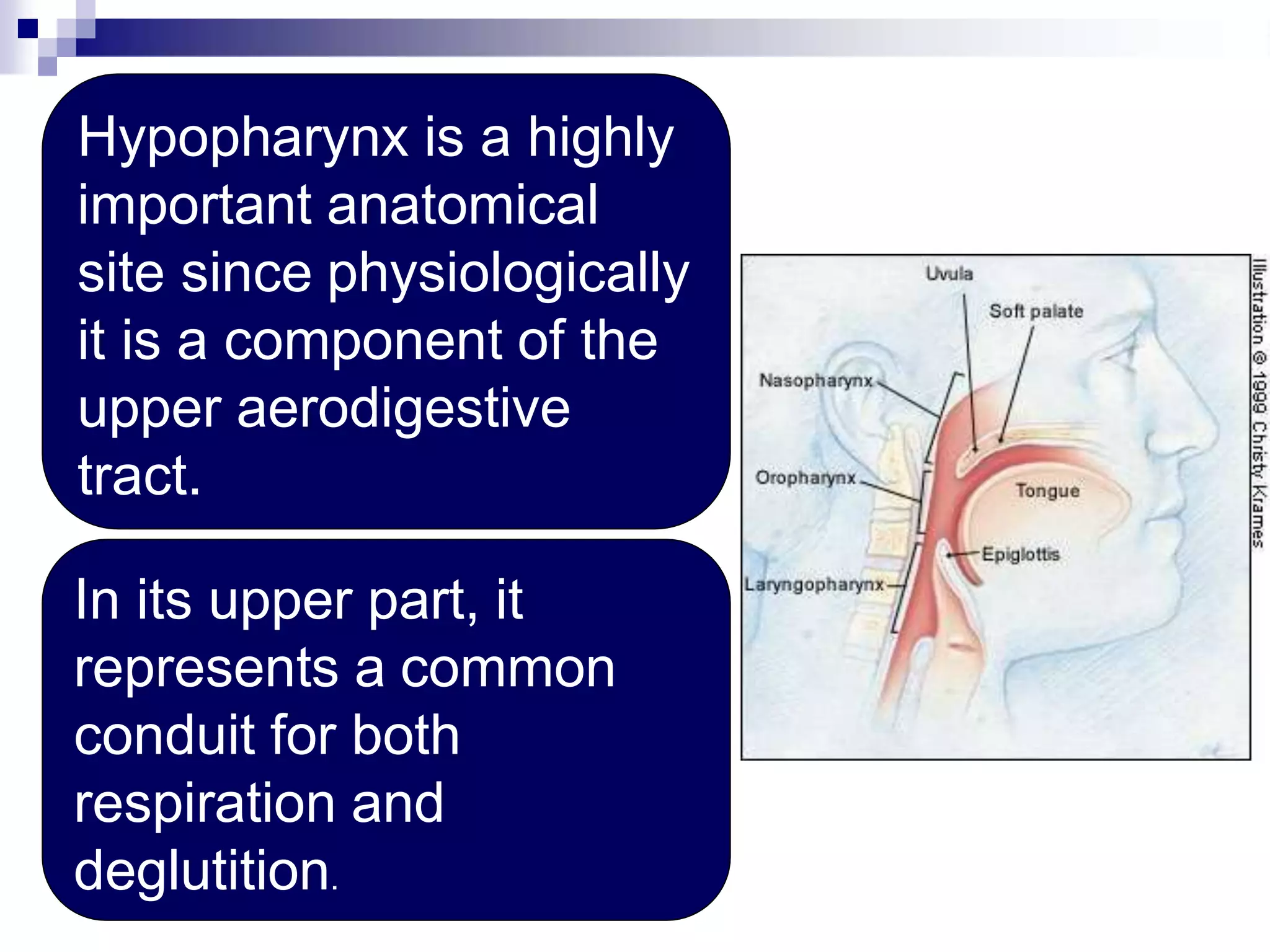
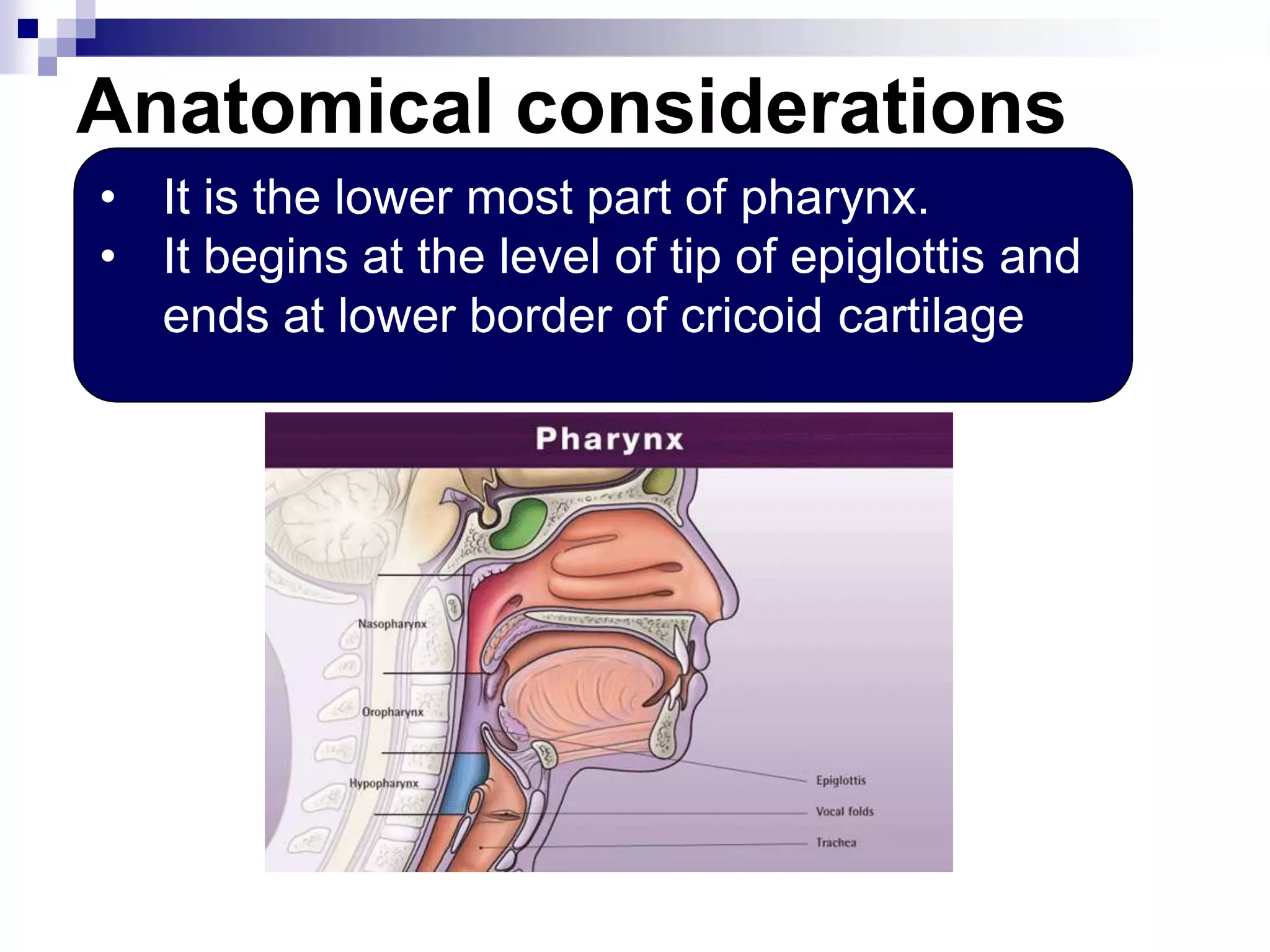
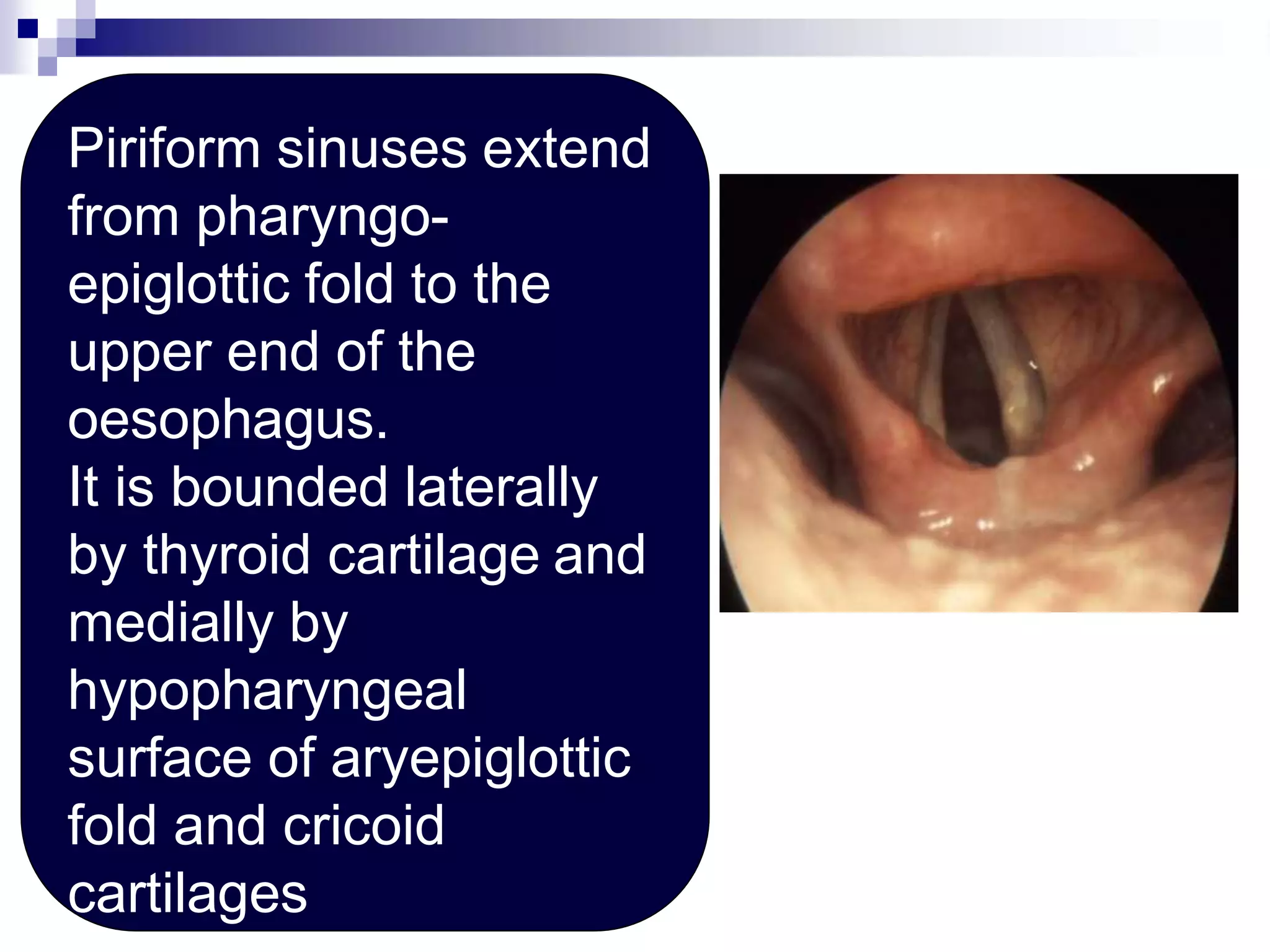
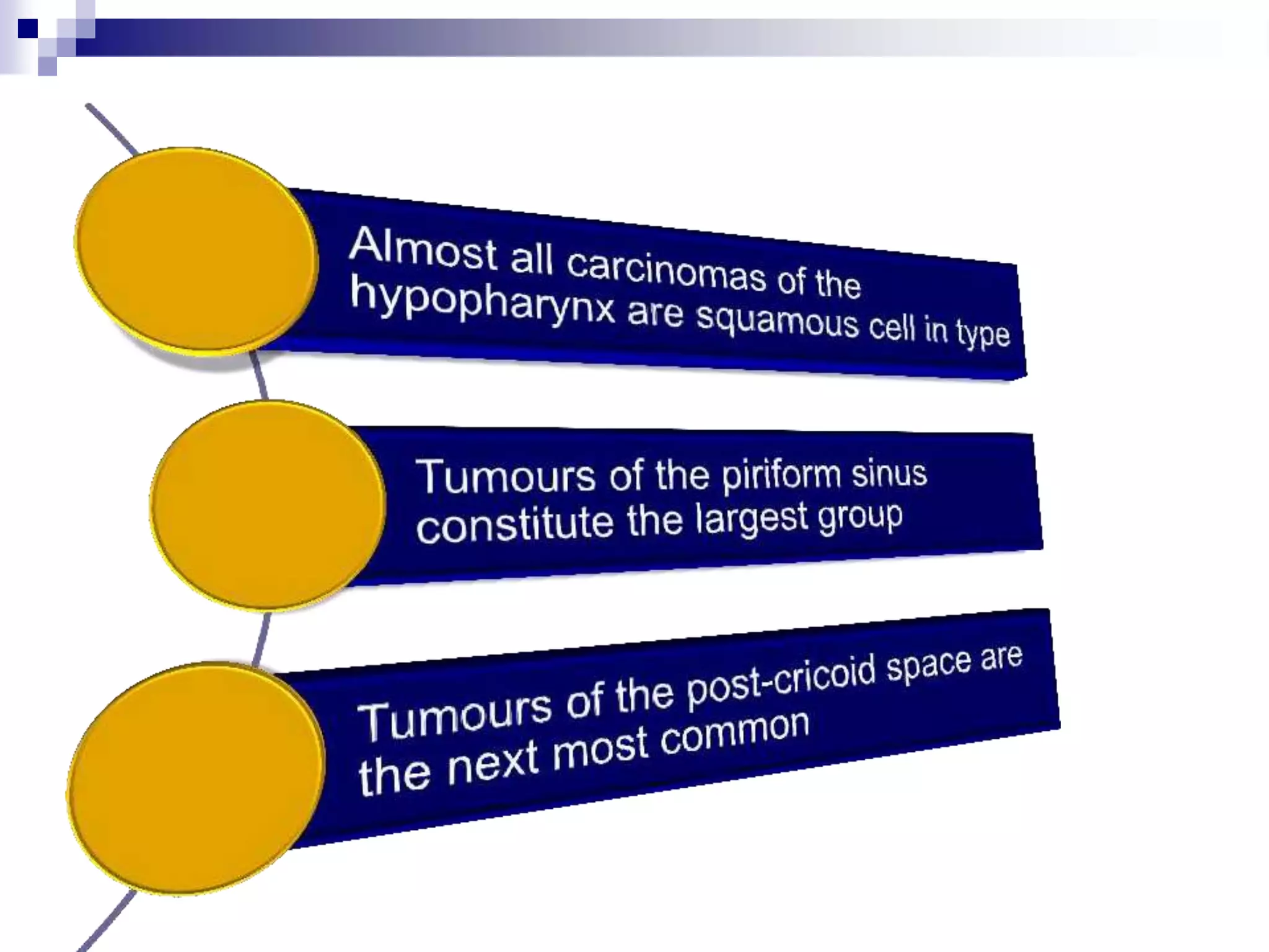
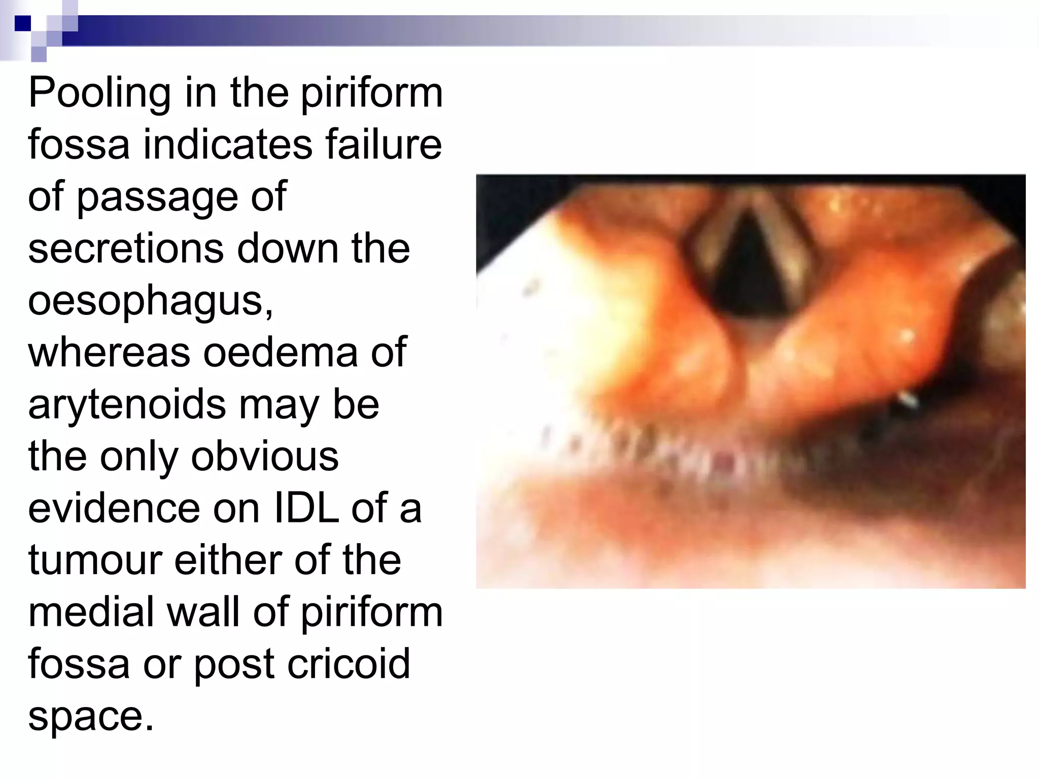
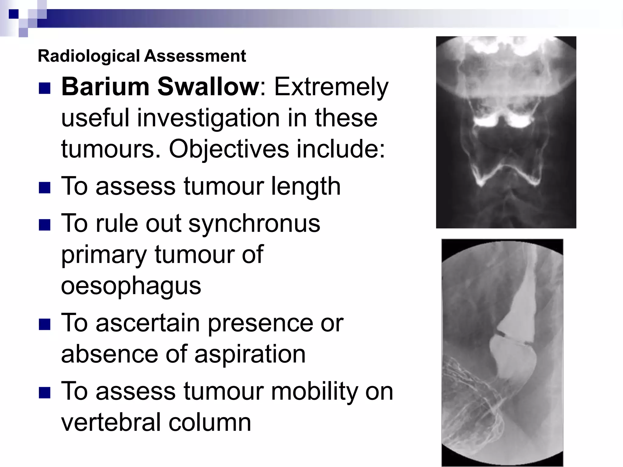
Carcinoma
Hypopharynx
Disclaimer: The pictures used in this presentation have been obtained from a
number of sources. Their use is purely for academic and teaching purposes. The
contents of this presentation do not have any intended commercial use. In case the
owner of any of the pictures has any objection and seeks their removal please
contact at dineshkumarsharma@gmail.com . These pictures will be removed
immediately.
Hypopharynx is a highly
important anatomical
site since physiologically
it is a component of the
upper aerodigestive
tract.
In its upper part, it
represents a common
conduit for both
respiration and
deglutition.
Anatomical considerations
• It is the lower most part of pharynx.
• It begins at the level of tip of epiglottis and
ends at lower border of cricoid cartilage
Tumours arising in this
region often present in
an advanced state
Key to cure lies in an
early & accurate
diagnosis, subsequent
staging & in majority
cases treatment by
curative intent using
surgery and post-
operative radiotherapy.
Two piriform sinuses-one on each side
Posterior pharyngeal wall
Post-cricoid region where aerodigestive tract
continues with cervical oesophagus
Pharyngo-oesophageal
junction (post-cricoid
area) extends from
level of arytenoid
cartilages and the
connecting folds to the
inferior border of cricoid
cartilage, thus forming
the anterior wall of the
hypopharynx..
Piriform sinuses extend
from pharyngo-
epiglottic fold to the
upper end of the
oesophagus.
It is bounded laterally
by thyroid cartilage and
medially by
hypopharyngeal
surface of aryepiglottic
fold and cricoid
cartilages
Posterior Pharyngeal wall extends
from superior level of the hyoid
bone to the level of border of
cricoid cartilage and from apex of
one piriform sinus to the other.
Hypopharynx is lined throughout by
squamous cell epithelium.
Physiologically the
hypopharynx acts as a conduit
for oral intake, participating in
second stage of deglutition
and a tumor in this region can
impair this activity either by
mass effect or by interference
with muscular coordination or
nervous innervation.
Lymphatics
Piriform sinus has rich underlying
network of lymphatics but this is
less extensive in other subsites
In general, the lymphatics in
this area drain to deep cervical
chain in level IV, but the inferior
part of piriform sinus and post-
cricoid area also drain to the
The posterior pharyngeal wall
also drains to the
retropharyngeal lymph nodes
Tumours on the posterior
hypopharyngeal wall are the least
common and constitute approx. 10%
of these tumours
Alcohol and tobacco remain the two
principal carcinogens implicated in
tumours of the upper aerodigestive tract.
Post cricoid carcinoma
It remains the only squamous cell
carcinoma common in women than men.
In relation to post cricoid carcinoma a
major dietary (iron deficiency) has
particularly been described particularly
with Plummer Vinson Syndrome.
This syndrome is also called Paterson
Brown Kelly Syndrome
History of
irradiation of
neck
Anaemia
Glossitis
Esophageal
web
Splenomegly
Koilonychia
Achlorhydria.
Paterson Brown Kelly Syndrome
Dysphagia
and/or
Odynophagia
Hoarseness
of Voice
Neck
mass
Haemoptysis
Symptoms
Symptoms suggesting a hypo-
pharyngeal tumour
Dysphagia: Often persistent and progressive. Patients
who complain of food sticking in throat need to be
investigated
Pain: Usually lateralized and prominent on swallowing,
may radiate to ipsilateral ear
Hoarseness: When it occurs in
association with dysphagia or
referred otalgia usually means
extension of tumour into larynx
Symptoms suggesting a
possible pharyngeal tumour
 Neck Mass: Likely to be due to nodal
metastasis, but may be due to direct
extension through thyrohyoid membrane.
 Haemoptysis: An unusual symptom, but
can occur with tumours of Piriform Sinus
or Post Pharyngeal wall.
 Weight loss: Often occurs in presence of
significant disease.
Examination
 Full head and neck and GPE
 Indirect Laryngoscopy (IDL)
 Direct Laryngoscopy (DL)
 Particular attention shall be paid
to obvious swelling or ulceration
and also presence of pooling of
secretions in the piriform fossa
(Chevalier Jackson’s sign) and
oedema of arytenoids.
Pooling in the piriform
fossa indicates failure
of passage of
secretions down the
oesophagus,
whereas oedema of
arytenoids may be
the only obvious
evidence on IDL of a
tumour either of the
medial wall of piriform
fossa or post cricoid
space.
Laboratory investigations
Following investigations are considered
essential:
 Full Blood count
 Iron Stores
 Urea and electrolytes
 LFT
 Serum Calcium
 Thyroid Function
Radiological Assessment
 Barium Swallow: Extremely
useful investigation in these
tumours. Objectives include:
 To assess tumour length
 To rule out synchronus
primary tumour of
oesophagus
 To ascertain presence or
absence of aspiration
 To assess tumour mobility on
vertebral column
Radiological Assessment
 CT and MRI
 To assess the extent of the
primary tumour and extensions.
 To rule out second primary and
distant metastasis
 To assess neck
 To look for cartilage invasion
Transverse CT image through the lower postcricoid region in a 62-year-old man with a
fungating mass in the right piriform sinus demonstrates definite involvement of the
lower postcricoid region by a tumor (T) on the right, which is seen as a thickening of the
pharyngeal wall and obliteration of the intramural fat planes.
Schmalfuss I M et al. Radiology2000;214:237-246
©2000 by Radiological Society of North America
Endoscopy
 Examination of
larynx, pharynx,
trachea and
esophagus
 Examination of
oral cavity
 Biopsy
Staging
 T1:Tumour limited to one subsite of
hypopharynx and 2 cm or less in greatest
dimension.
 T2; Tumour invades more than one subsite
or measures >2cm but < 4 cm without
fixation of hemilarynx.
 T3: Tumours > 4 cm or with fixation of
hemilarynx
 T4: Tumour invades adjacent structures
Treatment policy
Approximately 25% of patients
with hypopharyngeal carcinoma
are not treatable at presentation
due to advanced age, poor
general health, inoperability of the
tumor and extensive neck
disease. Such patients may be
considered for radiotherapy with a
palliative intent. Chemotherapy
has no established role.
Treatment policy
Long term results may be obtained
with surgery or radiotherapy but
are not encouraging. 5 year
survival rate with surgery and with
or w/o post operative radiotherapy
are 35%.
Treatment policy
Some rare Stage I and II tumours of PF
sinus and posterior pharyngeal wall can
be successfully treated with irradiation
with 50-90% 5 year survival rate. The
inclusion criteria for such patients
includes, vertical length of tumour shall
not exceed 5cm, mobile vocal cords and
N0 neck.
If the patient does not fulfill above criteria
he should be submitted for surgery. In
case of recurrence in a previously
irradiated patient , surgery is considered
The various surgical options include
• Endoscopic excision using
LASER or diathermy
• Partial pharyngectomy
• Partial pharyngectomy with
supraglottic laryngectomy
• Total laryngectomy with Partial
pharyngectomy
• Total pharyngectomy
• Total
laryngopharyngoesophagectomy

Throat_Pharynx_Carcinoma Hypopharynx_ENT_lectures-2.pptx

  • 1.
    Carcinoma Hypopharynx Disclaimer: The picturesused in this presentation have been obtained from a number of sources. Their use is purely for academic and teaching purposes. The contents of this presentation do not have any intended commercial use. In case the owner of any of the pictures has any objection and seeks their removal please contact at dineshkumarsharma@gmail.com . These pictures will be removed immediately.
  • 2.
    Hypopharynx is ahighly important anatomical site since physiologically it is a component of the upper aerodigestive tract. In its upper part, it represents a common conduit for both respiration and deglutition.
  • 3.
    Anatomical considerations • Itis the lower most part of pharynx. • It begins at the level of tip of epiglottis and ends at lower border of cricoid cartilage
  • 4.
    Tumours arising inthis region often present in an advanced state Key to cure lies in an early & accurate diagnosis, subsequent staging & in majority cases treatment by curative intent using surgery and post- operative radiotherapy.
  • 5.
    Two piriform sinuses-oneon each side Posterior pharyngeal wall Post-cricoid region where aerodigestive tract continues with cervical oesophagus
  • 6.
    Pharyngo-oesophageal junction (post-cricoid area) extendsfrom level of arytenoid cartilages and the connecting folds to the inferior border of cricoid cartilage, thus forming the anterior wall of the hypopharynx..
  • 7.
    Piriform sinuses extend frompharyngo- epiglottic fold to the upper end of the oesophagus. It is bounded laterally by thyroid cartilage and medially by hypopharyngeal surface of aryepiglottic fold and cricoid cartilages
  • 8.
    Posterior Pharyngeal wallextends from superior level of the hyoid bone to the level of border of cricoid cartilage and from apex of one piriform sinus to the other. Hypopharynx is lined throughout by squamous cell epithelium.
  • 9.
    Physiologically the hypopharynx actsas a conduit for oral intake, participating in second stage of deglutition and a tumor in this region can impair this activity either by mass effect or by interference with muscular coordination or nervous innervation.
  • 10.
    Lymphatics Piriform sinus hasrich underlying network of lymphatics but this is less extensive in other subsites In general, the lymphatics in this area drain to deep cervical chain in level IV, but the inferior part of piriform sinus and post- cricoid area also drain to the The posterior pharyngeal wall also drains to the retropharyngeal lymph nodes
  • 12.
    Tumours on theposterior hypopharyngeal wall are the least common and constitute approx. 10% of these tumours Alcohol and tobacco remain the two principal carcinogens implicated in tumours of the upper aerodigestive tract.
  • 13.
    Post cricoid carcinoma Itremains the only squamous cell carcinoma common in women than men. In relation to post cricoid carcinoma a major dietary (iron deficiency) has particularly been described particularly with Plummer Vinson Syndrome. This syndrome is also called Paterson Brown Kelly Syndrome
  • 14.
  • 15.
  • 16.
  • 17.
    Symptoms suggesting ahypo- pharyngeal tumour Dysphagia: Often persistent and progressive. Patients who complain of food sticking in throat need to be investigated Pain: Usually lateralized and prominent on swallowing, may radiate to ipsilateral ear Hoarseness: When it occurs in association with dysphagia or referred otalgia usually means extension of tumour into larynx
  • 18.
    Symptoms suggesting a possiblepharyngeal tumour  Neck Mass: Likely to be due to nodal metastasis, but may be due to direct extension through thyrohyoid membrane.  Haemoptysis: An unusual symptom, but can occur with tumours of Piriform Sinus or Post Pharyngeal wall.  Weight loss: Often occurs in presence of significant disease.
  • 19.
    Examination  Full headand neck and GPE  Indirect Laryngoscopy (IDL)  Direct Laryngoscopy (DL)  Particular attention shall be paid to obvious swelling or ulceration and also presence of pooling of secretions in the piriform fossa (Chevalier Jackson’s sign) and oedema of arytenoids.
  • 20.
    Pooling in thepiriform fossa indicates failure of passage of secretions down the oesophagus, whereas oedema of arytenoids may be the only obvious evidence on IDL of a tumour either of the medial wall of piriform fossa or post cricoid space.
  • 21.
    Laboratory investigations Following investigationsare considered essential:  Full Blood count  Iron Stores  Urea and electrolytes  LFT  Serum Calcium  Thyroid Function
  • 22.
    Radiological Assessment  BariumSwallow: Extremely useful investigation in these tumours. Objectives include:  To assess tumour length  To rule out synchronus primary tumour of oesophagus  To ascertain presence or absence of aspiration  To assess tumour mobility on vertebral column
  • 23.
    Radiological Assessment  CTand MRI  To assess the extent of the primary tumour and extensions.  To rule out second primary and distant metastasis  To assess neck  To look for cartilage invasion
  • 24.
    Transverse CT imagethrough the lower postcricoid region in a 62-year-old man with a fungating mass in the right piriform sinus demonstrates definite involvement of the lower postcricoid region by a tumor (T) on the right, which is seen as a thickening of the pharyngeal wall and obliteration of the intramural fat planes. Schmalfuss I M et al. Radiology2000;214:237-246 ©2000 by Radiological Society of North America
  • 25.
    Endoscopy  Examination of larynx,pharynx, trachea and esophagus  Examination of oral cavity  Biopsy
  • 26.
    Staging  T1:Tumour limitedto one subsite of hypopharynx and 2 cm or less in greatest dimension.  T2; Tumour invades more than one subsite or measures >2cm but < 4 cm without fixation of hemilarynx.  T3: Tumours > 4 cm or with fixation of hemilarynx  T4: Tumour invades adjacent structures
  • 27.
    Treatment policy Approximately 25%of patients with hypopharyngeal carcinoma are not treatable at presentation due to advanced age, poor general health, inoperability of the tumor and extensive neck disease. Such patients may be considered for radiotherapy with a palliative intent. Chemotherapy has no established role.
  • 28.
    Treatment policy Long termresults may be obtained with surgery or radiotherapy but are not encouraging. 5 year survival rate with surgery and with or w/o post operative radiotherapy are 35%.
  • 29.
    Treatment policy Some rareStage I and II tumours of PF sinus and posterior pharyngeal wall can be successfully treated with irradiation with 50-90% 5 year survival rate. The inclusion criteria for such patients includes, vertical length of tumour shall not exceed 5cm, mobile vocal cords and N0 neck. If the patient does not fulfill above criteria he should be submitted for surgery. In case of recurrence in a previously irradiated patient , surgery is considered
  • 30.
    The various surgicaloptions include • Endoscopic excision using LASER or diathermy • Partial pharyngectomy • Partial pharyngectomy with supraglottic laryngectomy • Total laryngectomy with Partial pharyngectomy • Total pharyngectomy • Total laryngopharyngoesophagectomy