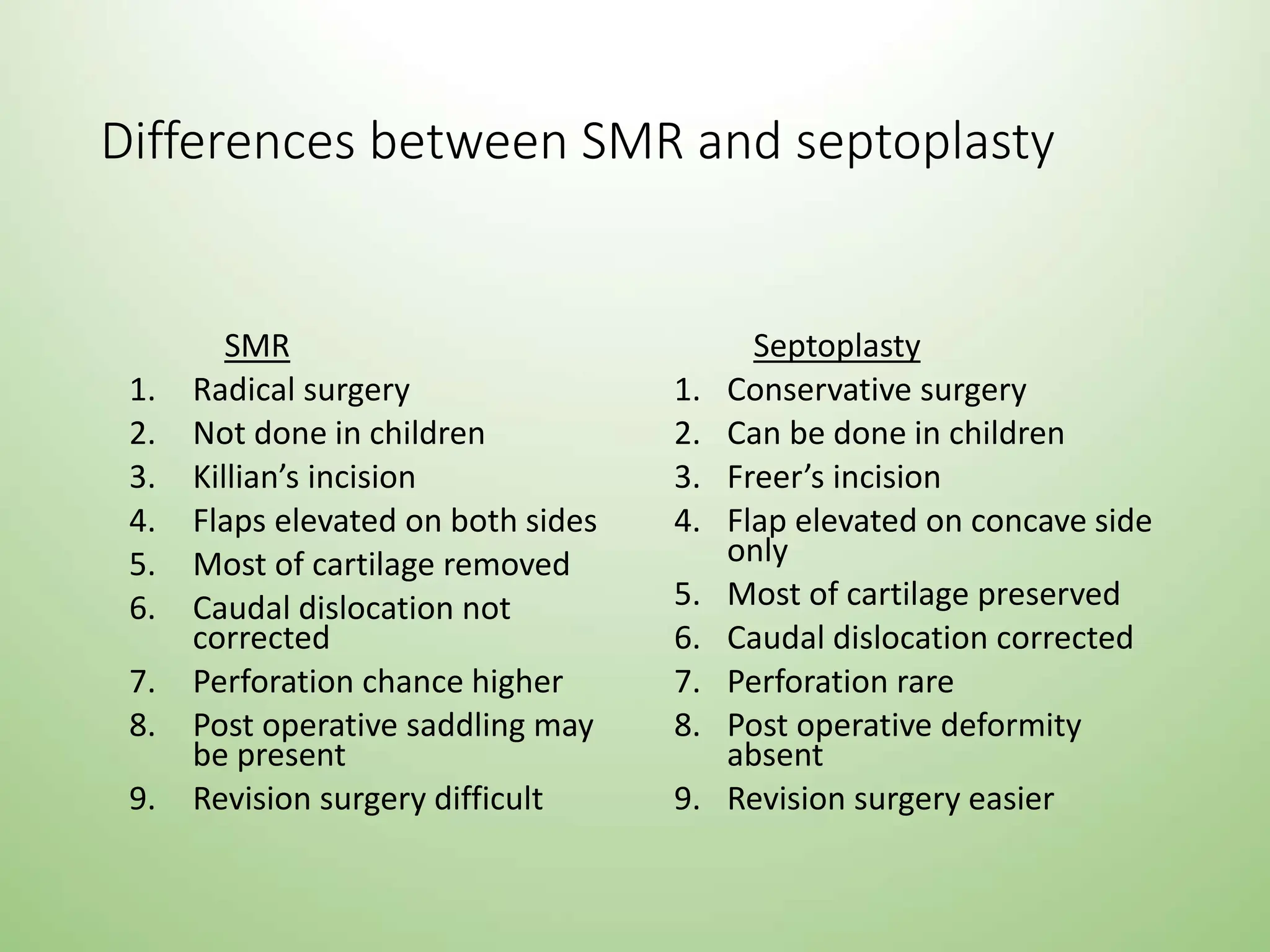
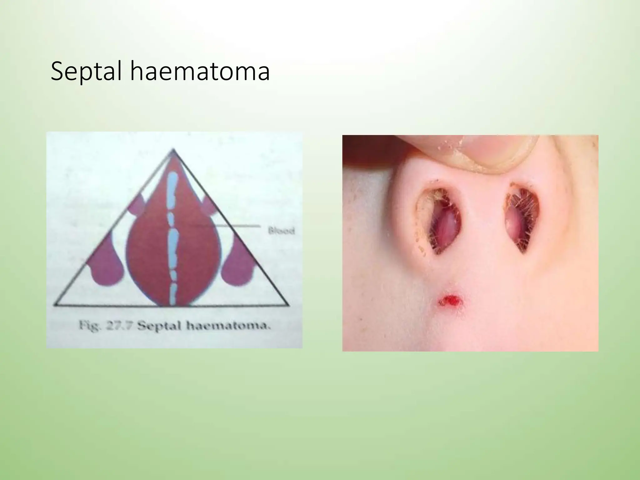
NASAL SEPTUM AND ITS
DISEASES
Dr. Srinivas Pennam
Nasal septum and its diseases
Anatomy of nasal septum:
Nasal septum consists of three parts:
1. Columellar septum
2. Membranous septum
3. Septum proper: principle constituents of septum proper are
a)perpendicular plate of ethmoid
b)vomer
c)septal(quadrilateral cartilage)
minor contributions from crest of nasal bone,nasal spine of frontal
bone,rostrum of sphenoid,crest of palatine and maxilla and
anterior nasal spine of maxilla.
Nasal septum and its diseases
Septal cartilage forms a partition between right and left nasal
cavities and provides support to tip and dorsum of cartilagenous
part of nose.
Septal destruction may occur in septal abscess, injuries,
tuberculosis, excess removal during SMR leads to depression of
lower part of nose and drooping of tip.
Septal cartilage lies in a groove in the anterior edge of vomer
and rests anteriorly on anterior nasal spine. during trauma, it
may get dislocated from nasal spine or vomer causing caudal
septal deviation and spur respectively.
BLOOD SUPPLY-NASAL SEPTUM
NERVE SUPPLY-NASAL SEPTUM
Fractures of nasal septum
• Aetiopathogenisis:
-Trauma inflicted from front, side or below. the septum may
buckle on itself, fracture vertically, horizontally or get
crushed.
-fracture of septal cartilage or its dislocation can occur
without nasal bones fracture in cases of trauma to lower
nose.
-septal injuries with mucosal tears cause profuse epistaxis
while with intact mucosa result in septal hematoma.
Fractures of nasal septum
Types :
1}Jarjavay fracture: result from blow from front. fracture line
starts just above the anterior nasal spine and runs
horizontally backwards just above the junction of septal
cartilage with the vomer.
2}Chevallet fracture: results from blow from below. it runs
vertically from anterior nasal spine upwards to the junction
of bony and cartilaginous dorsum of nose.
Fractures of nasal septum
Jarjavay fracture
Chevallet
Fractures of nasal septum
Treatment: -early recognition and treatment of septal injuries
is essential.
-dislocated or fractured fragments should be repositioned
and supported between mucoperichondrial flaps.
-haematomas should be drained.
Complications: a) deviation of cartilagenous nose.
b) asymmetry of nasal tip, columella, or
nostril.
DEVIATED NASAL SEPTUM
AETIOLOGY:
1) Trauma:
Lateral Blow - displacement of septal cartilage from vomer.
Blow From Front - buckling, fracture, duplication of septum with
telescoping of fragments.
2) Developmental: the septum should grow at the same rate as that
of face. if septum grows at faster rate it becomes buckled. unequal
growth between palate and base of skull may also cause buckling
(high arched palate)
3) Congenital: abnormal intrauterine posture cause compressing
forces acting on nose and upper jaw.
4) Hereditary
5) Racial: Caucasians are more affected
6) Secondary to a tumour, mass or polyp.
DEVIATED NASAL SEPTUM
Types:
1) Deviations: upper or lower, anterior or posterior, C
shaped, S shaped. nasal cavity on the concave side of
the septum will be wider and may show compensatory
hypertrophy of turbinates.
2) Anterior Dislocation: seen on tilting the patients head
backwards.
3) Spurs: shelf like projection at the junction of bone and
cartilage. may predispose for epistaxis and headache.
4) Thickening: it may be due to organized haematoma or
over-riding of dislocated septal fragments
Site
• Cartilage/ Bone/ Both
• High/Low,
• Anterior/Posterior
• Simple deviation (No nasal obst)
• Obstructed deviation (severe nasal obstruction improves on
vasoconstriction)
• Impacted deviation ( no improvement )
DEVIATED NASAL SEPTUM-types
Clinical features
• Nasal obstruction: depending on the type it may be unilateral or
bilateral. It is the most common symptom
• Headache- sluders neuralgia
• Recurrent attacks of cold
• Epistaxis
• Anosmia
• External deformity
• Middle ear infection
Clinical features
• Hyposmia – more common than anosmia
• Epistaxis – spur , crusting (dry air currents)
• Post nasal discharge – impeded mucociliary clearance
• Unpleasant taste – due to post nasal discharge
• Signs
• Vestibule – caudal dislocation
• Ant rhinoscopy – compensatory hypertrophy of turbinates, nasal
secretions, crusting, deviation
Cottle’s test
• Cottle’s test: used in nasal obstruction due to abnormality of nasal
valve.
• In this test cheek is drawn laterally while the patient breathes quietly.
If the nasal airway improves on test side the test is positive and
indicates abnormality of nasal valve
Differential diagnosis
• Polyps
• Septal haematoma
• Hypertrophied turbinates
Sequelae
• Sleep apnoea
• Atrophic rhinitis - due to inadequate humidification
• Middle ear infection – ET catarrh
• Asthma – DNS causes bronchospasm
• Recurrent infections of pharynx, oral cavity due to mouth breathing,
larynx infections
• Pulmonary effects
• Cardiac effects
Investigations
• X Ray – PNS (water’s, calwell’s) – septum, turbinates, sunuses
• CT Scan PNS – OMC, concha bullosa, sinuses
• DNE – septum, lateral wall, OMC
• CBC
• Bleeding and cooagulation profile
Surgical management
• Only if DNS causes recurrent nasal obstruction, recurrent sinusitis,
recurrent epistaxis, recurrent headache, recurrent otitis media,
hyposmia
• Other indications – as a approach for FESS, vidian neurectomy,
septorhinoplasty, hypophysectomy
• For using graft (septal cartilage or bone) for CSF leak repair or
rhinoplasty
• C/I
• Acute URTI, uncontrolled diabetes and HTN, bleeding diathesis, TB
• SMR is contraindicated before 17 years of age..as can interfere with
growth of nasal septum
• Surgeries – SMR (Submucosal resection), Septoplasty, Endoscopic
septoplasty, Septorhinoplasty
Treatment- surgery
• Submucous resection of nasal septum (SMR)
It is generally done in adults
It consists of elevating mucoperichondrial and mucoperiosteal flap
on either side of the septum,
 removing the deflected parts of bony and cartilagenous septum
 then repositioning the flaps
SMR
• Indications
Deviated nasal septum causing nasal obstruction and
recurrent headaches
Deviated nasal septum causing obstruction to ventilation of
paranasal sinuses and middle ear resulting in recurrent
infections
Recurrent epistaxis from septal spur
As a part of septorhinoplasty
Harvesting cartilage graft for tympanoplasty and rhinoplasty
As an approach to surgeries of sphenoidal sinus, vidian
nerve and pituitary gland
SMR
•Contraindications
Acute URTI
Patient below 17 yrs of age
Bleeding disorders
Uncontrolled hypertension and diabetes mellitus
SMR
• Anesthesia - Local anesthesia/ general anesthesia
• 4% lignocaine with adrenaline nasal packs for 10 minutes
• 2% lignocaine with 1:100000 adrenaline injection on either side of
septum in subperichondrial and subperiosteal plane with 27 G
needle
• Use oxymetazoline instead of adrenaline in cardiac patients
• Positioning: reclining position with head end of the table raised
SMR - STEPS
• Infiltration: subperichondrial infiltration with 2% xylocaine with
adrenaline
• Incision: killian’s incision- curvilinear incision 2-3mm behind the anterior
end of septal cartilage
• Elevation of flaps: the mucoperichondrial and mucoperiosteal flap is
elevated
• Incision of the cartilage- cartilage is incised just posterior to the first
incision
• Elevation of opposite mucoperichondrial and mucoperiosteal flap
SMR – STEPS (cont…)
•Removal of cartilage and bone - cartilage can be
removed with Ballinger swivel knife or luc’s forceps.
Bony spur is removed using gouge and hammer
•Preserve a strip of 1cm wide cartilage along the
dorsal and caudal borders (struts)
•Nasal packing
SMR – STEPS
complications
• Bleeding
• Septal haematoma
• Damage to surrounding structures
• Septal abscess
• Septal Perforation
• Depression of bridge
• Retraction of columella
• Synechiae
• Flapping septum
• Infection- sinus and middle ear
• CSF rhinorrhoea
Cottle’s line
• A vertical line between the
nasal process of frontal
bone and nasal spine of
maxillary crest. it divides
septum into anterior and
posterior segments
Septoplasty
• It is a conservative approach to septal surgery as much of the
septal framework is retained
• Indications:
Deviated nasal septum causing nasal obstruction and recurrent
headaches
Deviated nasal septum causing obstruction to ventilation of
paranasal sinuses and middle ear resulting in recurrent
infections
Recurrent epistaxis from septal spur
As a part of septorhinoplasty
As an approach to surgeries of sphenoidal sinus, vidian nerve
and pituitary gland
Septoplasty (cont…)
• Contraindications
Acute URTI
Bleeding disorders
Uncontrolled hypertension and diabetes mellitus
Septoplasty (cont…)
• Anesthesia: local or general anesthesia
• Position: same as SMR
• Steps :
Infiltration
Incision: Freer’s incision– a unilateral hemitransfixation
incision at the caudal border of the septum
Exposure: the mucoperichondrial and mucoperiosteal flap is
elevated on only one side
Septoplasty (cont…)
• flap elevation by 3 tunnels
• Anterior tunnel – mucoperichondrial elevation on incision side only
• Posterior tunnel – mucoperiosteal elevation on both sides
• Inferior tunnel – Expose crest of maxilla on both sides
• Columellar tunnel – if caudal dislocation
Separate septal cartilage from vomer and ethmoid plate
Inferior strip of cartilage is removed
Correct the bony septum by removing deformed parts
Minor deviations of cartilage are corrected by criss cross incision which
breaks spring action of cartilage
Nasal packing
• Endoscopic septoplasty – using 0 degree hopkins endoscope
• For minimal deviation, septal spur
• Better visualisation, illumination
• No need for nasal packing
• No need for extra unnewcessary resection
• Minimal trauma to surrounding mucosa
• Turbinoplasty – reduction of inferior or middle turbinate by bipolar
cautery, LASER or microdebrider or SMR of inferior turbinate
Post-operative complications
• Bleeding
• Septal haematoma
• Damage to surrounding structures
• Septal abscess
• Septal Perforation
• Depression of bridge
• Retraction of columella
• Synechiae
• Infection- sinus and middle ear
• CSF rhinorrhoea
• Patient in semi sitting position to prevent oozing of blood
• Soft diet for 1st 2 days
• Analgesics and antibiotics for 5 – 7 days
• Pack removal after 24-48 hours
• Decongestant nasal drops
• Steam inhalation
• Sutures if any removed after 5 – 7 days
• Avoid sternous exercises, nose blowing, trauma to nose for 3 weeks
Post operative care
Differences between SMR and septoplasty
SMR
1. Radical surgery
2. Not done in children
3. Killian’s incision
4. Flaps elevated on both sides
5. Most of cartilage removed
6. Caudal dislocation not
corrected
7. Perforation chance higher
8. Post operative saddling may
be present
9. Revision surgery difficult
Septoplasty
1. Conservative surgery
2. Can be done in children
3. Freer’s incision
4. Flap elevated on concave side
only
5. Most of cartilage preserved
6. Caudal dislocation corrected
7. Perforation rare
8. Post operative deformity
absent
9. Revision surgery easier
Septal haematoma
• It is collection of blood under the perichondrium or
periosteum of nasal septum
• Etiology: nasal trauma, post-operative, in bleeding disorder
• Clinical features:
Bilateral nasal obstruction
Frontal headache
Sense of pressure over nasal bridge
Examination reveals smooth rounded swelling of the
septum in both the nasal cavity. Palpation may show the
mass to be soft and fluctuant
Septal haematoma
• Investigations – BT, CT, PT, ESR, CBC, TLC, DLC
• X Ray PNS
• Complications – septal abscess, septal perforation, thickened
septum, depression of nasal dorsum
• Treatment: small haematomas can be aspirated with a wide bore
needle.
• larger haematomas are incised and drained. Excision of small piece
of mucosa from the edge of the incision gives better drainage.
• Nose is packed on both sides to prevent re-accumulation.
• Systemic antibiotics to prevent septal abscess
Septal haematoma
Septal abscess
• Collection of pus under mucoperichondrium or mucoperiosteum of
septum
• Etiology
Secondary infection of septal haematoma
Furuncle of the nasal vestibule
Clinical features
Severe bilateral nasal obstruction with pain and tenderness over bridge
of nose
Fever with chills
Frontal headache
Skin over the nose may be red and swollen
Smooth bilateral swelling of the nasal septum
Congested septal mucosa
Submandibular nodes may be enlarged and tender
Septal abscess
• Investigations - TLC, DLC (leukocytosis)
• Treatment
Abscess should be drained as early as possible
Pus and necrosed cartilage removed by suction
Incision may required to be re-opened daily for 2-3 days to drain
any pus or remove any necrosed piece of cartilage
Systemic antibiotics to be started as soon as possible and
continued for two weeks
Septal abscess
• Complications
Depression of the cartilagenous dorsum
Septal perforation,
columella retraction
Facial cellulitis,Meningitis and cavernous sinus thrombosis (rare)
Nasal septal perforation
• Hole in the septum through which there is direct communication
between two cavities
• Etiology
 Traumatic - post surgical, habitual nose picking, cauterization of septum
with chemicals or galvano-cautery for epistaxis,RTA
 Pathological perforation
a) Septal abscess
b) Nasal myasis
c) Rhinolith or neglected foreign body
d) Chronic granulomatous conditions like TB, lupus, leprosy, syphilis,
wegener’s
 Inhalant irritants- tobacco sniff and cocaine , industrial toxins
 Malignancy
 idiopathic
Nasal septal perforation
• Classification –
• small (<1cm),
• medium (1-2cm),
• large (>2cm)
• Anterior (more symptoms)/posterior
• C/F
• Small – whistling sound/hissing noise in anterior perforation during
nasal breathing
• Large – crusting, epistaxis, dryness of nose, nasal obstruction due
to crusts
• Diagnosis – biopsy from edge of perforation, Hb, serological tests
Nasal septal perforation
• Treatment
Treat the root cause
Inactive small perforation can be surgically closed by plastic flaps or
septal mucosal flaps
Septal buttons or obturator
Larger perforations are difficult to close: their treatment is aimed
to keep the nose crust free by alkaline nasal douch and application
of lubricants, silastic obturator may also be used
Nasal synechiae
• Adhesions between septum and lateral wall or middle turbinate and
lateral wall
• Etiology - Injury to opposing surfaces during nasal surgery
• Nasal packing
• C/F – persistent nasal obstruction, recurrent sinusitis and headache
Minor – asymptomatic
• Prevention – lubricate the nasal pack, proper post op cleaning
• Treatment – excise and release by bipolar cautery, diathermy and
LASER
• Prevent recurrence by placing gelfoam, silastic sheets, dental wax for
2 weeks
• TOXIC SHOCK SYNDROME
• Streptococci, staphylococci
• C/F – fever, nausea, vomiting, hypotension, rash
• Treatment – remove the nasal pack, hydrate the patient, IV
antibiotics, TPR chart
• FLAPPING OF SEPTUM – over removal septum, two side flaps move
with respiration
nasalseptumanditsdiseases................

nasalseptumanditsdiseases................

  • 1.
    NASAL SEPTUM ANDITS DISEASES Dr. Srinivas Pennam
  • 2.
    Nasal septum andits diseases Anatomy of nasal septum: Nasal septum consists of three parts: 1. Columellar septum 2. Membranous septum 3. Septum proper: principle constituents of septum proper are a)perpendicular plate of ethmoid b)vomer c)septal(quadrilateral cartilage) minor contributions from crest of nasal bone,nasal spine of frontal bone,rostrum of sphenoid,crest of palatine and maxilla and anterior nasal spine of maxilla.
  • 3.
    Nasal septum andits diseases Septal cartilage forms a partition between right and left nasal cavities and provides support to tip and dorsum of cartilagenous part of nose. Septal destruction may occur in septal abscess, injuries, tuberculosis, excess removal during SMR leads to depression of lower part of nose and drooping of tip. Septal cartilage lies in a groove in the anterior edge of vomer and rests anteriorly on anterior nasal spine. during trauma, it may get dislocated from nasal spine or vomer causing caudal septal deviation and spur respectively.
  • 4.
  • 5.
  • 6.
    Fractures of nasalseptum • Aetiopathogenisis: -Trauma inflicted from front, side or below. the septum may buckle on itself, fracture vertically, horizontally or get crushed. -fracture of septal cartilage or its dislocation can occur without nasal bones fracture in cases of trauma to lower nose. -septal injuries with mucosal tears cause profuse epistaxis while with intact mucosa result in septal hematoma.
  • 7.
    Fractures of nasalseptum Types : 1}Jarjavay fracture: result from blow from front. fracture line starts just above the anterior nasal spine and runs horizontally backwards just above the junction of septal cartilage with the vomer. 2}Chevallet fracture: results from blow from below. it runs vertically from anterior nasal spine upwards to the junction of bony and cartilaginous dorsum of nose.
  • 8.
    Fractures of nasalseptum Jarjavay fracture Chevallet
  • 9.
    Fractures of nasalseptum Treatment: -early recognition and treatment of septal injuries is essential. -dislocated or fractured fragments should be repositioned and supported between mucoperichondrial flaps. -haematomas should be drained. Complications: a) deviation of cartilagenous nose. b) asymmetry of nasal tip, columella, or nostril.
  • 10.
    DEVIATED NASAL SEPTUM AETIOLOGY: 1)Trauma: Lateral Blow - displacement of septal cartilage from vomer. Blow From Front - buckling, fracture, duplication of septum with telescoping of fragments. 2) Developmental: the septum should grow at the same rate as that of face. if septum grows at faster rate it becomes buckled. unequal growth between palate and base of skull may also cause buckling (high arched palate) 3) Congenital: abnormal intrauterine posture cause compressing forces acting on nose and upper jaw. 4) Hereditary 5) Racial: Caucasians are more affected 6) Secondary to a tumour, mass or polyp.
  • 11.
    DEVIATED NASAL SEPTUM Types: 1)Deviations: upper or lower, anterior or posterior, C shaped, S shaped. nasal cavity on the concave side of the septum will be wider and may show compensatory hypertrophy of turbinates. 2) Anterior Dislocation: seen on tilting the patients head backwards. 3) Spurs: shelf like projection at the junction of bone and cartilage. may predispose for epistaxis and headache. 4) Thickening: it may be due to organized haematoma or over-riding of dislocated septal fragments
  • 12.
    Site • Cartilage/ Bone/Both • High/Low, • Anterior/Posterior • Simple deviation (No nasal obst) • Obstructed deviation (severe nasal obstruction improves on vasoconstriction) • Impacted deviation ( no improvement )
  • 13.
  • 14.
    Clinical features • Nasalobstruction: depending on the type it may be unilateral or bilateral. It is the most common symptom • Headache- sluders neuralgia • Recurrent attacks of cold • Epistaxis • Anosmia • External deformity • Middle ear infection
  • 15.
    Clinical features • Hyposmia– more common than anosmia • Epistaxis – spur , crusting (dry air currents) • Post nasal discharge – impeded mucociliary clearance • Unpleasant taste – due to post nasal discharge • Signs • Vestibule – caudal dislocation • Ant rhinoscopy – compensatory hypertrophy of turbinates, nasal secretions, crusting, deviation
  • 16.
    Cottle’s test • Cottle’stest: used in nasal obstruction due to abnormality of nasal valve. • In this test cheek is drawn laterally while the patient breathes quietly. If the nasal airway improves on test side the test is positive and indicates abnormality of nasal valve
  • 18.
    Differential diagnosis • Polyps •Septal haematoma • Hypertrophied turbinates
  • 19.
    Sequelae • Sleep apnoea •Atrophic rhinitis - due to inadequate humidification • Middle ear infection – ET catarrh • Asthma – DNS causes bronchospasm • Recurrent infections of pharynx, oral cavity due to mouth breathing, larynx infections • Pulmonary effects • Cardiac effects
  • 20.
    Investigations • X Ray– PNS (water’s, calwell’s) – septum, turbinates, sunuses • CT Scan PNS – OMC, concha bullosa, sinuses • DNE – septum, lateral wall, OMC • CBC • Bleeding and cooagulation profile
  • 21.
    Surgical management • Onlyif DNS causes recurrent nasal obstruction, recurrent sinusitis, recurrent epistaxis, recurrent headache, recurrent otitis media, hyposmia • Other indications – as a approach for FESS, vidian neurectomy, septorhinoplasty, hypophysectomy • For using graft (septal cartilage or bone) for CSF leak repair or rhinoplasty • C/I • Acute URTI, uncontrolled diabetes and HTN, bleeding diathesis, TB • SMR is contraindicated before 17 years of age..as can interfere with growth of nasal septum • Surgeries – SMR (Submucosal resection), Septoplasty, Endoscopic septoplasty, Septorhinoplasty
  • 22.
    Treatment- surgery • Submucousresection of nasal septum (SMR) It is generally done in adults It consists of elevating mucoperichondrial and mucoperiosteal flap on either side of the septum,  removing the deflected parts of bony and cartilagenous septum  then repositioning the flaps
  • 23.
    SMR • Indications Deviated nasalseptum causing nasal obstruction and recurrent headaches Deviated nasal septum causing obstruction to ventilation of paranasal sinuses and middle ear resulting in recurrent infections Recurrent epistaxis from septal spur As a part of septorhinoplasty Harvesting cartilage graft for tympanoplasty and rhinoplasty As an approach to surgeries of sphenoidal sinus, vidian nerve and pituitary gland
  • 24.
    SMR •Contraindications Acute URTI Patient below17 yrs of age Bleeding disorders Uncontrolled hypertension and diabetes mellitus
  • 25.
    SMR • Anesthesia -Local anesthesia/ general anesthesia • 4% lignocaine with adrenaline nasal packs for 10 minutes • 2% lignocaine with 1:100000 adrenaline injection on either side of septum in subperichondrial and subperiosteal plane with 27 G needle • Use oxymetazoline instead of adrenaline in cardiac patients • Positioning: reclining position with head end of the table raised
  • 26.
    SMR - STEPS •Infiltration: subperichondrial infiltration with 2% xylocaine with adrenaline • Incision: killian’s incision- curvilinear incision 2-3mm behind the anterior end of septal cartilage • Elevation of flaps: the mucoperichondrial and mucoperiosteal flap is elevated • Incision of the cartilage- cartilage is incised just posterior to the first incision • Elevation of opposite mucoperichondrial and mucoperiosteal flap
  • 27.
    SMR – STEPS(cont…) •Removal of cartilage and bone - cartilage can be removed with Ballinger swivel knife or luc’s forceps. Bony spur is removed using gouge and hammer •Preserve a strip of 1cm wide cartilage along the dorsal and caudal borders (struts) •Nasal packing
  • 28.
  • 29.
    complications • Bleeding • Septalhaematoma • Damage to surrounding structures • Septal abscess • Septal Perforation • Depression of bridge • Retraction of columella • Synechiae • Flapping septum • Infection- sinus and middle ear • CSF rhinorrhoea
  • 30.
    Cottle’s line • Avertical line between the nasal process of frontal bone and nasal spine of maxillary crest. it divides septum into anterior and posterior segments
  • 31.
    Septoplasty • It isa conservative approach to septal surgery as much of the septal framework is retained • Indications: Deviated nasal septum causing nasal obstruction and recurrent headaches Deviated nasal septum causing obstruction to ventilation of paranasal sinuses and middle ear resulting in recurrent infections Recurrent epistaxis from septal spur As a part of septorhinoplasty As an approach to surgeries of sphenoidal sinus, vidian nerve and pituitary gland
  • 32.
    Septoplasty (cont…) • Contraindications AcuteURTI Bleeding disorders Uncontrolled hypertension and diabetes mellitus
  • 33.
    Septoplasty (cont…) • Anesthesia:local or general anesthesia • Position: same as SMR • Steps : Infiltration Incision: Freer’s incision– a unilateral hemitransfixation incision at the caudal border of the septum Exposure: the mucoperichondrial and mucoperiosteal flap is elevated on only one side
  • 34.
    Septoplasty (cont…) • flapelevation by 3 tunnels • Anterior tunnel – mucoperichondrial elevation on incision side only • Posterior tunnel – mucoperiosteal elevation on both sides • Inferior tunnel – Expose crest of maxilla on both sides • Columellar tunnel – if caudal dislocation Separate septal cartilage from vomer and ethmoid plate Inferior strip of cartilage is removed Correct the bony septum by removing deformed parts Minor deviations of cartilage are corrected by criss cross incision which breaks spring action of cartilage Nasal packing
  • 37.
    • Endoscopic septoplasty– using 0 degree hopkins endoscope • For minimal deviation, septal spur • Better visualisation, illumination • No need for nasal packing • No need for extra unnewcessary resection • Minimal trauma to surrounding mucosa • Turbinoplasty – reduction of inferior or middle turbinate by bipolar cautery, LASER or microdebrider or SMR of inferior turbinate
  • 38.
    Post-operative complications • Bleeding •Septal haematoma • Damage to surrounding structures • Septal abscess • Septal Perforation • Depression of bridge • Retraction of columella • Synechiae • Infection- sinus and middle ear • CSF rhinorrhoea
  • 39.
    • Patient insemi sitting position to prevent oozing of blood • Soft diet for 1st 2 days • Analgesics and antibiotics for 5 – 7 days • Pack removal after 24-48 hours • Decongestant nasal drops • Steam inhalation • Sutures if any removed after 5 – 7 days • Avoid sternous exercises, nose blowing, trauma to nose for 3 weeks Post operative care
  • 40.
    Differences between SMRand septoplasty SMR 1. Radical surgery 2. Not done in children 3. Killian’s incision 4. Flaps elevated on both sides 5. Most of cartilage removed 6. Caudal dislocation not corrected 7. Perforation chance higher 8. Post operative saddling may be present 9. Revision surgery difficult Septoplasty 1. Conservative surgery 2. Can be done in children 3. Freer’s incision 4. Flap elevated on concave side only 5. Most of cartilage preserved 6. Caudal dislocation corrected 7. Perforation rare 8. Post operative deformity absent 9. Revision surgery easier
  • 41.
    Septal haematoma • Itis collection of blood under the perichondrium or periosteum of nasal septum • Etiology: nasal trauma, post-operative, in bleeding disorder • Clinical features: Bilateral nasal obstruction Frontal headache Sense of pressure over nasal bridge Examination reveals smooth rounded swelling of the septum in both the nasal cavity. Palpation may show the mass to be soft and fluctuant
  • 42.
    Septal haematoma • Investigations– BT, CT, PT, ESR, CBC, TLC, DLC • X Ray PNS • Complications – septal abscess, septal perforation, thickened septum, depression of nasal dorsum • Treatment: small haematomas can be aspirated with a wide bore needle. • larger haematomas are incised and drained. Excision of small piece of mucosa from the edge of the incision gives better drainage. • Nose is packed on both sides to prevent re-accumulation. • Systemic antibiotics to prevent septal abscess
  • 43.
  • 44.
    Septal abscess • Collectionof pus under mucoperichondrium or mucoperiosteum of septum • Etiology Secondary infection of septal haematoma Furuncle of the nasal vestibule Clinical features Severe bilateral nasal obstruction with pain and tenderness over bridge of nose Fever with chills Frontal headache Skin over the nose may be red and swollen Smooth bilateral swelling of the nasal septum Congested septal mucosa Submandibular nodes may be enlarged and tender
  • 45.
    Septal abscess • Investigations- TLC, DLC (leukocytosis) • Treatment Abscess should be drained as early as possible Pus and necrosed cartilage removed by suction Incision may required to be re-opened daily for 2-3 days to drain any pus or remove any necrosed piece of cartilage Systemic antibiotics to be started as soon as possible and continued for two weeks
  • 46.
    Septal abscess • Complications Depressionof the cartilagenous dorsum Septal perforation, columella retraction Facial cellulitis,Meningitis and cavernous sinus thrombosis (rare)
  • 47.
    Nasal septal perforation •Hole in the septum through which there is direct communication between two cavities • Etiology  Traumatic - post surgical, habitual nose picking, cauterization of septum with chemicals or galvano-cautery for epistaxis,RTA  Pathological perforation a) Septal abscess b) Nasal myasis c) Rhinolith or neglected foreign body d) Chronic granulomatous conditions like TB, lupus, leprosy, syphilis, wegener’s  Inhalant irritants- tobacco sniff and cocaine , industrial toxins  Malignancy  idiopathic
  • 49.
    Nasal septal perforation •Classification – • small (<1cm), • medium (1-2cm), • large (>2cm) • Anterior (more symptoms)/posterior • C/F • Small – whistling sound/hissing noise in anterior perforation during nasal breathing • Large – crusting, epistaxis, dryness of nose, nasal obstruction due to crusts • Diagnosis – biopsy from edge of perforation, Hb, serological tests
  • 50.
    Nasal septal perforation •Treatment Treat the root cause Inactive small perforation can be surgically closed by plastic flaps or septal mucosal flaps Septal buttons or obturator Larger perforations are difficult to close: their treatment is aimed to keep the nose crust free by alkaline nasal douch and application of lubricants, silastic obturator may also be used
  • 52.
    Nasal synechiae • Adhesionsbetween septum and lateral wall or middle turbinate and lateral wall • Etiology - Injury to opposing surfaces during nasal surgery • Nasal packing • C/F – persistent nasal obstruction, recurrent sinusitis and headache Minor – asymptomatic • Prevention – lubricate the nasal pack, proper post op cleaning • Treatment – excise and release by bipolar cautery, diathermy and LASER • Prevent recurrence by placing gelfoam, silastic sheets, dental wax for 2 weeks
  • 54.
    • TOXIC SHOCKSYNDROME • Streptococci, staphylococci • C/F – fever, nausea, vomiting, hypotension, rash • Treatment – remove the nasal pack, hydrate the patient, IV antibiotics, TPR chart • FLAPPING OF SEPTUM – over removal septum, two side flaps move with respiration