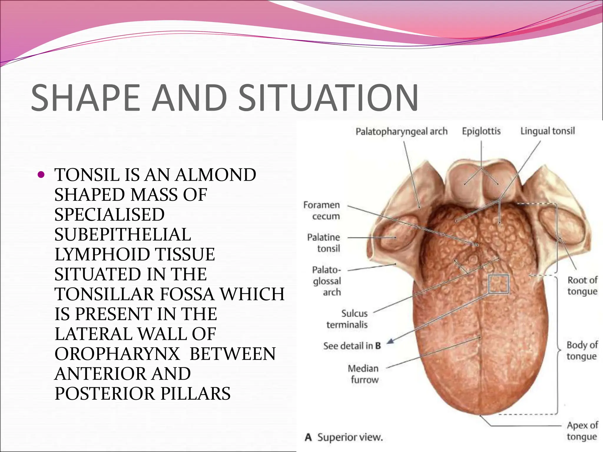
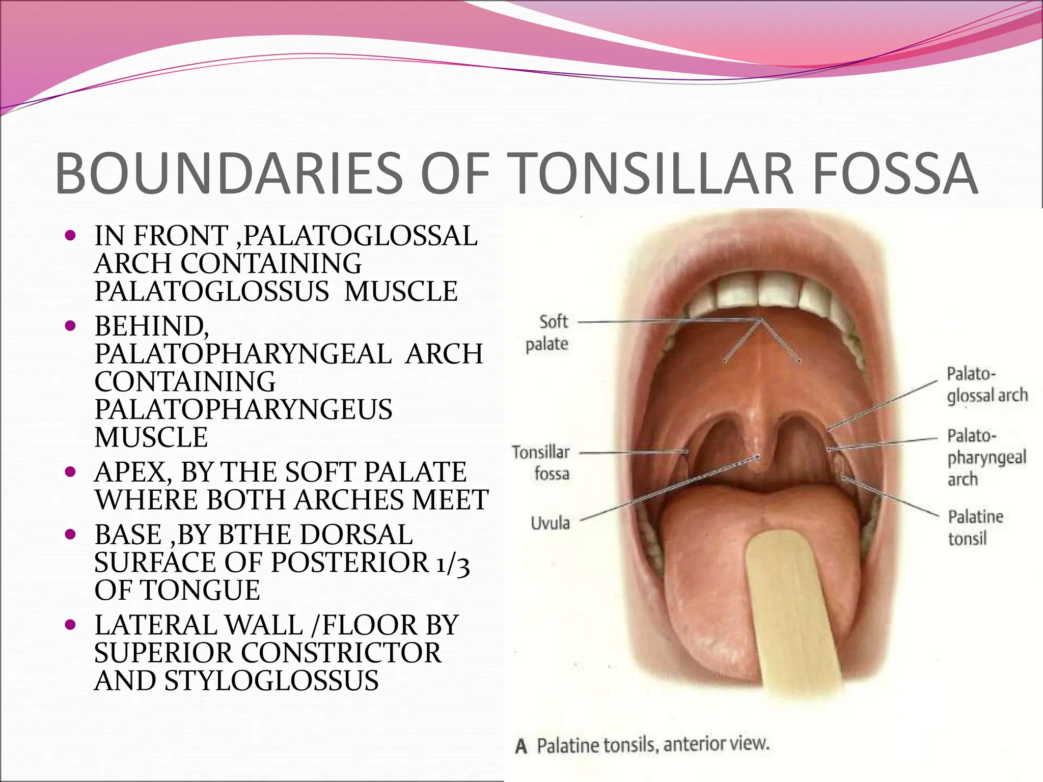
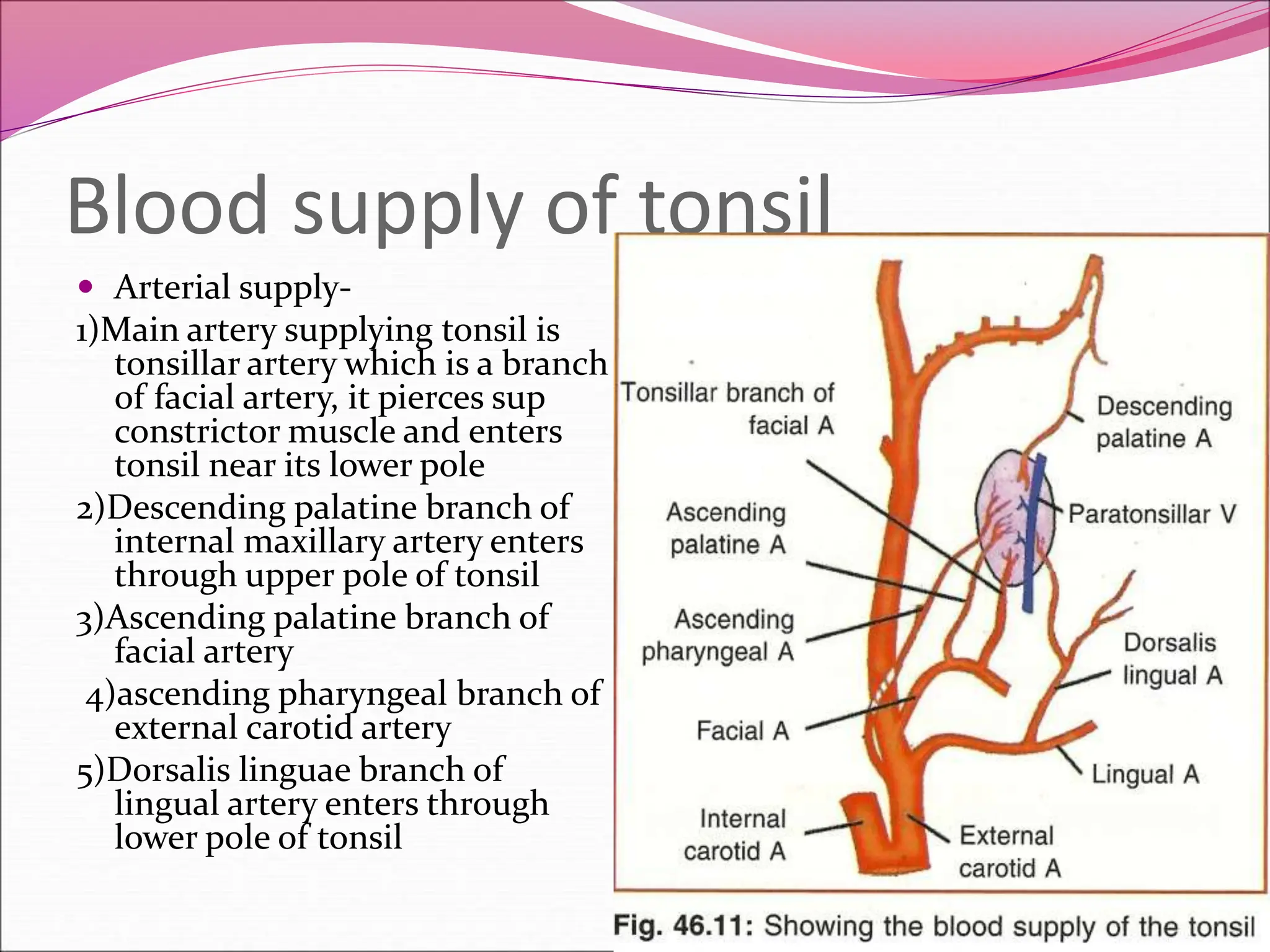
2. SHAPE AND SITUATION
TONSIL IS AN ALMOND
SHAPED MASS OF
SPECIALISED
SUBEPITHELIAL
LYMPHOID TISSUE
SITUATED IN THE
TONSILLAR FOSSA WHICH
IS PRESENT IN THE
LATERAL WALL OF
OROPHARYNX BETWEEN
ANTERIOR AND
POSTERIOR PILLARS
3. BOUNDARIES OF TONSILLAR FOSSA
IN FRONT ,PALATOGLOSSAL
ARCH CONTAINING
PALATOGLOSSUS MUSCLE
BEHIND,
PALATOPHARYNGEAL ARCH
CONTAINING
PALATOPHARYNGEUS
MUSCLE
APEX, BY THE SOFT PALATE
WHERE BOTH ARCHES MEET
BASE ,BY BTHE DORSAL
SURFACE OF POSTERIOR 1/3
OF TONGUE
LATERAL WALL /FLOOR BY
SUPERIOR CONSTRICTOR
AND STYLOGLOSSUS
4. IT HAS 2 SURFACES –MEDIAL AND LATERAL
2POLES –UPPER AND LOWER
5. MEDIAL SURFACE
SURFACE IS FREE AND HAS
1)TONSILLAR PITS –SMALL OPENINGS ABOUT 12 – 15 IN
NUMBER KNOWN AS TONSILLAR CRYPTS
2)INTRATONSILLAR CLEFT-ALSO KNOWN AS CRYPTA
MAGNAWHICH IS VERY LARGE DEEP SEMILUNAR
FISSURE AFFECTING UPPER PART OF TONSIL
3)EMBRYONIC FOLDS-
a)plica triangularis-is a triangular vestegeal fold of mucous
membrane at the lower part of palatoglossal arch
b)plica semilunaris- semilunar fold of mucous membrane at
the upper part of palatoglossal arch
Both these folds are replaced by lymphoid tissue after birth
6. Lateral surface
Surface is covered by a well defined fibrous capsule
Capule is an extension of pharyngobasilar fascia
Post superiorly ,capsule is attched loosely to muscular
wall of pharynx
Anteroinferiorly, it is firmly adherent to medial side of
tongue
Loose areolar tissue is present between capsule and
bed of tonsil
7. Relations of lateral surface
Within outwards
1.Loose areolar tissue containing paratonsillar vein
2.Pharyngobasillar fascia
3.Superior constrctor
4.Buccopharyngeal fascia
5.Facial artery with ascending palatine and tonsillar
branches
6. Ascending pharyngeal and internal carotid artery
8.
9. Bed of tonsil
With in outwards it is formed by
1.pharyngobasillar fascia
2. few fibers of palatopharyngeus muscle
3. superior constrictor muscle in post superior 2/3rd
4.,styloglossus muscle accompanied by
glossopharyngeal nerve in ant inferior 1/3rd
10. Upper pole of tonsil may extend upto the soft palate
Lower pole of tonsil may extend downwards beside the
base of tongue
Lymphoid tissue of tonsil is continuous with the
subepithelial lymphoid tissue on base of tongue called
lingual tonsil
Tonsillolingual sulcus separates the tonsil from base of
the tongue
11. DIFFERENCES B/W TONSILS
AND ADENOIDS
PALATINE TONSILS ADENOIDS
1.EPITHELIUM- stratified
squamous epithelium
2.SHAPE- almond shaped
3.AFFERENTS- only efferents
and no afferents
4.CRYPTS- tonsillar crypts
are present
5.present in lateral wall of
oropharynx
1.Ciliated columnar
epithelium
2.bunch of grapes
3.both afferents and efferents
are present
4.vertical ridges are
present.crypts are not seen
5.present in nasopharynx
12. Factors keeping tonsil in position
1. suspensory ligament of tonsil connecting it with
tongue
2. attachments of palatopharyngeus and palatoglossus
muscles to the fibrous capsule of tonsil
3.perivascular stalks
13. Blood supply of tonsil
Arterial supply-
1)Main artery supplying tonsil is
tonsillar artery which is a branch
of facial artery, it pierces sup
constrictor muscle and enters
tonsil near its lower pole
2)Descending palatine branch of
internal maxillary artery enters
through upper pole of tonsil
3)Ascending palatine branch of
facial artery
4)ascending pharyngeal branch of
external carotid artery
5)Dorsalis linguae branch of
lingual artery enters through
lower pole of tonsil
15. Lymphatic drainage
Lymphatics drain into jugulodigastric lymphnodes
situated at the junction of common facial and internal
juglar vein just below posterior belly of digastric
muscle
16. Nerve supply
Main sensory supply is by tonsillar branch of
glossopharngeal nerve
Greater and lesser palatine branches from
pterygopalatine ganglion supply upper part of tonsil
near the soft palate
17. Waldeyer ‘s ring
It is a scattered collection of
subepithelial lymphoid tissue
around aerodigestive tract
It consists of inner and outer
rings
Inner ring is formed by
1.Nasopharyngeal tonsils or
adenoids
2.Palatine tonsils
3. Lingual tonsils
4.Tubal tonsils
5. Lateral pharyngeal band
6.Discrete nodules in posterior
pharyngeal wall
18. Outer ring is formed by
1.Occipital lymphnodes
2.Post auricular lymph nodes
3.Parotid lymphnodes
4.Facial lymphnodes
5.Submental lymphnodes
19. Functins of waldeyer’s ring
1.helps in defensive mehanism of respiratory and
alimentary systems by destroying the entry of
microorganisms
2.formation of lymphocytes and plasma cells
3.body immunity and antibody formation in early
years of life
4.protection to lower respiratory tract by guarding the
entry of air passage
20. Developmental anatomy
Tonsils are developed from ventral part of second
pharyngeal pouch
Intra tonsillar cleft is a remnant of second pharyngeal
pouch
Lymphocytes are mesodermal in origin.they appear
during 3rd month.
Tosillar crypts develop as solid ingrowths from the
surface epithelium by 3rd to 6th month.
Tosillar capsule is formed from mesenchyme by 5th
month.
21. Pillars are formed from 2nd & 3rd branchial arches
through dorsal extension of mesenchyme into the
forming soft palate
Nasopharyngeal tonsils or adenoids develop during
4th to 6th month of intrauterine life
22. Histology of tonsil
Tonsil is lined by nonkeratinised stratified squamous
epithelium
Consists of numerous lymphatic follicles which
surround the tonsillar crypt
Each follicle presents a germinal centre composed of
lymphoblasts
Multiplication of lymphocytes occur in germinal
center.
23. Applied anatomy
Surgical removal of tonsils is indicated in recurrent
tonsillitis
Reffered pain in the ear following tonsillectomy is due
to common nerve supply by glossopharyngeal nerve
Sometimes patient may complain of loss of taste
sensation after tonsillectomy due to involvement of
glosspharyngeal nerve
During removal ,the paratonsillar vein may be
damaged producing excessive venous haemorrhage
24. A penetrating oropharyngeal injury directed
posterolaterally travels the tonsillar bed and superior
constrictor enters the parapharyngeal space
Stuctures that can be injured are
1.internal jugular vein
2.internal carotid artery
3.nerves associated with carotid sheath
4.sympathetic trunk