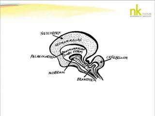
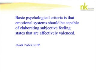
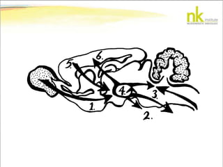
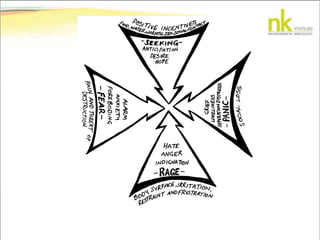
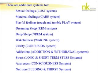
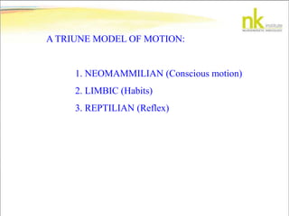
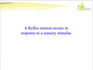
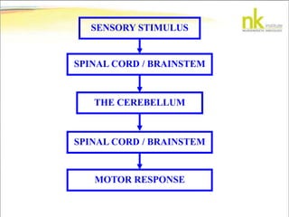
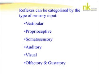
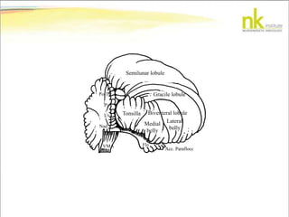
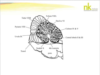
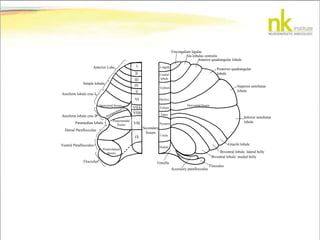
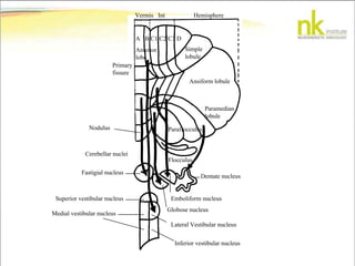
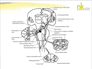
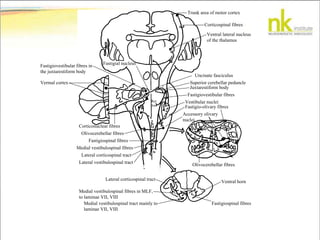
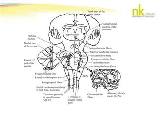
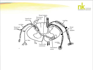
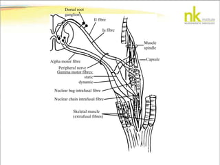
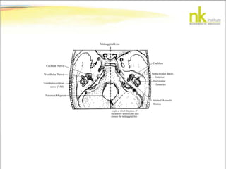
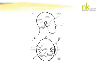
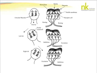
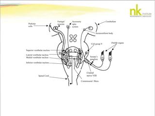
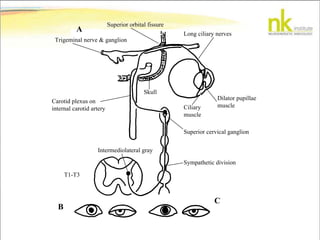
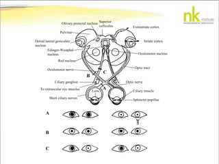
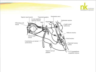
THE TRIUNE BRAIN, EMOTION &
MOTION
Basic psychological criteria is that
emotional systems should be capable
of elaborating subjective feeling
states that are affectively valenced.
JAAK PANKSEPP
The SEEKING system in what Jaak Panksepp renames the
"appetitive motivational system", encourages animals to search
for resources such as food, water, warmth etc. Another
designator that he used to highlight its function was
curiosity/interest/foraging/anticipation/craving/expectancy
system.
The RAGE system energises the body to
angrily defend its territory and resources.
The FEAR system is the brain system that
includes a major form of trepidation that
commonly leads to freezing or flight.
The PANIC system is the one that generates feelings
of loneliness and separation distress. This is not to be
confused with the term panic meaning intense states
of fear. The existing evidence on the disorder known
as panic attacks involves this circuitry.
There are additional systems for:
Sexual feelings (LUST system)
Maternal feelings (CARE system)
Playful feelings (rough and tumble PLAY system)
Dreaming Sleep (REM system)
Deep Sleep (NREM system)
Wakefullness (WAKING system)
Clarity (CONFUSION system)
Addictions (ADDICTION & WITHDRAWAL systems)
Stress (LONG & SHORT TERM STESS Systems)
Awareness (CONSCIOUSNESS Systems)
Nutriton (FEEDING & THIRST Systems)
A TRIUNE MODEL OF MOTION:
1. NEOMAMMILIAN (Conscious motion)
2. LIMBIC (Habits)
3. REPTILIAN (Reflex)
What is a Reflex?
A Reflex motion occurs in
response to a sensory stimulus
SENSORY STIMULUS
SPINAL CORD / BRAINSTEM
THE CEREBELLUM
SPINAL CORD / BRAINSTEM
MOTOR RESPONSE
Reflexes can be categorised by the
type of sensory input:
•Vestibular
•Proprioceptive
•Somatosensory
•Auditory
•Visual
•Olfactory & Gustatory
Semilunar lobule
Gracile lobule
Biventeral lobule
Lateral
belly
Medial
belly
Tonsilla
Fo-Tu
Py
Uv
Nad
VMA Flo
Acc. Paraflocc
pons
Central lobule II & III
Culmen IV & V
Declive VI
4th ventricle
Folium VIIA
Tuber VIIB
Pyramis VIII
Uvula IX
Nodule X
Tonsil
I
II
III
IV
V
VI
VIIA
VIIB
VIII
IX
Secondary
fissure
Lingula
Central
lobule
Culmen
Declive
Folium
Tuber
Pyramis
Uvula
Nodule
Horizontal fissureIntercrural fissure
Ansoparamed f.
Prepyramidal
fissure
Posterolateral
fissure
Tonsilla
Vincingulum ligulae
Ala lobulus centralis
Anterior quadrangular lobule
Posterior quadrangular
lobule
Superior semilunar
lobule
Inferior semilunar
lobule
Gracile lobule
Biventral lobule: lateral belly
Biventral lobule: medial belly
Flocculus
Accessory paraflocculus
Anterior Lobe
Simple lobule
Ansiform lobule crus I
Ansiform lobule crus II
Paramedian lobule
Dorsal Paraflocculus
Ventral Paraflocculus
Flocculus
Vermis Int Hemisphere
Primary
fissure
A B C1 C2 C3 D
Simple
lobule
Ansiform lobule
Paramedian
lobule
Paraflocculus
Flocculus
Cerebellar nuclei
Anterior
lobe
Fastigial nucleus
Dentate nucleus
Emboliform nucleus
Globose nucleus
Lateral Vestibular nucleus
Superior vestibular nucleus
Medial vestibular nucleus
Inferior vestibular nucleus
Nodulus
Extremity areas of the motor
cortex
Corticospinal fibres
Ventral lateral nucleus of the
thalamus
Interpositothalamic fibres
Red nucleus
Decussation of superior cerebellar
peduncle
Interposito-olivary fibres
Accessory olivary
nuclei
Olivocerebellar fibres
Interpositospinal fibres
Ventral horn
Rubrospinal fibres
Lateral corticospinal tract
Interpositospinal tract
Rubrospinal tract
Olivocerebellar fibres
Posterior interposed (globose) nucleus
Intermediate
zone
Corticonuclear
fibres
Anterior interposed
(emboliform) nucleus
Hypothalamus
Ret.
for
Trunk area of motor cortex
Corticospinal fibres
Ventral lateral nucleus
of the thalamus
Accessory olivary
nuclei
Fastigio-olivary fibres
Vestibular nuclei
Fastigiovestibular fibres
Juxtarestiform body
Superior cerebellar peduncle
Uncinate fasciculus
Olivocerebellar fibres
Ventral horn
Fastigiospinal fibres
Lateral corticospinal tract
Medial vestibulospinal fibres in MLF,
to laminae VII, VIII
Medial vestibulospinal tract mainly to
laminae VII, VIII
Corticonuclear fibres
Olivocerebellar fibres
Fastigiospinal fibres
Medial vestibulospinal fibres
Lateral corticospinal tract
Lateral vestibulospinal tract
Vermal cortex
Fastigial nucleus
Fastigiovestibular fibres in
the juxtarestiform body
Ret.
for.
Trunk area of the
motor cortex
Ventral lateral
nucleus of the
thalamus
hypothalamus
Fastigiothalamic fibres
Superior cerebellar peduncle
Juxtarestiform body
Fastigiovestibular fibres
Vestibular nuclei
Fastigio-olivary fibres
Accessory olivary
nuclei (AON)
Olivocerebellar
fibresTerminate in
medial ventral
horn
Terminate primarily
in spinal laminae
VII, VII
Medial vestibulospinal fibres
in med. long. fasciculus
Fastigiospinal fibres
Lateral vestibulospinal tract
Flocculonodular lobe
Lateral
part of the
vermis
Medial part
of the vermis
Fastigial
nucleus
Ret.
For.
AON
C2
C3
C4
C5
C6
C5
C3
C2
C4
C2
C2-C3
C3-C4
C6
C8
C8
C7
T1
T1
T2
T2
T3
T5
T7
T9
T12
T10
T8
T6
T4
C5
C6
L1
L2
L3
L5
L4
S1
S2
L5
L1
L2
S3
S3
S4
Cx
V1-ophthalmic division
V
2-m
axillary
division
V3-mandibulardivision
Golgi tendon
organ (Ib)
Muscle spindle
afferent fibre
Extrafusal muscle
fibres
Intrafusal
muscle fibres
Hair follicle
receptors
Free endings
Meissner’s corpuscle
Merkel cells
Epidermis
Dermis
A- fibre
A- fibre (SA)
A- fibre (RA)
A- fibre (SA)
A- fibre
(RA)
C fibreA- fibre
(RA,SA)
Ruffini endings
Nuclear
bag fibre
Nuclear chain fibre
Muscle spindle
Secondary ending
fibre (II)
Primary ending fibre (Ia)
Tendon
Gamma motor
fibres
Dorsal columns
Dorsolateral
fasciculusLateral root
entry zone
A- fibre
Dorsal root
ganglion
C fibre
Ventral white
commissure
Anterolateral
system
A- fibre
A-α fibre
Dorsal
root
ganglion
Dorsal root
ganglion
II fibre
Ia fibre
Muscle
spindle
Capsule
Skeletal muscle
(extrafusal fibres)
Nuclear chain intrafusal fibre
Nuclear bag intrafusal fibre
Gamma motor fibres:
static
dynamic
Peripheral nerve
Alpha motor fibre
Midsaggital Line
Angle at which the plane of
the anterior semicircular duct
crosses the midsaggital line
Cochlear
Anterior
Semicircular ducts:
Horizontal
Posterior
Internal Acoustic
Meatus
Cochlear Nerve
Vestibular Nerve
Vestibulocochlear
nerve (VIII)
Foramen Magnum
A
B
Anterior
Semicircular
Canal
Posterior
Semicircular
Canal
Horizontal
Semicircular
Canal
Plane of Saccule
Plane of
horizontal canal
and utricle
Naso-
occipital
plane
60
30
90
Horizontal
semicircular
canal
Anterior
semicircular
canal
Posterior
semicircular
canal
Foramen
Magnum
Temporal
bone
Anterior
canal
plane
Posterior
canal
plane
Striola
Otoconia
Otolith membrane
Receptor cell
Kinocilium
Gravity
Utricular Maculae
Resting Resting
ExcitationInhibition
InhibitionExcitation
Left tilt
Right tilt
Ocular
counteroll
Cerebellum
Juxtarestiform body
Accessory
optic
system
Fastigal
neuronsPurkinje
cells
Superior vestibular nucleus
Lateral vestibular nucleus
Medial vestibular nucleus
Inferior vestibular nucleus
Spinal Cord
Commissural fibres
Cranial
nerve VIII
Scarpa’s
ganglion
Cell group Y
Otolith organs
Superior orbital fissure
Long ciliary nerves
Trigeminal nerve & ganglion
Carotid plexus on
internal carotid artery
Skull
Dilator pupillae
muscleCiliary
muscle
Superior cervical ganglion
Sympathetic division
Intermediolateral gray
T1-T3
B
C
A
Extrastriate cortex
Striate cortex
Superior
colliculus
Olivary pretectal nucleus
Pulvinar
Dorsal lateral geniculate
nucleus
Edinger-Westphal
nucleus
Red nucleus
Oculomotor nucleus
Optic tract
C
B
A
Oculomotor nerve
Ciliary ganglion
To extraocular eye muscles
Short ciliary nerves
Ciliary muscle
Optic nerve
Lens
Sphincter pupillae
A
B
C
Hypothalamus
Oculomotor nerve
Oculomotor nucleus
Caudal central
subdivision
Abducens nucleus
Facial nucleus
Trigeminal sensory
nucleus
T1-T3 preganglionic
sympathetic neurons
Trigeminal ganglion
Skull
Levator palpebrae
superioris muscle
Superior tarsal muscle
Orbicularis oculi
muscle
Inferior tarsal
muscle
Carotid plexus on internal
carotid artery
Superior cervical
ganglion
P/L ISSUE
Sensory receptor
Find dermatome & challenge receptor
Cranial nerve format
A or C fibred receptors (ALS) Other fibre typed receptors (DCML)
Spinal trigeminal nucleus Sensory nucleus of the trigeminal nerve
Cerebellum Pons +
GV15 + alarm point
CV GV Rest
Fastigial or Lat
Vestib nuclei
Emboliform, Dentate
or Globose nuclei
Periaquaductal
grey matter
Nucleus raphe magnus
Spinal trigeminal
nucleus
Go to flow chart 4
Ventral posterior nucleus of the thalamus
Cerebral cortex
Specific Muscle, Test muscle,
then correct circuit
Pontine nucleus
The Triune Brain, Emotion & Motion

The Triune Brain, Emotion & Motion

  • 1.
    THE TRIUNE BRAIN,EMOTION & MOTION
  • 3.
    Basic psychological criteriais that emotional systems should be capable of elaborating subjective feeling states that are affectively valenced. JAAK PANKSEPP
  • 6.
    The SEEKING systemin what Jaak Panksepp renames the "appetitive motivational system", encourages animals to search for resources such as food, water, warmth etc. Another designator that he used to highlight its function was curiosity/interest/foraging/anticipation/craving/expectancy system.
  • 7.
    The RAGE systemenergises the body to angrily defend its territory and resources.
  • 8.
    The FEAR systemis the brain system that includes a major form of trepidation that commonly leads to freezing or flight.
  • 9.
    The PANIC systemis the one that generates feelings of loneliness and separation distress. This is not to be confused with the term panic meaning intense states of fear. The existing evidence on the disorder known as panic attacks involves this circuitry.
  • 10.
    There are additionalsystems for: Sexual feelings (LUST system) Maternal feelings (CARE system) Playful feelings (rough and tumble PLAY system) Dreaming Sleep (REM system) Deep Sleep (NREM system) Wakefullness (WAKING system) Clarity (CONFUSION system) Addictions (ADDICTION & WITHDRAWAL systems) Stress (LONG & SHORT TERM STESS Systems) Awareness (CONSCIOUSNESS Systems) Nutriton (FEEDING & THIRST Systems)
  • 11.
    A TRIUNE MODELOF MOTION: 1. NEOMAMMILIAN (Conscious motion) 2. LIMBIC (Habits) 3. REPTILIAN (Reflex)
  • 12.
    What is aReflex?
  • 13.
    A Reflex motionoccurs in response to a sensory stimulus
  • 14.
    SENSORY STIMULUS SPINAL CORD/ BRAINSTEM THE CEREBELLUM SPINAL CORD / BRAINSTEM MOTOR RESPONSE
  • 15.
    Reflexes can becategorised by the type of sensory input: •Vestibular •Proprioceptive •Somatosensory •Auditory •Visual •Olfactory & Gustatory
  • 17.
    Semilunar lobule Gracile lobule Biventerallobule Lateral belly Medial belly Tonsilla Fo-Tu Py Uv Nad VMA Flo Acc. Paraflocc
  • 18.
    pons Central lobule II& III Culmen IV & V Declive VI 4th ventricle Folium VIIA Tuber VIIB Pyramis VIII Uvula IX Nodule X Tonsil
  • 19.
    I II III IV V VI VIIA VIIB VIII IX Secondary fissure Lingula Central lobule Culmen Declive Folium Tuber Pyramis Uvula Nodule Horizontal fissureIntercrural fissure Ansoparamedf. Prepyramidal fissure Posterolateral fissure Tonsilla Vincingulum ligulae Ala lobulus centralis Anterior quadrangular lobule Posterior quadrangular lobule Superior semilunar lobule Inferior semilunar lobule Gracile lobule Biventral lobule: lateral belly Biventral lobule: medial belly Flocculus Accessory paraflocculus Anterior Lobe Simple lobule Ansiform lobule crus I Ansiform lobule crus II Paramedian lobule Dorsal Paraflocculus Ventral Paraflocculus Flocculus
  • 20.
    Vermis Int Hemisphere Primary fissure AB C1 C2 C3 D Simple lobule Ansiform lobule Paramedian lobule Paraflocculus Flocculus Cerebellar nuclei Anterior lobe Fastigial nucleus Dentate nucleus Emboliform nucleus Globose nucleus Lateral Vestibular nucleus Superior vestibular nucleus Medial vestibular nucleus Inferior vestibular nucleus Nodulus
  • 21.
    Extremity areas ofthe motor cortex Corticospinal fibres Ventral lateral nucleus of the thalamus Interpositothalamic fibres Red nucleus Decussation of superior cerebellar peduncle Interposito-olivary fibres Accessory olivary nuclei Olivocerebellar fibres Interpositospinal fibres Ventral horn Rubrospinal fibres Lateral corticospinal tract Interpositospinal tract Rubrospinal tract Olivocerebellar fibres Posterior interposed (globose) nucleus Intermediate zone Corticonuclear fibres Anterior interposed (emboliform) nucleus Hypothalamus Ret. for
  • 22.
    Trunk area ofmotor cortex Corticospinal fibres Ventral lateral nucleus of the thalamus Accessory olivary nuclei Fastigio-olivary fibres Vestibular nuclei Fastigiovestibular fibres Juxtarestiform body Superior cerebellar peduncle Uncinate fasciculus Olivocerebellar fibres Ventral horn Fastigiospinal fibres Lateral corticospinal tract Medial vestibulospinal fibres in MLF, to laminae VII, VIII Medial vestibulospinal tract mainly to laminae VII, VIII Corticonuclear fibres Olivocerebellar fibres Fastigiospinal fibres Medial vestibulospinal fibres Lateral corticospinal tract Lateral vestibulospinal tract Vermal cortex Fastigial nucleus Fastigiovestibular fibres in the juxtarestiform body Ret. for.
  • 23.
    Trunk area ofthe motor cortex Ventral lateral nucleus of the thalamus hypothalamus Fastigiothalamic fibres Superior cerebellar peduncle Juxtarestiform body Fastigiovestibular fibres Vestibular nuclei Fastigio-olivary fibres Accessory olivary nuclei (AON) Olivocerebellar fibresTerminate in medial ventral horn Terminate primarily in spinal laminae VII, VII Medial vestibulospinal fibres in med. long. fasciculus Fastigiospinal fibres Lateral vestibulospinal tract Flocculonodular lobe Lateral part of the vermis Medial part of the vermis Fastigial nucleus Ret. For. AON
  • 24.
  • 25.
    Golgi tendon organ (Ib) Musclespindle afferent fibre Extrafusal muscle fibres Intrafusal muscle fibres Hair follicle receptors Free endings Meissner’s corpuscle Merkel cells Epidermis Dermis A- fibre A- fibre (SA) A- fibre (RA) A- fibre (SA) A- fibre (RA) C fibreA- fibre (RA,SA) Ruffini endings Nuclear bag fibre Nuclear chain fibre Muscle spindle Secondary ending fibre (II) Primary ending fibre (Ia) Tendon Gamma motor fibres
  • 26.
    Dorsal columns Dorsolateral fasciculusLateral root entryzone A- fibre Dorsal root ganglion C fibre Ventral white commissure Anterolateral system A- fibre A-α fibre Dorsal root ganglion
  • 27.
    Dorsal root ganglion II fibre Iafibre Muscle spindle Capsule Skeletal muscle (extrafusal fibres) Nuclear chain intrafusal fibre Nuclear bag intrafusal fibre Gamma motor fibres: static dynamic Peripheral nerve Alpha motor fibre
  • 28.
    Midsaggital Line Angle atwhich the plane of the anterior semicircular duct crosses the midsaggital line Cochlear Anterior Semicircular ducts: Horizontal Posterior Internal Acoustic Meatus Cochlear Nerve Vestibular Nerve Vestibulocochlear nerve (VIII) Foramen Magnum
  • 29.
    A B Anterior Semicircular Canal Posterior Semicircular Canal Horizontal Semicircular Canal Plane of Saccule Planeof horizontal canal and utricle Naso- occipital plane 60 30 90 Horizontal semicircular canal Anterior semicircular canal Posterior semicircular canal Foramen Magnum Temporal bone Anterior canal plane Posterior canal plane
  • 30.
    Striola Otoconia Otolith membrane Receptor cell Kinocilium Gravity UtricularMaculae Resting Resting ExcitationInhibition InhibitionExcitation Left tilt Right tilt Ocular counteroll
  • 31.
    Cerebellum Juxtarestiform body Accessory optic system Fastigal neuronsPurkinje cells Superior vestibularnucleus Lateral vestibular nucleus Medial vestibular nucleus Inferior vestibular nucleus Spinal Cord Commissural fibres Cranial nerve VIII Scarpa’s ganglion Cell group Y Otolith organs
  • 32.
    Superior orbital fissure Longciliary nerves Trigeminal nerve & ganglion Carotid plexus on internal carotid artery Skull Dilator pupillae muscleCiliary muscle Superior cervical ganglion Sympathetic division Intermediolateral gray T1-T3 B C A
  • 33.
    Extrastriate cortex Striate cortex Superior colliculus Olivarypretectal nucleus Pulvinar Dorsal lateral geniculate nucleus Edinger-Westphal nucleus Red nucleus Oculomotor nucleus Optic tract C B A Oculomotor nerve Ciliary ganglion To extraocular eye muscles Short ciliary nerves Ciliary muscle Optic nerve Lens Sphincter pupillae A B C
  • 34.
    Hypothalamus Oculomotor nerve Oculomotor nucleus Caudalcentral subdivision Abducens nucleus Facial nucleus Trigeminal sensory nucleus T1-T3 preganglionic sympathetic neurons Trigeminal ganglion Skull Levator palpebrae superioris muscle Superior tarsal muscle Orbicularis oculi muscle Inferior tarsal muscle Carotid plexus on internal carotid artery Superior cervical ganglion
  • 35.
    P/L ISSUE Sensory receptor Finddermatome & challenge receptor Cranial nerve format A or C fibred receptors (ALS) Other fibre typed receptors (DCML) Spinal trigeminal nucleus Sensory nucleus of the trigeminal nerve Cerebellum Pons + GV15 + alarm point CV GV Rest Fastigial or Lat Vestib nuclei Emboliform, Dentate or Globose nuclei Periaquaductal grey matter Nucleus raphe magnus Spinal trigeminal nucleus Go to flow chart 4 Ventral posterior nucleus of the thalamus Cerebral cortex Specific Muscle, Test muscle, then correct circuit Pontine nucleus