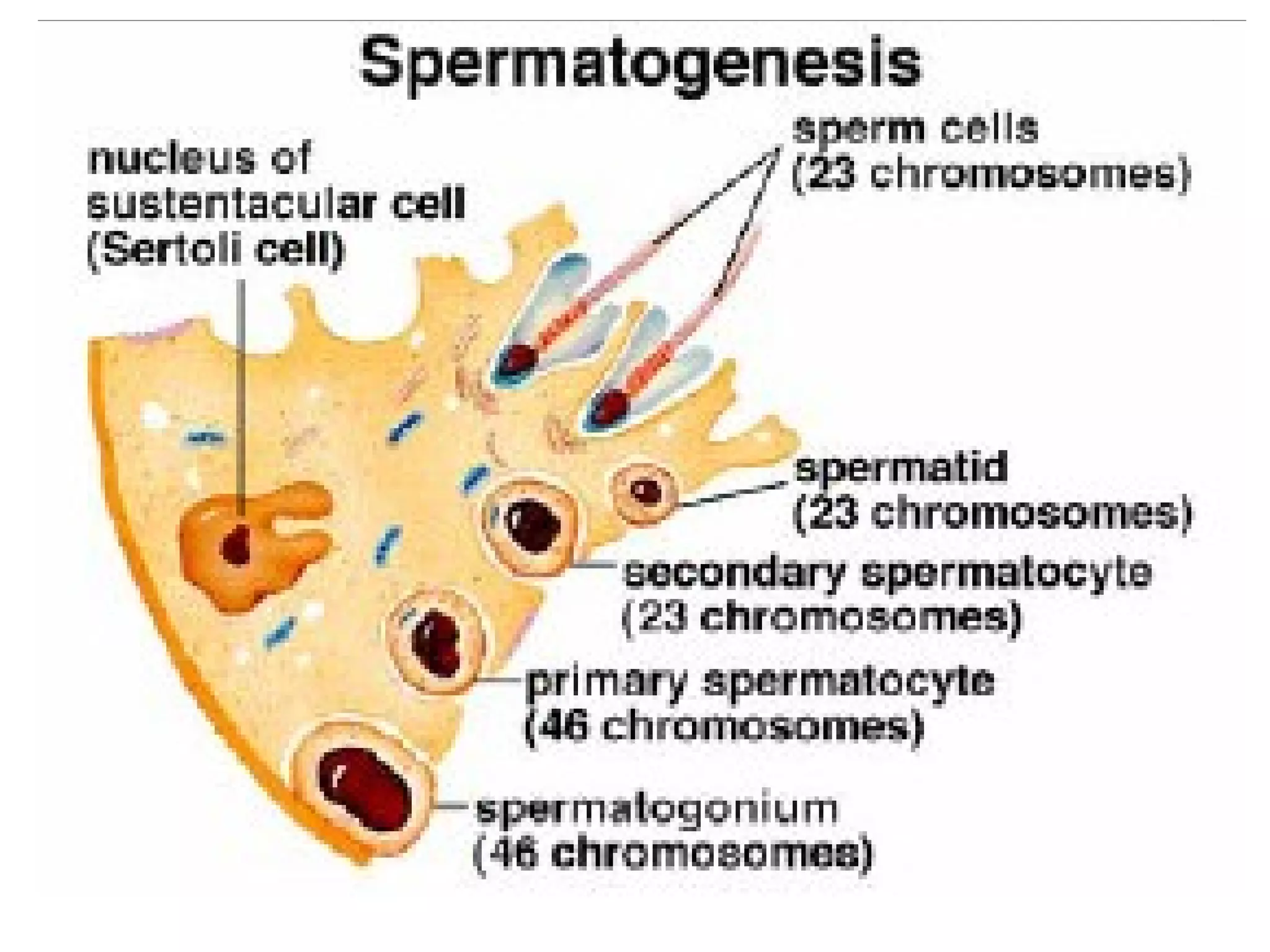
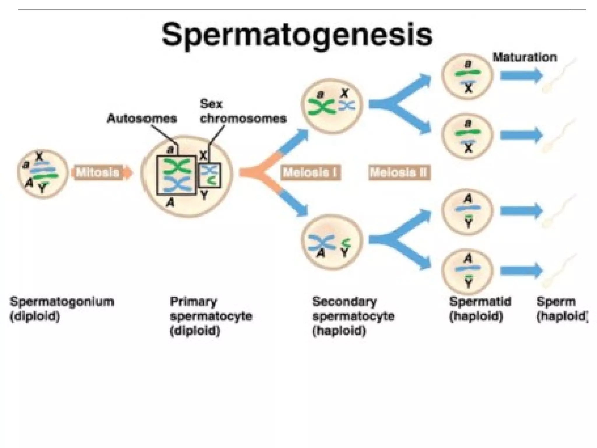
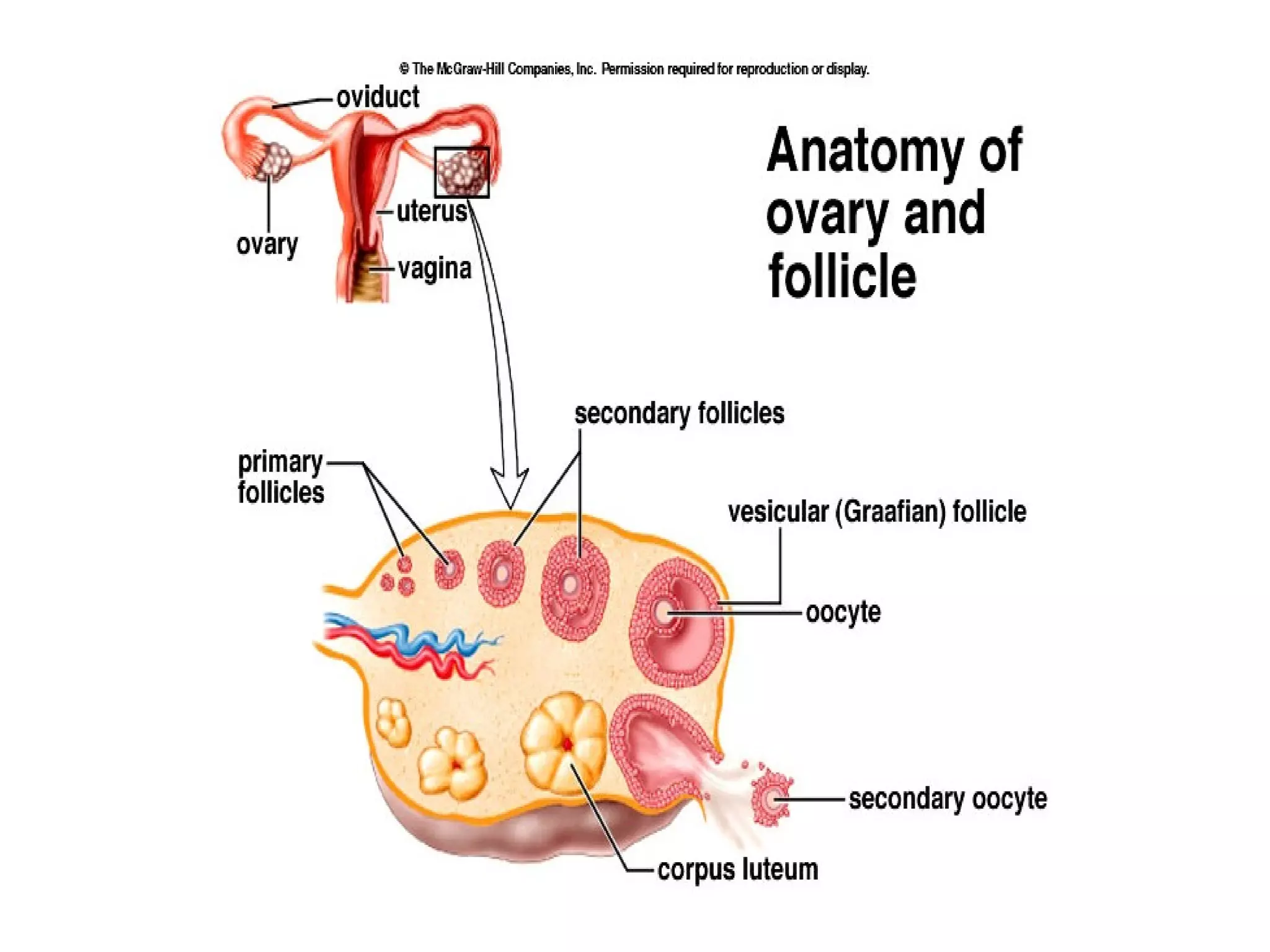
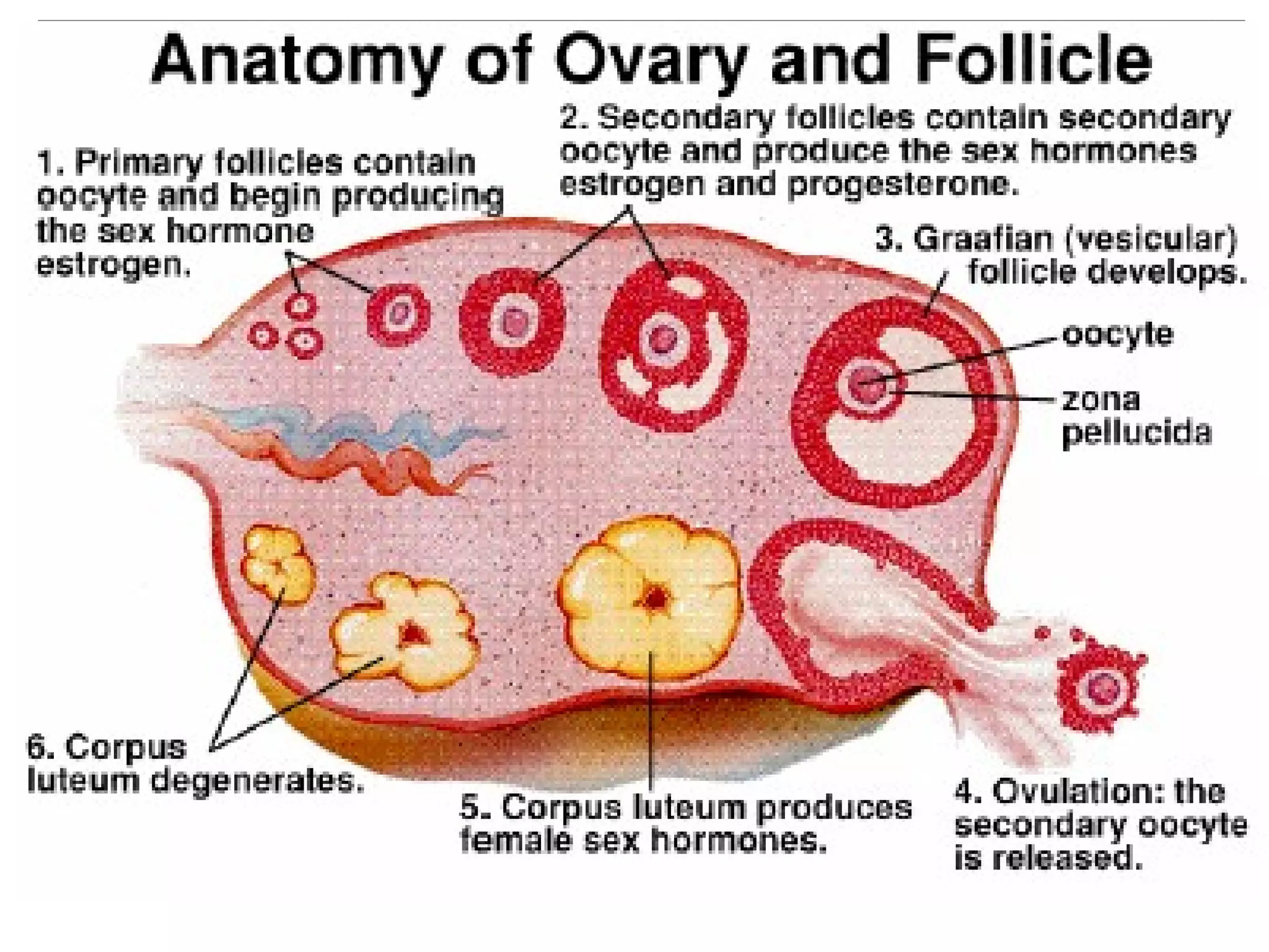
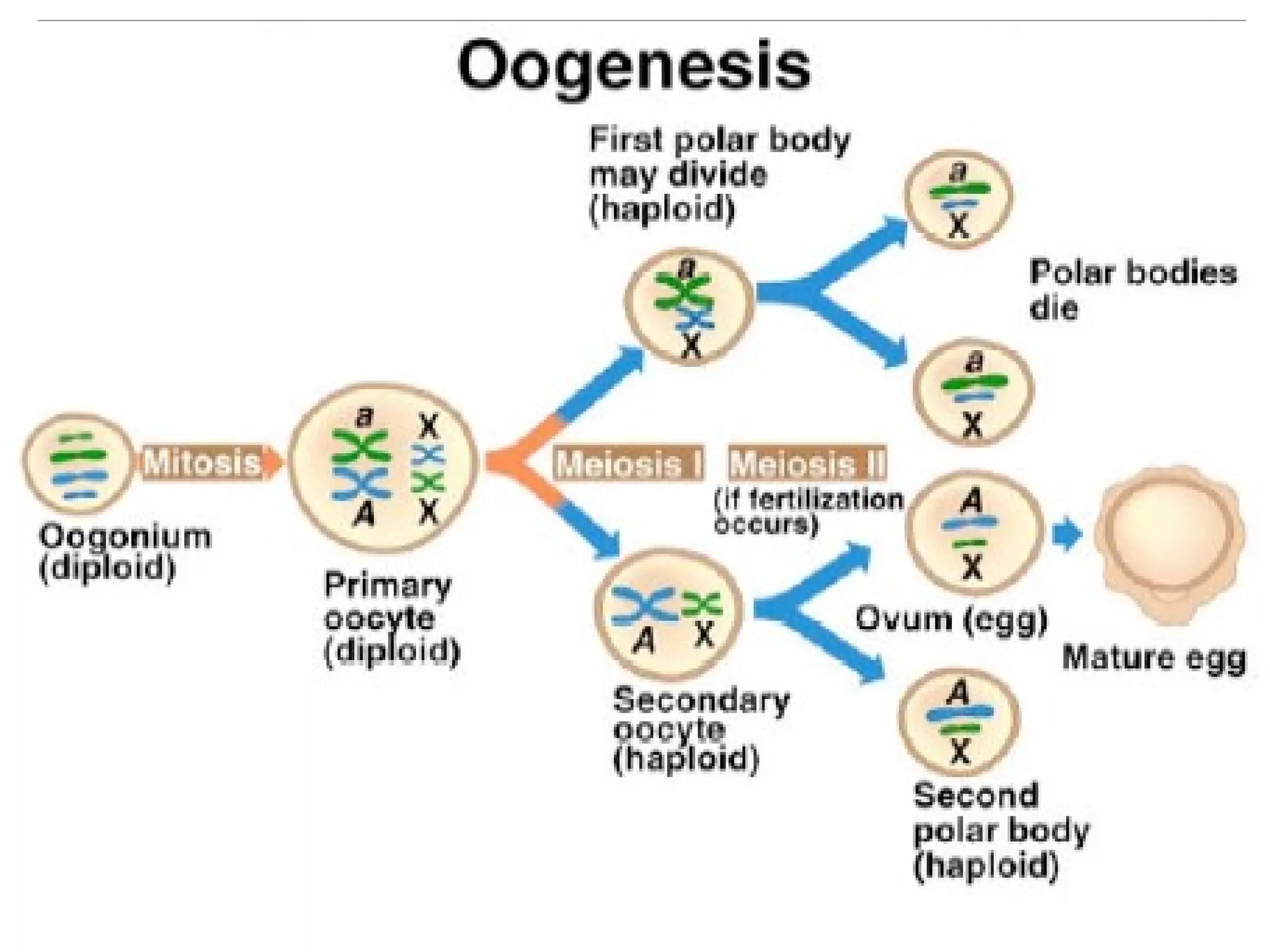
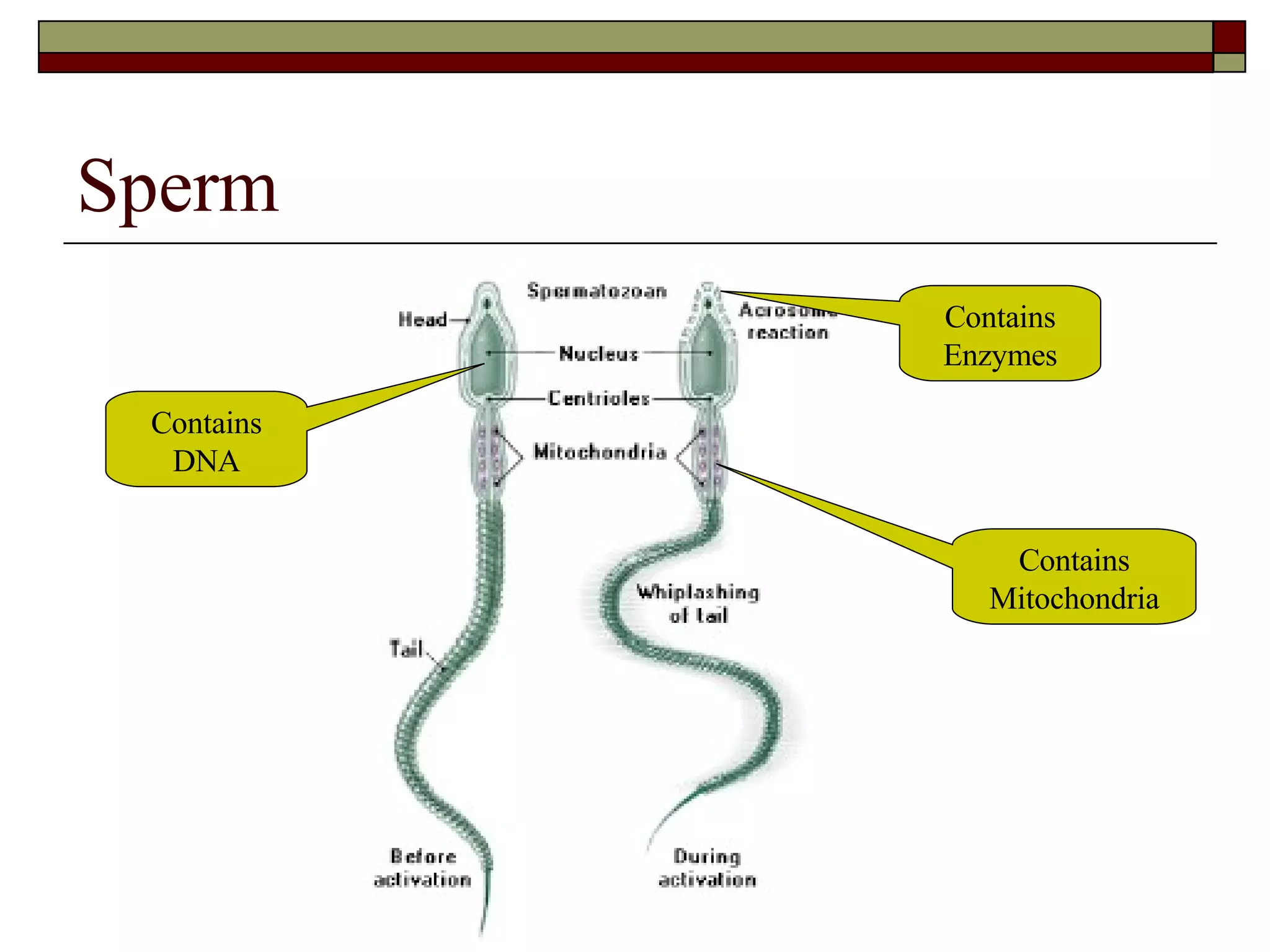
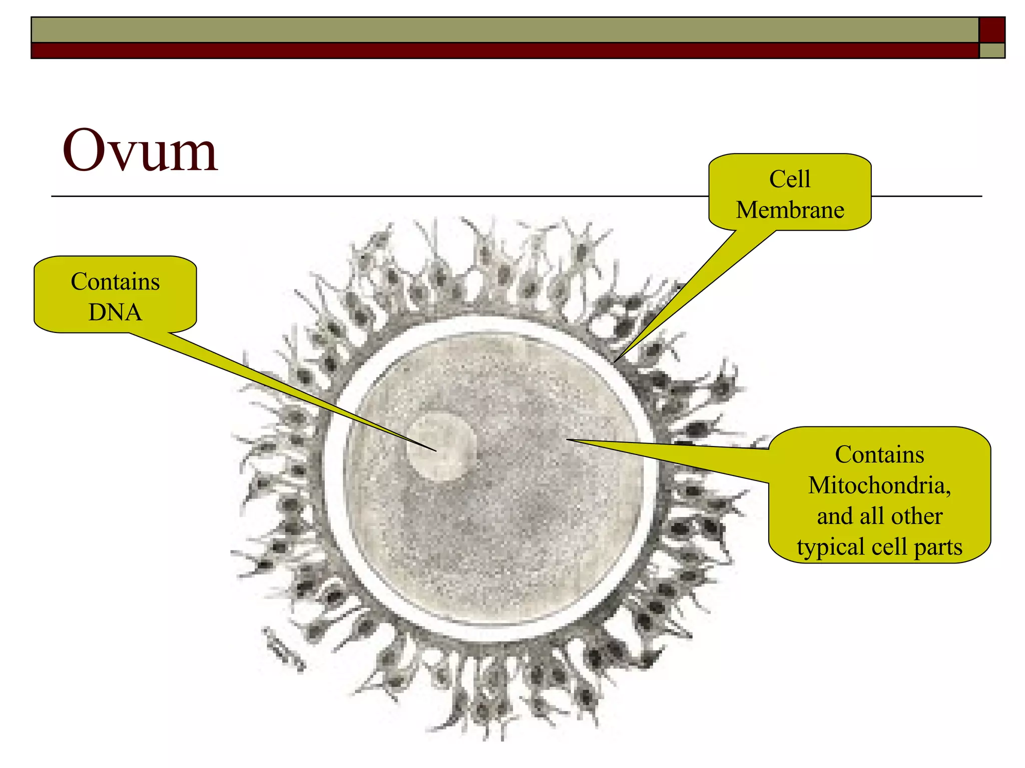
The Reproductive System Chapter 21
Overview Males and females have been created with specialized structures that become “operational” in puberty The key component of these structures, the gonads, have the ability to make unique cells called gametes The fusion of male and female gametes provides the genetic mixture of a new life!
The Gonads The gonads are organs that make gametes Male gonads: testicles…make spermatozoa Female gonads: ovaries…make ova Gametes are formed by a special type of cell division called meiosis Spermatogenesis occurs in the testicles’ seminiferous tubules Oogenesis occurs in the ovaries’ follicles


The Gametes Male and female gametes both carry genetic material – 23 chromosomes each Gametes are developed at different rates Male – up to 1 billion sperm made per day Female – born with ~2 million follicles; reduced to ~350,000 at puberty; ~400 ovum released in lifetime (1 per ~28 day cycle) Male and female gametes have different structures…
Sperm Contains Enzymes Contains Mitochondria Contains DNA
Ovum Contains DNA Contains Mitochondria, and all other typical cell parts Cell Membrane
Gamete Transport Both sperm and ova move through pathways Sperm… Have motility due to flagella action Moved by cilia in the epididymus Moved by muscle contractions in the penis  Ovum… Bursts from follicle into gap Moves into oviduct through contractile motion of fimbriae Moves through oviduct by contractions and cilia action
Sperm Pathway
Ova Pathway
Fluids Male… Tissue fluid in testicles Seminal vesicle fluids Fructose – used to make ATP Prostaglandins – cause vaginal/uterine contractions; suppress immune response in vagina Proteins – help coagulation in female system Basic solution – counteract acidic vagina
Fluids Prostate gland fluids Basic solution (more so than seminal vesicles) Increases sperm motility Milky appearance…only released upon ejaculation Cowper’s gland fluids Secreted during sexual arousal (pre-ejaculate fluid) Mucus-like lubricant for intercourse Possible function may be to cleanse the urethra Overall, seminal fluids provide nutrition, motility, protection, and lubrication
Fluids Female… Tissue fluids throughout (ovaries, oviducts, etc.) Vaginal secretions Droplets of fluid released with sexual arousal (lubricating function) Cervical/Vagina mucus (consistency and color changes throughout menstrual cycle…used in “fertility awareness” methods)
Hormones In both male and female, the hypothalamus secretes GnRF or GnRH (gonadotropic releasing factor or hormone) into the anterior pituitary gland This factor/hormone stimulates the pituitary to secrete two hormones: FSH – follicle stimulating hormone LH – luteininizing hormone
Male Hormone Regulation In the male… FSH targets the seminiferous tubules and promotes spermatogenesis LH targets the interstitial tissue and promotes the production of testosterone The production of testosterone… Stimulates growth of sex organs; matures sperm Increases sex drive and promotes secondary sexual characteristics (hair, voice, muscle, etc.)
 
Female Hormone Regulation The production of estrogen and progesterone: Stimulates growth of uterus/vagina; matures ova Promotes secondary characteristics (hair, fat deposits, hips, etc.) In the female’s Ovarian Cycle… Follicular phase  (~day 1-14)  FSH targets the ovaries to develop follicles which triggers the release of estrogen Luteal phase  (~day 15-28)  LH also targets the ovaries to develop the corpus luteum which triggers the release of progesterone
Note:  Progesterone has its own control mechanism – similar to the one shown here for estrogen
 
Female Hormone Regulation In the female’s Uterine Cycle… Menstrual period  (~day 1-5)  Low levels of estrogen and progesterone cause the endometrium (uterus lining) to disintegrate be released out the vagina Proliferative phase  (~day 6-13) Estrogen levels peak; endometrium thickens Secretory Phase  (~day 15-28) Hormone levels peak; endometrium doubles thickness; uterine glands mature and secretes mucus in preparation for implantation
 
A few more details… Estrogen stimulates endometrial growth Progesterone makes endometrium thick with blood vessels Menopause occurs in females between the ages of 45-55 Ovaries no longer responsive to FSH/LH and stop producing estrogen/progesterone Hormone therapy sometimes needed to combat hot flashes, dizziness, headaches, and insomnia There is no menopausal parallel in males
Pregnancy Of the 400 million sperm that begin the journey in the female reproductive system, only a few thousand meet the released ova in the oviducts Upon meeting, the sperm collectively break down the coating surrounding the mature ova and attempt entry Entry and fusion of nuclei = fertilization
 
 
Pregnancy Once implantation has occurred, the outer cells of the embryo produce HCG (human chorionic gonadotropic hormone) – which prevents the usual degeneration of the corpus luteum in the ovary HCG, estrogen, and progesterone levels are maintained to sustain the endometrium until the placenta is fully developed As such, menstruation ceases and new follicle growth is inhibited
Stage 1: Fertilization 0.1 - 0.15 mm 1 day post-ovulation 1 Egg, 300 Million Sperm Fertilization begins when a sperm penetrates an an egg  and it ends with the creation of the zygote. Fertilization takes about  24 hours .
Stage 2: Division 0.1 - 0.2 mm 1.5 - 3 days post-ovulation First Cell Division When cell division produces sixteen cells, the zygote becomes mulberry shaped. It  leaves  the fallopian tube and three to four days after fertilization. IUD / RU486 – “morning after pill”
Stage 3: Early Blastocyst 0.1 - 0.2 mm 4 days post-ovulation About four days after fertilization, the egg enters the uterine cavity. Cell division continues, forming a  cavity  in the center of the egg. Cells flatten and compact on the inside of the cavity. The entire structure is now called a  blastocyst .
Stage 4: Implantation Begins 0.1 - 0.2 mm 5 - 6 days post-ovulation The blastocyst "hatches" around the sixth day The implantation site becomes swollen with new capillaries, and  blood circulation begins
Stage 5: Implantation Completed 0.1 - 0.2 mm 7-12 days post-ovulation The inner cell mass divides, rapidly forming a  two-layered disc . The top layer of cells will become the embryo and amniotic cavity, while the lower cells become the yolk sac. Placenta  begins forming
Stage 6: Gastrulation Begins 0.2 mm 13 days post-ovulation The formation of  blood and blood vessels  of the embryo begins Embryo is attached by a connecting stalk (which will later become part of the  umbilical cord ), to placenta Narrow line  of cells appears on the surface
Stage 7: Gastrulation Completed 0.4 mm 16 days post-ovulation Gastrulation changes the two-layered disc into a  three-layered disc .  The three layers of the will eventually give rise to the:  Endoderm  (lungs, tongue, etc.),  Mesoderm  (muscles, bones, etc.),  Ectoderm  (skin, nails, etc.)
Stage 8: Neurulation 1.0 - 1.5 mm 17-19 days post-ovulation The ectoderm thickens to form the  neural plate The edges of this plate rise and form a concave area known as the  neural groove  - the precursor of the embryo's  nervous system
Stage 9: Appearance of Somites 1.5 - 2.5 mm 19 - 21 days post-ovulation Somites  appear on either side of the neural groove A head fold rises on either side of the primitive middle line Heart muscle  cells begin to form into two heart tubes
Stage 10: Appearance of Eyes 1.5 - 3.0 mm 21 - 23 days post-ovulation Embryo becomes longer and yolk sac expands The cells which become the  eyes appear  as thickened circles  Cardiac (heart) muscle  contraction  begins
Stage 11: Heart Beats 2.5 - 3.0 mm 23 - 25 days post-ovulation Thirteen to twenty pairs of somites are present in and the embryo is shaped in a modified S curve A primitive S-shaped  heart is beating
Stage 12: Face and Neck Form 3.0 - 5.0 mm 25 - 27 days post-ovulation The embryo curves into a C shape The arches that form the  face and neck  become evident under the enlarging brain The beginning cells of the  liver  form
Stage 13: Mouth and Tongue Visible 4 - 6 mm 27-29 days post-ovulation The  brain divides  into the three main parts Primitive  mouth  with a  tongue  is recognizable Lung  buds continue to form
Stage 14: Throat Systems Formed 5 - 7 mm 4 to 8 weeks post fertilization Nasal plate  can be detected Esophagus  (the tube through which food is swallowed) forms  Right and left lung sacs formed Limb  stubs visible
Stage 15: Intestinal Tracts Form 7 - 9 mm 6 to 8 weeks post fertilization The  intestinal  tracts form and lengthen
Stage 16: Urinary Tract Formed 9 - 11 mm 6 to 8 weeks post fertilization Ureter , the tube that will convey urine from the kidney to the bladder, lengthens The thigh, leg, and foot areas distinguished in the lower limb buds
Stage 17: Four-Chamber Heart 10 - 13 mm 41 postovulatory days Jaw and  facial muscles  are now developing The heart begins to  separate into four chambers The  diaphragm , the tissue that separates the chest cavity from the abdomen, forms
Stage 18: Eyelids Develop 11 - 14 mm Eyes are pigmented and  eyelids  begin to develop and may fold Kidneys begin to produce  urine The wrist is clearly visible; hands already have ridges or notches indicating the future separation of the  fingers  and the  thumbs
Stage 19: First Brain Waves 13 - 18 mm 47-48 post ovulatory days Brain has the first detectable  brain waves The  gonads  form. In about a week, the sex of the embryo will be recognizable in the form of testes or ovaries.
Stage 20: Sex Recognizable 15 - 20 mm Nasal openings  and the tip of the nose are fully formed The upper limbs become longer and continue to bend at the elbows and extend forward  Testes or ovaries  are distinguishable
Stage 21: Fingers Separate 17 - 22 mm 52 postovulatory days Tongue  development finishes Fingers lengthen while  distinct grooves  (digital rays) form between the fingers
Stage 22: Brain Controls Muscles 19 - 24 mm The  brain  can move muscles Feet  lengthen and become more defined
Stage 23: Senses Completed 23 - 26 mm 56 - 57 post-ovular days External ear  is completely developed The  eyelids  begin to unite and are only half closed Taste buds  begin to form  Tail  has disappeared Abortion still “legal” up to 12 weeks after this point!
From Zygote to Embryo to Fetus...   Once fertilized the  ovum  becomes a  zygote Once division begins (replication),  zygote  becomes  embryo Once an  embryo  reaches stage 23 (or 8-10 weeks), it becomes a  fetus And finally, once a  fetus  reaches “full term” (at around 40 weeks), it is delivered as a (hopefully) healthy  baby
The Birthing Process Once the fetus is matured, the hormone oxytocin is released from the posterior pituitary into the woman’s body to: 1.  Loosen  pelvic bone connections 2. Help open the  cervix 3. Cause the  muscular  walls of the uterus to  contract
The Birth Canal When the cervix is sufficiently opened, the uterus’ contractions  force the baby out of the uterus, through the cervix and out the vagina.
Birth Control and Contraception
What is Birth Control? One of the functions of sexual intercourse is the continuation of our species.  However , it is also a means of personal ( and mutual )  expression, intimacy, enjoyment , and symbolizes the oneness of the marital union. Birth control  refers to the many ways in which people control their reproductive capacity ( how many babies they produce !).
What is Contraception? The definition of  contraception  has various interpretations. The two main interpretations are: 1. The prevention of a fertilized egg (zygote)  implanting  itself in the wall of the uterus. 2. The prevention of sperm’s  fertilization  of the egg to form the first offspring cell.
Modern Birth Control Methods Modern birth control methods use  one or both  of these mechanisms to prevent pregnancy. There are  at least 20  different methods in use today…some of which are used in combination with one another. The following slides are meant to give you a  brief  look at how these methods work...
Barrier Methods Barrier methods of birth control all function by imposing a  physical barrier  between the female’s unfertilized egg and the male’s sperm. The following are “Barrier Methods”...
1. Male Condoms Condoms reportedly were used by the ancient Egyptians as early as 1350 BC Early condoms were made of snake skin or linen, whereas modern condoms are mostly made of latex Condoms are best used with other forms of birth control The male condom is a sheath that is placed over the penis to prevent ejaculated semen from entering the vagina.
2. Diaphragms The diaphragm was the first widely available form of effective birth control for women. Diaphragms need to be fitted by a physician   Provides a cup-shaped holder for spermicide near the opening of the cervix Diaphragms are placed along the back wall of the vagina prior to intercourse. It  creates an obstacle  for sperm trying to enter the uterus.
3. Cervical Caps Early cervical caps, called “pessaries”, contained acacia gum Pessaries of crocodile dung and fermented dough date back to 1850 BC! A cervical cap also needs to be fitted by a physician The cervical cap functions similarly to the diaphragm. It attaches to the neck of the uterus via suction. This essentially  covers the opening  in the cervix.
4. Female Condoms This is a relatively new method of birth control Can be inserted in the vagina up to 8 hours before intercourse Not intended to be used in combination with male condoms The female condom has two ends. The closed end has a flexible ring that is fixed in the vagina, while the open end remains partly outside the vagina. It functions much like its male counterpart.
Hormonal Methods All hormonal forms of birth control function by  simulating pregnancy  in a woman. Because a woman does not ovulate and is unable to conceive while she is pregnant, hormonal forms of birth control are very effective.  The following are “ Hormonal Methods ”...
1. Oral “Contraceptives” “ The Pill” uses a combination of the two hormones, while “The Mini-Pill” uses only progesterone Currently, oral contraceptives are available by prescription only There is evidence that “The Pill”  also prevents  implantation  of fertilized eggs “ The Pill” and “The Mini-Pill” use the hormones estrogen and progesterone to  mimic pregnancy  - thus preventing ovulation.
2. Implants Norplant provides “protection” for up to five years; Depo-Provera provides three years Both methods prevent  fertilization and implantation Both methods may prevent pregnancy for prolonged periods after discontinued use Norplant, a set of tiny rods, is implanted in a woman’s arm by a  surgical  procedure.  Depo-Provera is an  injection . Both release the same hormones as “The Pill” for the same overall effect.
Spermicidal Methods Spermicidal methods involve the placing of a chemical that is  hostile to sperm  directly in the vagina. In theory, spermicides eliminate any mobile sperm before they reach an unfertilized egg. The following are “Spermicidal Methods”...
1. Foams, Creams, and Jellies Some forms of spermicide, such as Nonoxynol-9, also act as  viricide , helping to protect the user from infections such as HIV Contraceptive foam is a light mixture of spermicide and air, which is inserted into the vagina with an applicator. Spermicidal creams and jellies are identical to foam except in texture.
2. Contraceptive Sponge The sponge works by releasing spermicidal gel over the vaginal mucus while the sponge forms a barrier to  kill or immobilize sperm The sponge can be inserted several hours before intercourse and can be left in place up to 12 hours after sex The contraceptive sponge, which looks similar to a diaphragm, is a synthetic sponge coated with spermicide.
Intrauterine Devices (IUDs) They mainly  prevent the implantation  of a fertilized egg on the uterus wall, but may also prevent fertilization There are two types of IUD available, one which protects against pregnancy for 10 years, and one which must be replaced every year Intrauterine devices, or IUDs, are small T-shaped devices inserted into a woman’s uterus by a physician.
Fertility Awareness Methods Fertility Awareness methods are contingent on correctly predicting the  time of ovulation . A woman is to avoid intercourse during the several-day period before and following ovulation. In doing so she greatly reduces her chances of becoming pregnant. Using symptoms such as  temperature  and  vaginal mucus , a woman can learn to read her “fertile” period with a great deal of accuracy.
The Abstinence Method One of the oldest and most reliable forms of birth control is  choosing  not to have sexual intercourse. By postponing sex until one is ready to produce offspring or settle into a monogamous relationship (I.e. one partner), one decreases his/her risk of STDs and eliminates the risk of an unplanned pregnancy.
Surgical Methods Surgical methods of  ensuring  contraception are available for both men and women. These surgical procedures are, however, irreversible.
For Men... Sometimes known as vasectomy, this surgical procedure involves the cutting or tying off of the  vas deferens , which prevents sperm from reaching the ejaculatory duct.  Note:   Male sterilization is not castration and in no way interferes with ejaculation.
For Women... Female sterilization, which is not to be confused with  hysterectomy ,  does not  involve the removal of the  uterus ,  Fallopian tubes, or ovaries .  The procedure, sometimes known as  tubal ligation , instead involves the  tying of the Fallopian tubes  so that sperm cannot reach an unfertilized egg.
So Which Method is Best? No  one method is best (or even works!) for everyone.  Personal preference  and  moral convictions  play into one’s decisions about birth control choices. Clearly, there are many available today. Some are more effective than others, some are more expensive, some more risky…

The Reproductive System

  • 1.
  • 2.
    Overview Males andfemales have been created with specialized structures that become “operational” in puberty The key component of these structures, the gonads, have the ability to make unique cells called gametes The fusion of male and female gametes provides the genetic mixture of a new life!
  • 3.
    The Gonads Thegonads are organs that make gametes Male gonads: testicles…make spermatozoa Female gonads: ovaries…make ova Gametes are formed by a special type of cell division called meiosis Spermatogenesis occurs in the testicles’ seminiferous tubules Oogenesis occurs in the ovaries’ follicles
  • 4.
  • 5.
  • 6.
  • 7.
  • 8.
  • 9.
  • 10.
    The Gametes Maleand female gametes both carry genetic material – 23 chromosomes each Gametes are developed at different rates Male – up to 1 billion sperm made per day Female – born with ~2 million follicles; reduced to ~350,000 at puberty; ~400 ovum released in lifetime (1 per ~28 day cycle) Male and female gametes have different structures…
  • 11.
    Sperm Contains EnzymesContains Mitochondria Contains DNA
  • 12.
    Ovum Contains DNAContains Mitochondria, and all other typical cell parts Cell Membrane
  • 13.
    Gamete Transport Bothsperm and ova move through pathways Sperm… Have motility due to flagella action Moved by cilia in the epididymus Moved by muscle contractions in the penis Ovum… Bursts from follicle into gap Moves into oviduct through contractile motion of fimbriae Moves through oviduct by contractions and cilia action
  • 14.
  • 15.
  • 16.
    Fluids Male… Tissuefluid in testicles Seminal vesicle fluids Fructose – used to make ATP Prostaglandins – cause vaginal/uterine contractions; suppress immune response in vagina Proteins – help coagulation in female system Basic solution – counteract acidic vagina
  • 17.
    Fluids Prostate glandfluids Basic solution (more so than seminal vesicles) Increases sperm motility Milky appearance…only released upon ejaculation Cowper’s gland fluids Secreted during sexual arousal (pre-ejaculate fluid) Mucus-like lubricant for intercourse Possible function may be to cleanse the urethra Overall, seminal fluids provide nutrition, motility, protection, and lubrication
  • 18.
    Fluids Female… Tissuefluids throughout (ovaries, oviducts, etc.) Vaginal secretions Droplets of fluid released with sexual arousal (lubricating function) Cervical/Vagina mucus (consistency and color changes throughout menstrual cycle…used in “fertility awareness” methods)
  • 19.
    Hormones In bothmale and female, the hypothalamus secretes GnRF or GnRH (gonadotropic releasing factor or hormone) into the anterior pituitary gland This factor/hormone stimulates the pituitary to secrete two hormones: FSH – follicle stimulating hormone LH – luteininizing hormone
  • 20.
    Male Hormone RegulationIn the male… FSH targets the seminiferous tubules and promotes spermatogenesis LH targets the interstitial tissue and promotes the production of testosterone The production of testosterone… Stimulates growth of sex organs; matures sperm Increases sex drive and promotes secondary sexual characteristics (hair, voice, muscle, etc.)
  • 21.
  • 22.
    Female Hormone RegulationThe production of estrogen and progesterone: Stimulates growth of uterus/vagina; matures ova Promotes secondary characteristics (hair, fat deposits, hips, etc.) In the female’s Ovarian Cycle… Follicular phase (~day 1-14) FSH targets the ovaries to develop follicles which triggers the release of estrogen Luteal phase (~day 15-28) LH also targets the ovaries to develop the corpus luteum which triggers the release of progesterone
  • 23.
    Note: Progesteronehas its own control mechanism – similar to the one shown here for estrogen
  • 24.
  • 25.
    Female Hormone RegulationIn the female’s Uterine Cycle… Menstrual period (~day 1-5) Low levels of estrogen and progesterone cause the endometrium (uterus lining) to disintegrate be released out the vagina Proliferative phase (~day 6-13) Estrogen levels peak; endometrium thickens Secretory Phase (~day 15-28) Hormone levels peak; endometrium doubles thickness; uterine glands mature and secretes mucus in preparation for implantation
  • 26.
  • 27.
    A few moredetails… Estrogen stimulates endometrial growth Progesterone makes endometrium thick with blood vessels Menopause occurs in females between the ages of 45-55 Ovaries no longer responsive to FSH/LH and stop producing estrogen/progesterone Hormone therapy sometimes needed to combat hot flashes, dizziness, headaches, and insomnia There is no menopausal parallel in males
  • 28.
    Pregnancy Of the400 million sperm that begin the journey in the female reproductive system, only a few thousand meet the released ova in the oviducts Upon meeting, the sperm collectively break down the coating surrounding the mature ova and attempt entry Entry and fusion of nuclei = fertilization
  • 29.
  • 30.
  • 31.
    Pregnancy Once implantationhas occurred, the outer cells of the embryo produce HCG (human chorionic gonadotropic hormone) – which prevents the usual degeneration of the corpus luteum in the ovary HCG, estrogen, and progesterone levels are maintained to sustain the endometrium until the placenta is fully developed As such, menstruation ceases and new follicle growth is inhibited
  • 32.
    Stage 1: Fertilization0.1 - 0.15 mm 1 day post-ovulation 1 Egg, 300 Million Sperm Fertilization begins when a sperm penetrates an an egg and it ends with the creation of the zygote. Fertilization takes about 24 hours .
  • 33.
    Stage 2: Division0.1 - 0.2 mm 1.5 - 3 days post-ovulation First Cell Division When cell division produces sixteen cells, the zygote becomes mulberry shaped. It leaves the fallopian tube and three to four days after fertilization. IUD / RU486 – “morning after pill”
  • 34.
    Stage 3: EarlyBlastocyst 0.1 - 0.2 mm 4 days post-ovulation About four days after fertilization, the egg enters the uterine cavity. Cell division continues, forming a cavity in the center of the egg. Cells flatten and compact on the inside of the cavity. The entire structure is now called a blastocyst .
  • 35.
    Stage 4: ImplantationBegins 0.1 - 0.2 mm 5 - 6 days post-ovulation The blastocyst "hatches" around the sixth day The implantation site becomes swollen with new capillaries, and blood circulation begins
  • 36.
    Stage 5: ImplantationCompleted 0.1 - 0.2 mm 7-12 days post-ovulation The inner cell mass divides, rapidly forming a two-layered disc . The top layer of cells will become the embryo and amniotic cavity, while the lower cells become the yolk sac. Placenta begins forming
  • 37.
    Stage 6: GastrulationBegins 0.2 mm 13 days post-ovulation The formation of blood and blood vessels of the embryo begins Embryo is attached by a connecting stalk (which will later become part of the umbilical cord ), to placenta Narrow line of cells appears on the surface
  • 38.
    Stage 7: GastrulationCompleted 0.4 mm 16 days post-ovulation Gastrulation changes the two-layered disc into a three-layered disc . The three layers of the will eventually give rise to the: Endoderm (lungs, tongue, etc.), Mesoderm (muscles, bones, etc.), Ectoderm (skin, nails, etc.)
  • 39.
    Stage 8: Neurulation1.0 - 1.5 mm 17-19 days post-ovulation The ectoderm thickens to form the neural plate The edges of this plate rise and form a concave area known as the neural groove - the precursor of the embryo's nervous system
  • 40.
    Stage 9: Appearanceof Somites 1.5 - 2.5 mm 19 - 21 days post-ovulation Somites appear on either side of the neural groove A head fold rises on either side of the primitive middle line Heart muscle cells begin to form into two heart tubes
  • 41.
    Stage 10: Appearanceof Eyes 1.5 - 3.0 mm 21 - 23 days post-ovulation Embryo becomes longer and yolk sac expands The cells which become the eyes appear as thickened circles Cardiac (heart) muscle contraction begins
  • 42.
    Stage 11: HeartBeats 2.5 - 3.0 mm 23 - 25 days post-ovulation Thirteen to twenty pairs of somites are present in and the embryo is shaped in a modified S curve A primitive S-shaped heart is beating
  • 43.
    Stage 12: Faceand Neck Form 3.0 - 5.0 mm 25 - 27 days post-ovulation The embryo curves into a C shape The arches that form the face and neck become evident under the enlarging brain The beginning cells of the liver form
  • 44.
    Stage 13: Mouthand Tongue Visible 4 - 6 mm 27-29 days post-ovulation The brain divides into the three main parts Primitive mouth with a tongue is recognizable Lung buds continue to form
  • 45.
    Stage 14: ThroatSystems Formed 5 - 7 mm 4 to 8 weeks post fertilization Nasal plate can be detected Esophagus (the tube through which food is swallowed) forms Right and left lung sacs formed Limb stubs visible
  • 46.
    Stage 15: IntestinalTracts Form 7 - 9 mm 6 to 8 weeks post fertilization The intestinal tracts form and lengthen
  • 47.
    Stage 16: UrinaryTract Formed 9 - 11 mm 6 to 8 weeks post fertilization Ureter , the tube that will convey urine from the kidney to the bladder, lengthens The thigh, leg, and foot areas distinguished in the lower limb buds
  • 48.
    Stage 17: Four-ChamberHeart 10 - 13 mm 41 postovulatory days Jaw and facial muscles are now developing The heart begins to separate into four chambers The diaphragm , the tissue that separates the chest cavity from the abdomen, forms
  • 49.
    Stage 18: EyelidsDevelop 11 - 14 mm Eyes are pigmented and eyelids begin to develop and may fold Kidneys begin to produce urine The wrist is clearly visible; hands already have ridges or notches indicating the future separation of the fingers and the thumbs
  • 50.
    Stage 19: FirstBrain Waves 13 - 18 mm 47-48 post ovulatory days Brain has the first detectable brain waves The gonads form. In about a week, the sex of the embryo will be recognizable in the form of testes or ovaries.
  • 51.
    Stage 20: SexRecognizable 15 - 20 mm Nasal openings and the tip of the nose are fully formed The upper limbs become longer and continue to bend at the elbows and extend forward Testes or ovaries are distinguishable
  • 52.
    Stage 21: FingersSeparate 17 - 22 mm 52 postovulatory days Tongue development finishes Fingers lengthen while distinct grooves (digital rays) form between the fingers
  • 53.
    Stage 22: BrainControls Muscles 19 - 24 mm The brain can move muscles Feet lengthen and become more defined
  • 54.
    Stage 23: SensesCompleted 23 - 26 mm 56 - 57 post-ovular days External ear is completely developed The eyelids begin to unite and are only half closed Taste buds begin to form Tail has disappeared Abortion still “legal” up to 12 weeks after this point!
  • 55.
    From Zygote toEmbryo to Fetus... Once fertilized the ovum becomes a zygote Once division begins (replication), zygote becomes embryo Once an embryo reaches stage 23 (or 8-10 weeks), it becomes a fetus And finally, once a fetus reaches “full term” (at around 40 weeks), it is delivered as a (hopefully) healthy baby
  • 56.
    The Birthing ProcessOnce the fetus is matured, the hormone oxytocin is released from the posterior pituitary into the woman’s body to: 1. Loosen pelvic bone connections 2. Help open the cervix 3. Cause the muscular walls of the uterus to contract
  • 57.
    The Birth CanalWhen the cervix is sufficiently opened, the uterus’ contractions force the baby out of the uterus, through the cervix and out the vagina.
  • 58.
    Birth Control andContraception
  • 59.
    What is BirthControl? One of the functions of sexual intercourse is the continuation of our species. However , it is also a means of personal ( and mutual ) expression, intimacy, enjoyment , and symbolizes the oneness of the marital union. Birth control refers to the many ways in which people control their reproductive capacity ( how many babies they produce !).
  • 60.
    What is Contraception?The definition of contraception has various interpretations. The two main interpretations are: 1. The prevention of a fertilized egg (zygote) implanting itself in the wall of the uterus. 2. The prevention of sperm’s fertilization of the egg to form the first offspring cell.
  • 61.
    Modern Birth ControlMethods Modern birth control methods use one or both of these mechanisms to prevent pregnancy. There are at least 20 different methods in use today…some of which are used in combination with one another. The following slides are meant to give you a brief look at how these methods work...
  • 62.
    Barrier Methods Barriermethods of birth control all function by imposing a physical barrier between the female’s unfertilized egg and the male’s sperm. The following are “Barrier Methods”...
  • 63.
    1. Male CondomsCondoms reportedly were used by the ancient Egyptians as early as 1350 BC Early condoms were made of snake skin or linen, whereas modern condoms are mostly made of latex Condoms are best used with other forms of birth control The male condom is a sheath that is placed over the penis to prevent ejaculated semen from entering the vagina.
  • 64.
    2. Diaphragms Thediaphragm was the first widely available form of effective birth control for women. Diaphragms need to be fitted by a physician Provides a cup-shaped holder for spermicide near the opening of the cervix Diaphragms are placed along the back wall of the vagina prior to intercourse. It creates an obstacle for sperm trying to enter the uterus.
  • 65.
    3. Cervical CapsEarly cervical caps, called “pessaries”, contained acacia gum Pessaries of crocodile dung and fermented dough date back to 1850 BC! A cervical cap also needs to be fitted by a physician The cervical cap functions similarly to the diaphragm. It attaches to the neck of the uterus via suction. This essentially covers the opening in the cervix.
  • 66.
    4. Female CondomsThis is a relatively new method of birth control Can be inserted in the vagina up to 8 hours before intercourse Not intended to be used in combination with male condoms The female condom has two ends. The closed end has a flexible ring that is fixed in the vagina, while the open end remains partly outside the vagina. It functions much like its male counterpart.
  • 67.
    Hormonal Methods Allhormonal forms of birth control function by simulating pregnancy in a woman. Because a woman does not ovulate and is unable to conceive while she is pregnant, hormonal forms of birth control are very effective. The following are “ Hormonal Methods ”...
  • 68.
    1. Oral “Contraceptives”“ The Pill” uses a combination of the two hormones, while “The Mini-Pill” uses only progesterone Currently, oral contraceptives are available by prescription only There is evidence that “The Pill” also prevents implantation of fertilized eggs “ The Pill” and “The Mini-Pill” use the hormones estrogen and progesterone to mimic pregnancy - thus preventing ovulation.
  • 69.
    2. Implants Norplantprovides “protection” for up to five years; Depo-Provera provides three years Both methods prevent fertilization and implantation Both methods may prevent pregnancy for prolonged periods after discontinued use Norplant, a set of tiny rods, is implanted in a woman’s arm by a surgical procedure. Depo-Provera is an injection . Both release the same hormones as “The Pill” for the same overall effect.
  • 70.
    Spermicidal Methods Spermicidalmethods involve the placing of a chemical that is hostile to sperm directly in the vagina. In theory, spermicides eliminate any mobile sperm before they reach an unfertilized egg. The following are “Spermicidal Methods”...
  • 71.
    1. Foams, Creams,and Jellies Some forms of spermicide, such as Nonoxynol-9, also act as viricide , helping to protect the user from infections such as HIV Contraceptive foam is a light mixture of spermicide and air, which is inserted into the vagina with an applicator. Spermicidal creams and jellies are identical to foam except in texture.
  • 72.
    2. Contraceptive SpongeThe sponge works by releasing spermicidal gel over the vaginal mucus while the sponge forms a barrier to kill or immobilize sperm The sponge can be inserted several hours before intercourse and can be left in place up to 12 hours after sex The contraceptive sponge, which looks similar to a diaphragm, is a synthetic sponge coated with spermicide.
  • 73.
    Intrauterine Devices (IUDs)They mainly prevent the implantation of a fertilized egg on the uterus wall, but may also prevent fertilization There are two types of IUD available, one which protects against pregnancy for 10 years, and one which must be replaced every year Intrauterine devices, or IUDs, are small T-shaped devices inserted into a woman’s uterus by a physician.
  • 74.
    Fertility Awareness MethodsFertility Awareness methods are contingent on correctly predicting the time of ovulation . A woman is to avoid intercourse during the several-day period before and following ovulation. In doing so she greatly reduces her chances of becoming pregnant. Using symptoms such as temperature and vaginal mucus , a woman can learn to read her “fertile” period with a great deal of accuracy.
  • 75.
    The Abstinence MethodOne of the oldest and most reliable forms of birth control is choosing not to have sexual intercourse. By postponing sex until one is ready to produce offspring or settle into a monogamous relationship (I.e. one partner), one decreases his/her risk of STDs and eliminates the risk of an unplanned pregnancy.
  • 76.
    Surgical Methods Surgicalmethods of ensuring contraception are available for both men and women. These surgical procedures are, however, irreversible.
  • 77.
    For Men... Sometimesknown as vasectomy, this surgical procedure involves the cutting or tying off of the vas deferens , which prevents sperm from reaching the ejaculatory duct. Note: Male sterilization is not castration and in no way interferes with ejaculation.
  • 78.
    For Women... Femalesterilization, which is not to be confused with hysterectomy , does not involve the removal of the uterus , Fallopian tubes, or ovaries . The procedure, sometimes known as tubal ligation , instead involves the tying of the Fallopian tubes so that sperm cannot reach an unfertilized egg.
  • 79.
    So Which Methodis Best? No one method is best (or even works!) for everyone. Personal preference and moral convictions play into one’s decisions about birth control choices. Clearly, there are many available today. Some are more effective than others, some are more expensive, some more risky…