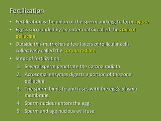
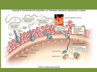
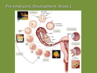
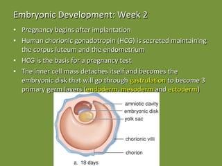
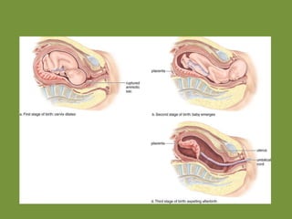
Human Development Chapter 17
Fertilization Fertilization is the union of the sperm and egg to form  zygote Egg is surrounded by an outer matrix called the  zona of pellucida Outside this matrix has a few layers of follicular cells collectively called the  corona radiata Steps of fertilization: Several sperm penetrate the corona radiata  Acrosomal enzymes digests a portion of the zona pellucida The sperm binds to and fuses with the egg’s plasma membrane Sperm nucleus enters the egg Sperm and egg nucleus will fuse
 
What prevents more than one sperm from entering the egg? The egg’s plasma membrane changes to prevent other sperm from binding Vesicles within the egg release enzymes that cause the zona pellucida to become impenetrable and sperm cannot bind
Embryonic Development Cleavage  – cells undergo division (2, 4, 8 etc) without the embryo increasing in size Growth  – cells undergo division as well as increase in size Morphogenesis  – the embryo begins to take shape as cells migrate Differentiation  – when cells take on specific structure and function (the nervous system is the first visible system)
Extra-embryonic Membranes Chorion  – fetal half of the placenta, the organ that provides the embryo with nourishment and gets rid of wastes Allantois  – gives rise to the bladder and the blood vessels of the umbilical cord that carry blood to and from the fetus Yolk sac  – contains many blood vessels and where blood cells first form (little yolk in humans) Amnion  – contains amniotic fluid that cushions and protects the embryo
Stages of Development Pre-embryonic development  - 1st week of development after fertilization Embryonic development  – 2nd week after fertilization until the end of the 2nd month Fetal development  – the 3rd through the 9th months of development Development after birth  – stages of life including infancy, childhood, adolescence and adulthood
Pre-embryonic Development: Week 1 Cleavage  – cell division that increases the number of cells Morula  – compact ball of embryonic cells Early blastocyst  – inner cell mass that becomes the embryo covered by a layer of cells that becomes the chorion Implantation  – embryo embeds into the uterus around day 6
Pre-embryonic Development: Week 1
Embryonic Development: Week 2 Pregnancy begins after implantation Human chorionic gonadotropin (HCG) is secreted maintaining the corpus luteum and the endometrium  HCG is the basis for a pregnancy test The inner cell mass detaches itself and becomes the embryonic disk that will go through  gastrulation  to become 3 primary germ layers ( endoderm ,  mesoderm  and  ectoderm )


Embryonic Development: Week 3 Nervous system begins to develop The posterior neural tube will become the spinal cord and brain Development of the heart begins
Embryonic Development: Weeks 4 and 5 4th week: Embryo is slightly larger than the height of the print in your book Chorionic villi form Umbilical cord forms Limb buds form (later develop into legs and arms) Hands and feet are apparent 5th week: Head enlarges Eyes, ears and nose become prominent
Week 5
Embryonic Development: Weeks 6 to 8 Embryo begins to look like a human being Reflex actions occur All organ systems have been established 38mm by the end of this period and weight ~ the same as an aspirin tablet
Fetal Development During fetal development organs continue to develop and grow Fetal development begins in the eighth week of pregnancy Birth defects include structural and functional defects that appear in the newborn About 31 % of all successful fertilizations end in a miscarriage About 10-12 % of all newborns have birth defects  Birth defects can arise from chemical, biological, and physical agents known as teratogens The effect of teratogenic agents is related to the time of exposure, the nature of the agent, and the dose
Fetal Development: 3 to 4 Months Hair develops The head slows in growth so that the body size can catch up Cartilage begins to be replaced by bone Able to distinguish female from male (month 3) Can hear heartbeat with the stethoscope (month 4) By the end of this period the fetus is ~6 inches and ~6 ounces
Fetal Development: 5 to 7 Months Fetal movement can be felt by the mother Fetus is in fetal position Eyelids are fully open Fetus size has increased to ~12 inches and ~3 pounds
Fetal Development: 8 to 9 Months Weight gain is about a pound per week Fetus rotates so that the head is pointed towards the cervix At the end of fetal development the fetus weighs ~7.5 pounds and ~20.5 inches
Fetal Circulation Note: Fetal and maternal blood do not mix because exchange of materials between the two occurs at the chorionic villi.
Development of Sex Organs Sex of an individual is determined at conception (XX is female and XY is male) If the  SRY  (the sex determining region on the Y chromosome) gene is present at ~ week 6 then the embryo develops into a male Anti-Mullerian hormone  secreted by the testes prevents the development of female sex organs At 14 weeks primitive testes and ovaries with gametes are already developing The development of the external organs is dependent on the presence or absence of  dihydrotestosterone (DHT)  produced by the testes
 
Abnormal Sex Organ Development XY female syndrome  - an individual develops into a male because piece of the Y chromosome containing the SRY gene is missing XX male syndrome  – an individual develops into a male because the same small piece of the Y containing the SRY gene is present on an X chromosome
Ambiguous Sex Determination Results from the absence of testosterone, Anti-Mullerian hormone and/or DHT Androgen insensitivity syndrome : all hormones are made but testosterone receptors on cells are ineffective thus the individual has testes that do not descend and outwardly appear to be female Male pseudo-hermaphroditism : an individual appears female until puberty when anti-Mullerian hormone is produced but the testes never produce testosterone or DTH
Birth True labor is characterized by uterine contraction that occur every 15-20 minutes and last for at least 40 seconds 3 stages: 1st:  effacement occurs in which the cervical canal slowly disappears and the babies head acts as a wedge to cause cervical dilation 2nd:  uterine contractions every 1-2 minutes lasting for ~1 minute each an incision is made to the opening to help the baby as its head reaches the exterior; once the baby is born the umbilical cord is cut and tied 3rd:  The afterbirth is delivered usually about 15 minutes after the birth of the baby
Birth Childbirth occurs in three stages  The dilation stage - cervix dilates The expulsion stage - delivery of the child Uterine muscle contractions are stimulated by a change in hormonal levels.  It’s believed that fetal oxytocin stimulates the release of prostaglandins by the placenta Oxytocin and prostaglandins stimulate contractions in the sensitized uterine musculature The placental stage - placenta is expelled
 

Development

  • 1.
  • 2.
    Fertilization Fertilization isthe union of the sperm and egg to form zygote Egg is surrounded by an outer matrix called the zona of pellucida Outside this matrix has a few layers of follicular cells collectively called the corona radiata Steps of fertilization: Several sperm penetrate the corona radiata Acrosomal enzymes digests a portion of the zona pellucida The sperm binds to and fuses with the egg’s plasma membrane Sperm nucleus enters the egg Sperm and egg nucleus will fuse
  • 3.
  • 4.
    What prevents morethan one sperm from entering the egg? The egg’s plasma membrane changes to prevent other sperm from binding Vesicles within the egg release enzymes that cause the zona pellucida to become impenetrable and sperm cannot bind
  • 5.
    Embryonic Development Cleavage – cells undergo division (2, 4, 8 etc) without the embryo increasing in size Growth – cells undergo division as well as increase in size Morphogenesis – the embryo begins to take shape as cells migrate Differentiation – when cells take on specific structure and function (the nervous system is the first visible system)
  • 6.
    Extra-embryonic Membranes Chorion – fetal half of the placenta, the organ that provides the embryo with nourishment and gets rid of wastes Allantois – gives rise to the bladder and the blood vessels of the umbilical cord that carry blood to and from the fetus Yolk sac – contains many blood vessels and where blood cells first form (little yolk in humans) Amnion – contains amniotic fluid that cushions and protects the embryo
  • 7.
    Stages of DevelopmentPre-embryonic development - 1st week of development after fertilization Embryonic development – 2nd week after fertilization until the end of the 2nd month Fetal development – the 3rd through the 9th months of development Development after birth – stages of life including infancy, childhood, adolescence and adulthood
  • 8.
    Pre-embryonic Development: Week1 Cleavage – cell division that increases the number of cells Morula – compact ball of embryonic cells Early blastocyst – inner cell mass that becomes the embryo covered by a layer of cells that becomes the chorion Implantation – embryo embeds into the uterus around day 6
  • 9.
  • 10.
    Embryonic Development: Week2 Pregnancy begins after implantation Human chorionic gonadotropin (HCG) is secreted maintaining the corpus luteum and the endometrium HCG is the basis for a pregnancy test The inner cell mass detaches itself and becomes the embryonic disk that will go through gastrulation to become 3 primary germ layers ( endoderm , mesoderm and ectoderm )
  • 11.
  • 12.
  • 13.
  • 14.
  • 15.
    Embryonic Development: Week3 Nervous system begins to develop The posterior neural tube will become the spinal cord and brain Development of the heart begins
  • 16.
    Embryonic Development: Weeks4 and 5 4th week: Embryo is slightly larger than the height of the print in your book Chorionic villi form Umbilical cord forms Limb buds form (later develop into legs and arms) Hands and feet are apparent 5th week: Head enlarges Eyes, ears and nose become prominent
  • 17.
  • 18.
    Embryonic Development: Weeks6 to 8 Embryo begins to look like a human being Reflex actions occur All organ systems have been established 38mm by the end of this period and weight ~ the same as an aspirin tablet
  • 19.
    Fetal Development Duringfetal development organs continue to develop and grow Fetal development begins in the eighth week of pregnancy Birth defects include structural and functional defects that appear in the newborn About 31 % of all successful fertilizations end in a miscarriage About 10-12 % of all newborns have birth defects Birth defects can arise from chemical, biological, and physical agents known as teratogens The effect of teratogenic agents is related to the time of exposure, the nature of the agent, and the dose
  • 20.
    Fetal Development: 3to 4 Months Hair develops The head slows in growth so that the body size can catch up Cartilage begins to be replaced by bone Able to distinguish female from male (month 3) Can hear heartbeat with the stethoscope (month 4) By the end of this period the fetus is ~6 inches and ~6 ounces
  • 21.
    Fetal Development: 5to 7 Months Fetal movement can be felt by the mother Fetus is in fetal position Eyelids are fully open Fetus size has increased to ~12 inches and ~3 pounds
  • 22.
    Fetal Development: 8to 9 Months Weight gain is about a pound per week Fetus rotates so that the head is pointed towards the cervix At the end of fetal development the fetus weighs ~7.5 pounds and ~20.5 inches
  • 23.
    Fetal Circulation Note:Fetal and maternal blood do not mix because exchange of materials between the two occurs at the chorionic villi.
  • 24.
    Development of SexOrgans Sex of an individual is determined at conception (XX is female and XY is male) If the SRY (the sex determining region on the Y chromosome) gene is present at ~ week 6 then the embryo develops into a male Anti-Mullerian hormone secreted by the testes prevents the development of female sex organs At 14 weeks primitive testes and ovaries with gametes are already developing The development of the external organs is dependent on the presence or absence of dihydrotestosterone (DHT) produced by the testes
  • 25.
  • 26.
    Abnormal Sex OrganDevelopment XY female syndrome - an individual develops into a male because piece of the Y chromosome containing the SRY gene is missing XX male syndrome – an individual develops into a male because the same small piece of the Y containing the SRY gene is present on an X chromosome
  • 27.
    Ambiguous Sex DeterminationResults from the absence of testosterone, Anti-Mullerian hormone and/or DHT Androgen insensitivity syndrome : all hormones are made but testosterone receptors on cells are ineffective thus the individual has testes that do not descend and outwardly appear to be female Male pseudo-hermaphroditism : an individual appears female until puberty when anti-Mullerian hormone is produced but the testes never produce testosterone or DTH
  • 28.
    Birth True laboris characterized by uterine contraction that occur every 15-20 minutes and last for at least 40 seconds 3 stages: 1st: effacement occurs in which the cervical canal slowly disappears and the babies head acts as a wedge to cause cervical dilation 2nd: uterine contractions every 1-2 minutes lasting for ~1 minute each an incision is made to the opening to help the baby as its head reaches the exterior; once the baby is born the umbilical cord is cut and tied 3rd: The afterbirth is delivered usually about 15 minutes after the birth of the baby
  • 29.
    Birth Childbirth occursin three stages The dilation stage - cervix dilates The expulsion stage - delivery of the child Uterine muscle contractions are stimulated by a change in hormonal levels. It’s believed that fetal oxytocin stimulates the release of prostaglandins by the placenta Oxytocin and prostaglandins stimulate contractions in the sensitized uterine musculature The placental stage - placenta is expelled
  • 30.