
Recommended
Recommended
More Related Content
What's hot
What's hot (20)
Similar to THE AMAZING WORLD OF INVERTEBRATES
Similar to THE AMAZING WORLD OF INVERTEBRATES (20)
Recently uploaded
Recently uploaded (20)
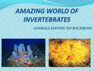
THE AMAZING WORLD OF INVERTEBRATES
- 1. ANIMALS HAVING NO BACKBONE
- 2. What is an Invertebrate? Invertebrates are animals that do not have backbones. 97% of the animal kingdom is made up of invertebrates. Some can be found in ponds, oceans, and other water environments. .
- 3. Animal Characteristics Many-celled organisms sharing similar features and that are made of different kinds of cells. Animal cells have a nucleus and organelles surrounded by a membrane – EUKARYOTIC. Cannot make their own food – HETEROTROPHIC – digest their food. Can move from place to place to find food, shelter, and mates, and to escape from predators.
- 4. Symmetry Symmetry: arrangement of the individual parts of an object Radial: body parts arranged in a circle around a central point Bilateral: parts are mirror images of each other Asymmetrical: bodies cannot be divided into matching halves
- 5. Symmetry
- 6. Major Phyla INVERTEBRATE Porifera Helminthes CnedariaProtozoa Echinoderm Arthropod Molluscs Annelids
- 8. 1.They are unicellular with some colonial and multicellular stages. 2. Most are microscopic. 3. All symmetries are present within members of the group. 4. No germ layers are present. 5. No organs or tissues are formed, but specialized organelles serve many of these functions.
- 9. 6.They include free-living, mutualistic, commensal and parasitic forms. 7.They move by pseudopodia, flagella, cilia and they can direct cell movements. 8.Most are naked, but some have a simple endoskeleton or exoskeleton.
- 10. PARAMECIUM Paramecium is a small unicellular organism. It is plentiful in freshwater ponds.
- 12. Paramecium Movement The outer surface of the cell is covered with many hundreds of tiny hair-like structures called cilia. These act like microscopic oars to push through the water, enabling the organism to swim. If Paramecium comes across an obstacle, it stops, reverses the beating of the cilia, swims backwards, turns through an angle and moves forward again on a slightly different course. It moves so quickly that we have to add a thickening agent or quieting solution to the slide to slow it down to study it.
- 13. Paramecium Feeding Paramecium has a permanent feeding mechanism, consisting of an oral groove and a funnel-shaped gullet into which food is drawn by the combined action of cilia which cover the body and other cilia lining the oral groove and the gullet. As it moves through the water it rotates on its axis and small particles of debris and food are collected and swept into the gullet. They feed on small organisms such as bacteria, yeasts, algae and even other smaller protozoa.
- 14. Paramecium Reproduction In favourable conditions the cell divides in two by a process called binary fission (asexual reproduction). This forms two new cells, each of which rapidly grows any new structures required and increases in size. This whole process may take place two or three times a day if conditions were right.
- 15. Paramecium Reproduction This is a more complicated method called conjugation (sexual reproduction). It involves two cells coming together to exchange nuclear material. The two cells then separate and continue to reproduce by simple division. It is similar in some ways to sexual reproduction in more complex animals.
- 16. Paramecium Excretion Food waste left in a food vacuole is excreted through the anal pore (the vacuole and pore fuse. Other wastes left over from cellular activity (metabolic waste) simply diffuse through the pellicle. Excess water and some metabolic wastes are excreted through the contractile vacuole.
- 18. Porifera Characteristics They live in water. (Most are found in the ocean.) They look like plants but they are animals. Sponges stay fixed in one place - SESSILE. Their bodies are full of pores and their skeleton is made of spiky fibers (spicules) or rubbery spongin Sponges are divided into classes according to the type of spicule they have – 5,000 species identified! Water flows through the pores of their body, aided by flagella, which enables them to catch food – FILTER FEEDERS
- 19. Porifera Characteristics Sponges can reproduce asexually through budding ~ GEMMULES; a new sponge grows from pieces of an old sponge Most sponges that reproduce sexually are hermaphrodites, meaning they have both eggs and sperm Sperm is released into water Sperm floats until they are drawn into another sponge where they fertilize an egg Larva develops in sponge, leaves sponge, and settles to the bottom where it grows into an adult
- 20. 12-20 Sessile Sponges - do not move
- 21. General Morphology • The internal cavity is called the atrium or spongocoel • Water is drawn into it through a series of incurrent pores or dermal ostia present in the body wall into a central cavity and then flows out of the sponge through a large opening at the top called the osculum
- 22. Body layers 1. The pinacoderm - an outer layer of flattened cells called pinacocytes 2. An inner lining containing flagellated cells (choanocytes) - draw water in through the pores and move out through the osculum; also trap food particles • The water current is also used for gas exchange, removal of wastes, and release of the gametes 3. Between the pinacodern and the choanocytes is a gelatinous material called mesenchyme; Archaeocytes are amoeboid cells and they can also undergo differentiation to form other cells(Totipotency)
- 23. The Skeleton In the mesenchyme is the skeleton composed of tiny pointed structures made of silica or calcium carbonate called spicules. These structures act as an internal scaffolding, but also function in protection Among some sponges the skeleton consist of spongin fibers made of collagenous material; found in many of the commercial sponges
- 24. 12-24
- 25. Types of Canal Systems ASCON TYPE • Simple vaselike structure • This stucture puts limitations on size; (increase in volume without a corresponding increase in the surface area of the choanocytes)
- 26. SYCON TYPE • The flagellated choanocyte layer has undergone folding forming finger like projections • There is a single osculum but the body wall is more complex, with water being received through incurrent canals, which pass it along to radial canals through to the spongocoel • Results in an increase in the surface area which allowed sponges to increase in the size
- 27. LEUCON TYPE • No atrium; several small chambers in which choanocytes are located • There is a whole series of incurrent canals leading to the choanocyte chambers; water is discharges through excurrent canals • The leuconoid sponges exhibit a significant increase in surface area and are, therefore, among the largest sponges
- 29. Types of Porifera Cells 12-29
- 30. 12-30 Food Trapping by Sponge Cells
- 31. MODE OF REPRODUCTION Asexual reproduction can occur by bud formation External buds Small individuals that break off after attaining a certain size Internal buds or gemmules Formed by archaeocytes that collect in mesenchyme Coated with tough spongin and spicules Survive harsh environmental conditions 12-31
- 33. Sexual Reproduction Most are monoecious (have both sexes) Sperm and eggs sometimes arise from choanocytes or archaeocytes 12-33
- 35. Cnidaria: Corals, Hydras, and Jellyfish
- 36. Basic Characteristics A. Tissue level 1. Sac – like body with 3 layers a. epidermis b. mesoglea c. gastrodermis 2. Gastrovascular cavity – hollow internal body cavity B. Nervous system 1. Nerve net – nerves evenly spaced 2. Statocysts – structures for balance (hollow ball of cells with a grain of sand) 3. Ocelli – light sensitive structure
- 37. Taxonomic Characteristics Diploblastic Epiderm & hypoderm Tissues
- 38. C. Tentacles 1. Capture food 2. Cnidoblast/cnidocyte – cell that contains the stinging organelle 3. Nematocyst – stinging organelle a. capsule with coiled “harpoon” containing toxins b. Operculum – flap that holds the coil inside c. Stimulated by touch and chemicals
- 40. D. Habitat 1. Mostly shallow, marine 2. Pelagic – open water 3. Benthic – bottom dweller 4. Symbiosis a. on other animal’s shells b. with algae that provide energy from photosynthesis
- 41. E. Reproduction 1. Polymorphism “many shapes” 2. Polyp – sessile, tentacles up 3. Medusa – floating, tentacles down 4. Many alternate forms 5. Asexual reproduction a. budding b. regeneration 6. Sexual reproduction a. mostly dioecious
- 43. Obelia – polymorphic life cycle
- 45. Class Scyphozoa – “cup animal” A. genus Aurelia – common jellyfish B. Thick mesoglea C. Tentacles can be up to 70 m D. Dioecious, polymorphic life cycle
- 46. Dimorphic Life Cycle and Reproductive Modes Asexual Budding Medusa buds Polyp buds Sexual Gonadal tissue Gametes Fertilization, embryogenesis Planula larvae
- 48. Jelly Fish: Sting scars
- 50. THE FLAT WORMS
- 51. General Characteristics • They exhibit bilateral symmetry: anterior and posterior ends are different; so are the dorsal (top) and ventral (bottom) surfaces •The platyhelminths also exhibit some degree of cephalization Commonly referred to as the 'flatworms' because their bodies are dorsoventrally flattened. •They are acoelomates •This phylum (and all remaining phyla) possess 3 germ layers (=triploblastic) •The mesoderm (third germ layer) gives rise to muscles, various organ systems, and the parenchyma, a form of solid tissue containing cells and fibers
- 52. Outer Body Covering • The body of some platyhelminthes (e.g., turbellarians) is covered by a ciliated epidermis • Epidermal cells contain rod-shaped structures called rhabdites that when released into the surrounding water, expand and form a protective mucous coat around the animal • The outer body covering of other platyhelminthes (e.g., parasitic forms) is a non-ciliated tegument • The tegument is referred to as a syncytial epithelium
- 53. Organ Systems of the Platyhelminthes Digestive System • Some of the flatworms possess a digestive system, with a mouth, pharynx, and a branching intestine from which the nutrients are absorbed • The intestine, with only one opening, is a blind system
- 54. Excretory System (osmoregulation) • A network of water collecting tubules adjacent to flame cells or a protonephridia • When cilia beat they move water into the tubules and out the body through pores called nephridiopores
- 55. Taxonomic Summary Phylum Platyhelminthes (Flatworms) Class Turbellaria Class Cestoda Class Trematoda Class Monogeneans Phylum Platyhelminthes 55
- 56. Cestoda The Tapeworms Endoparasites Body consists of proglottids and scolex Proglottids snapshots of development Scolex has structures for attachment (Hooks, suckers and rostellum) No digestive system
- 58. Class Cestoda General Morphology • Nonciliated tegument composed of glycoprotein • The anterior region is called a scolex; often armed with suckers and hooks • Extending from the neck is a series of proglottids; contain the sex organs and eggs; no digestive system • Mature eggs released through an opening in the proglottid or leave the host when the proglottids are separated from the main body of the worm.
- 59. Taenia scolex
- 61. TaeniaEntire – can get to 30+ feet long
- 63. Trematoda :The Flukes Endoparasites Cuticle covering body Oral sucker surrounds mouth Ventral sucker used for attachment Complex life cycles
- 64. Fasciola hepatica Fasciola hepatica, also known as the common liver fluke or sheep liver fluke. Is a parasitic flatworm of the class Trematoda, phylum Platyhelminthes that infects liver of various mammals, including humans. The disease caused by the fluke is called fascioliasis (also known as fasciolosis). F. hepatica is world-wide distributed and causes great economic losses in sheep and cattle. 64
- 66. Trematoda Lifecycle The lifecycle is complex with up to 4 different hosts and several larval types Phylum Platyhelminthes 66
- 67. 67
- 68. Sheep liver infected with Fasciola hepatica
- 69. HIGHLIGHTS OF PRESENTATION • Helpful in understanding the trend of evolution. • For better understanding the living conditions and behaviour of animals,specially Jellyfish,Taenia and Fasciola. • Their economic status.
- 70. Dr Sameer Mishra
Editor's Notes
- Fig. 13.9
- Fig. 13.Fig. 13
- Fig. 13.18