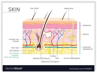
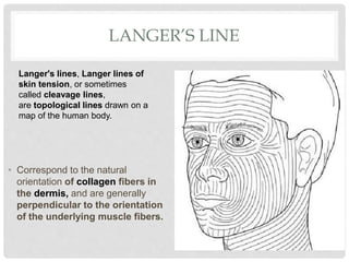
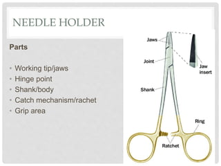
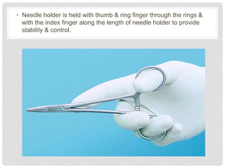
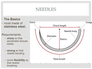
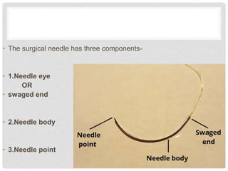
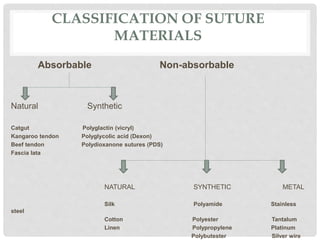
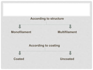
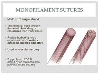
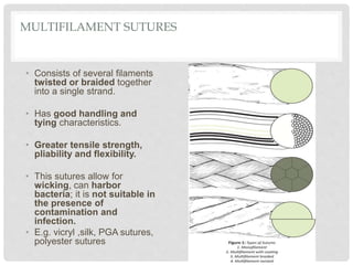
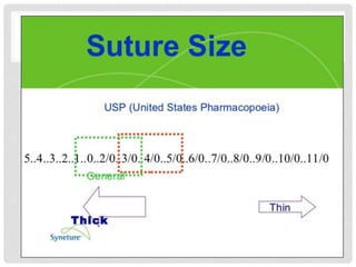
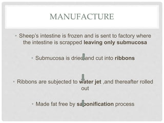
D R . A M R U TA S A R D E S H M U K H ( 1 S T Y E A R M D S )
SUTURE MATERIALS
AND TECHNIQUES
CONTENTS
• Anatomy of skin
• Definition
• Goals of suturing
• Ideal requirement of sutures
• Armamentarium of suturing
• Suture material
• Principle of suturing
• Surgical knots
• Suturing techniques
• Possible complications
• Alternatives to sutures
LANGER’S LINE
• Correspond to the natural
orientation of collagen fibers in
the dermis, and are generally
perpendicular to the orientation
of the underlying muscle fibers.
Langer's lines, Langer lines of
skin tension, or sometimes
called cleavage lines,
are topological lines drawn on a
map of the human body.
• Incisions made parallel to Langer's lines may heal better and
produce less scarring than those that cut across.
• Keloids are more common when incision is given across
Langer's lines
• Wound closure methods impact the mechanics of the healing
wound and the resulting performance.
• Perpandicular injuries to the langer’s line will gape because
they are pulled by tension.
• This can have important implication of surgeries, particularly
in cosmetic procedures where the goal is to minimize
scarring.
DEFINITIONS
• Suture material is an artificial fibre used to keep
wound together until they hold sufficiently well by
themselves by natural fibre (collagen) which is
synthesized and woven into a stronger scar.
• Suture is a stitches/ series of stitches made to secure
apposition of the edges of a surgical/traumatic wound
(Wilkins)
• Any strand of material utilised to ligate blood vessels or
approximate tissues (Silverstein L. H. 1999)
• Time Frame for Closing the Wound
• American College of Emergency Physicians policy is no
more than 8 to 12 hours from the time of injury.
• Wounds that are at low risk for infection, safely
approximated upto 12 hours after the time of injury.
• Likewise, wounds that are at moderate risk or infection
within a 6 to 10 hour period.
Contraindications to Suturing
• Redness
• Edema of the wound margins
• Infection
• Fever
• Puncture wounds
• Animal bites
• Tendon, nerve, or vessel involvement
• Wound more than 12 hours old (body) and 24 hrs (face)
REQUISITES OF AN IDEAL SUTURE
• TENSILE STRENGTH
• Adequate material strength will prevent suture breakdown & use of
proper knots for the material used will prevent untying or knot
slippage.
• TISSUE BIOCOMPATIBILITY
• Sutures made from organic material will evoke a higher tissue
response than synthetic sutures
• Tissue reaction is directly proportional amount & size of suture
material.
• Low capillarity
• Multifilament type soak up tissue fluid by capillary action providing a
rich medium for microbes increasing chances of inflammation an
infection.
• Good handling and knotting properties
• Ease of tying &a thread type that permits minimal knot slippage also
influence thread selecion.
• Sterilization without deterioration of properties
Most sutures available in packages are sterilized by dry heat and
ethylene oxide gas.
• Non allergic, non electrolytic and non carcinogenic.
• Its use should be possible in any operation.
• Low cost
• It should not fray, should slide through tissues readily & knot
should not slip after tying.
• Should be readily visualized , should not shrink and
should not be extruded from the wound.
• On break down, it should not release toxic agents.
GOALS OF SUTURING
Suturing is performed to –
• Provide adequate tension
• Maintain hemostasis
• Provide support for tissue margins
• Reduce post-op pain
• Prevent bone exposure
• Permit proper flap position
• Produce aesthetically pleasing scar by approximation skin edges.
ARMAMENTARIUM OF SUTURING
• Needle holder
• A suture material
• Tissue forcep
• Suture needle
• Suture cutting scissor
NEEDLE HOLDER
Parts
• Working tip/jaws
• Hinge point
• Shank/body
• Catch mechanism/rachet
• Grip area
• Needle holder is held with thumb & ring finger through the rings &
with the index finger along the length of needle holder to provide
stability & control.
NEEDLES
• The Basics
• most made of
stainless steel.
Requirements
• sharp so that
penetrates tissues
easily.
• strong so that
resists bending.
• some flexibility so
that resists
breaking.
• The surgical needle has three components-
• 1.Needle eye
OR
• swaged end
• 2.Needle body
• 3.Needle point
CLASSIFICATION OF NEEDLES
• According to shape
1. Straight
2. Curved
• According to eye
1. Eyed needle/Traumatic needle
2.Eyeless needle/Atraumatic needle
• According to edge
1.Round body
2.Cutting body- Conventional
Reverse cutting
• According to its tip
1. Triangular
2.Round
3.Blunt
EYE/SWAGED NEEDLE
• Suture material attached to
needle in 2 basic ways:
• Eyed needles:
• suture threaded through eye during
surgery from inside curve toward
outside
• reusable
• economical
• less sharp if reused
• less efficient (time spent threading
needle)
• more traumatic (bigger hole because
double strand of suture pulled through
tissue)
• avoid threading eye twice to prevent
suture pulling out as creates more
tissue trauma
• Swaged needles:
• suture permanently
attached to the needle
• consistently sharp with
known shape and
durability (not damaged
by previous use)
• convenient as threading
not required
• more efficient as suture
doesn’t pull out while
suturing
NEEDLE BODY
• Straight needles
• used near surface (primarily skin)
• inserted through tissues with fingers not needle holders
• usually combined with hand ties
• Curved needles
• manipulated with needle holders
• rotating wrist in arc similar to that of needle is easiest and most efficient
• curvature is described by amount of the circumference of a circle (e.g. 1/4,
3/8, 1/2, 5/8, etc.)
• most common curves are 3/8 and 1/2
• wider curves useful when suturing thick tissues or in deep or poorly
accessible locations (easier to retrieve tip).
• Size of the needle
• Needles come in various lengths and diameters
• considerations when selecting needle size:
• tissue to be sutured (larger for thick dense tissue, smaller for delicate
tissue)
• size and location of wound
• size of suture material needed to support incision during healing (bigger
needle for larger suture)
• selected needle should be
• smallest that will do the job (minimize tissue trauma)
• large enough not to bend during insertion or extraction
• long enough to reach both sides of incision in a single pass
TIP
• tip’s design affects its sharpness and how
easily it penetrates tissue
• Taper or non-cutting needles
• have a round body with a sharp pointed tip
• generally used for viscera, muscle and light
fascia
• penetrates tissue, without cutting, creating a
round hole
• should NOT be used for dense tissue like
skin because the extra force needed to
penetrate the tissue causes extra trauma or
bends the needle
• taper cut tip a newer design that combines
the round body with a cutting tip so can be
used for both delicate and dense tissue
• Traditional cutting needle
• triangular shaped point with 2-3 cutting edges to
facilitate penetration of dense tissue
• cutting edge is on the inside of the curve (concave
surface)
• cut edge is where the tension is on the tied suture so this
type of needle predisposes the suture to cutting through the
tissue
• use has generally been replaced by the reverse cutting
needle
• Reverse cutting needle
• cutting edge on outer surface of the curve (convex
surface)
• more efficiently uses the cutting surface when curve
wrist during insertion
• more resistant to suture cutting through tissue because
the cut edge is opposite to the direction of tension on the
tied suture
• preferred by most surgeons
SUTURE MATERIALS
CLASSIFICATION OF SUTURE
MATERIALS
Absorbable Non-absorbable
Natural Synthetic
Catgut Polyglactin (vicryl)
Kangaroo tendon Polyglycolic acid (Dexon)
Beef tendon Polydioxanone sutures (PDS)
Fascia lata
NATURAL SYNTHETIC METAL
Silk Polyamide Stainless
steel
Cotton Polyester Tantalum
Linen Polypropylene Platinum
Polybutester Silver wire
According to structure
Monofilament Multifilament
According to coating
Coated Uncoated
MONOFILAMENT SUTURES
• Made up of single strand.
• This material goes through
tissue with less drag or
resistance than multifilament.
• Resists harboring micro-
organisms hence resists
infection and ties smoothly.
• Used in vascular surgery
• E.g prolene , PDS II,
catgut,nylon,stainless steel,
polydioxanone sutures
MULTIFILAMENT SUTURES
• Consists of several filaments
twisted or braided together
into a single strand.
• Has good handling and
tying characteristics.
• Greater tensile strength,
pliability and flexibility.
• This sutures allow for
wicking, can harbor
bacteria; it is not suitable in
the presence of
contamination and
infection.
• E.g. vicryl ,silk, PGA sutures,
polyester sutures
BARBED SUTURE.
• Lesser known suture type.
• The suture has many small
barbs cut into its monofilament
core along its length, and is
available in several absorbable
and nonabsorbable polymers,
including polydioxanone, PGA-
PCL, and polypropylene.
• The barbs ensure that the
suture stays in place and
approximates the tissue without
the need to tie knots, ensuring
even distribution wound tension
and decreased closure time.
It is used extensively in plastic surgery and also has
applications in other specialties as well such as
orthopedics and general surgery.
If the suture breaks during suturing, the surgeon leaves it
in place and begins again rather than removing it to
prevent tissue damage.
COATED SUTURES
• Usually coated with a biologically inert non-resorbable
compound.
• Acts as lubricant.
• Reduces surface friction of the braid and helps the
thread in passing more easily through the tissue.
SIZE OF THE SUTURE
• The larger the suture diameter, the relatively stronger it is
• Measured in metric units (tenths of a millimeter) or by a numeric
scale standardized by USP regulations.
• USP scale runs from 7 (largest) to 11-0 (smallest)
• zeros are written as 2-0 for 00 and 3-0 for 000, etc. for
convenience and clarity
• From 0 to 11-0, each extra zero corresponds to a
unit decrease in diameter (e.g. 0, 00, 000, etc. until the smallest
size of eleven 0s is reached)
• ie.more zeros means smaller (and weaker)
PRINCIPLES OF SUTURE MATERIAL
SELECTION
Rate of healing of tissues: When a wound has reached
maximal strength, sutures are no longer required. Hence,
• Surgeon should select a suture that will lose its tensile strength
at about the same rate that the tissue gain strength.
• Tissues that heal slowly such as skin, fascia and tendons
should be closed with non absorbable sutures.
• Tissues that heal rapidly such as muscles , periosteum may be
closed with absorbable sutures
Tissue contamination:
• Foreign bodies in potentially contaminated tissues may
convert contamination to infection.
• Hence, monofilament absorbable or non-absorbable
sutures are used in potentially contaminated wounds
• Cosmetic results:
• Use the smallest, inert monofilament suture material
CATGUT
• Catgut is prepared from the submucosa of the sheep’s
intestine.
• It has rich content of elastic tissue which accounts for
high tensile strength of the catgut.
MANUFACTURE
• Sheep’s intestine is frozen and is sent to factory where
the intestine is scrapped leaving only submucosa
• Submucosa is dried and cut into ribbons
• Ribbons are subjected to water jet ,and thereafter rolled
out
• Made fat free by saponification process
• For even and predictable absorption, these ribbons are
treated with chromic acid
• Then treated in electronic spinning process to
produce extremely smooth and highly uniform suture
Sterilization
• Should not be sterilized or boiled as it destroys tensile
strength.
• Catgut is sterilized during its preparation and kept in
preservative solution ( ethicon fluid containing 2.5%v/v
formaldehyde plus 87.5% v/v denatured alcohol).
• Packing
• It was previously
available in glass spool
with Ethicon fluid as a
preservative.
• Presently available as
presterilized, in
aluminium coated
sterile foil which
overwraps packs with
Ethicon fluid as a
preservative
• Colour
• Plain catgut- yellow
• Chromic catgut- tan
• Absorption
• Absorbed by proteolytic digestive enzymes released
from inflammatory cells collected around the catgut.
• This is why in the presence of infection catgut is rapidly
absorbed.
• Duration of Catgut in body tissues
• Plain catgut retains its tensile strength approximately for
10 days and chromic catgut for 20 days
USES
For ligation of small blood vessels near the surface of the
skin.
To suture subcutaneous tissue.
SILK
• Natural, non-absorbable, polyfilament suture.
• Obtained from cocoon of silkworm.
• Colour - black
• Sizes-
No. 2 is the thickest.
The thickness decreases as the number of the size
increases, the size No. 80 being the thinnest.
• Manufacture
Natural silk filaments are obtained from silkworm cocoon.
Comprises of protein fibres approximately 70% and extraneous
material or gum 30%.
Process of degumming- allows the silk to retain its natural body and
elasticity.
Appropriate number of filaments for each size is tightly braided.
The suture is chromicized for non-capillarity.
Availability-
In sterile foil overwrap pack
as eyeless needled sutures.
As Sutupack- precut lengths
of sterile sutures, in a pack of
two and six pieces of sutures
material, without a needle.
As nonsterile on reels.
• Types
According to preparation:
a. Perma-Hand surgical silk suture.
b.Virgin silk suture material which is prepared from the
glands of silkworm before their pupae stage.
According to fibre pattern:
a.Braided
b.Twisted
c.Floss
• Sterilization
Autoclaving
• Advantages-
It does not soak up fluids; it never becomes limp or
brittle.
Ties down smoothly and securely, and its natural elasticity
it an extensibility that signals when optimum knot
placement has been achieved.
• Disadvantages
Stitch granuloma.
Infection rate is high as compared to synthetic materials.
• Uses-
To ligate blood vessels.
To repair hernia.
To suture nerves.
To suture grafts in vascular surgery.
To suture tensons.
Skin suturing.
For fixing the skin grafts
NYLON
• Synthetic, non-absorbable suture.
• Monofliament/multifilament.
• Thickness varies from 1 N to 8 N.
• Advantages-
Less irritant.
High tensile strength.
Cheaper.
• Disadvantages-
Knot is slippery, so 5-7 knots should be applied.
Infection due to crevices in braided nylon.
Too smooth and stiff knots are likely to slip.
• Uses-
To suture skin. (monofilament)
To repair hernia. (braided)
LINEN
• Natural, non-absorbable,
polyfilament suture material of
vegetable origin, made from
jute fibres.
• Manufacture
It is spun from long staple fax
fibres, especially selected for
surgical use, and then twisted
into a tight and uniform strand
without any slubs, fuzziness and
waviness.
• Colour- Natural linen colour.
• Sizes
the thickest is No. 20 and the thinnest is No. 80.
• Advantages
Easily handled, knots tie down smoothly, and tie securely.
• Uses
It is used almost exclusively in gastrointestinal surgeries, in
transfixation of hernial sacs, and for skin closure.
VICRYL (POLYGLACTIN)
• It is absorbable, synthetic suture material.
• Polyglactin 910 is a copolymer of glycoline and lactide.
• Polyfilament braided suture manufacured by the process of extrusion.
• Colour- Violet
• Sizes – 7-0 to 1-0
• Length-
• 7-0 : 30 cms
• 6-0 to 3-0 : 45 cms
• 2-0 to 1-0 : 90 cms
• Absorption
Disintegrated by hydrolysis and then the pieces of filaments are
phagocytosed by PMNs and other macrophages.
Due to this ,there is least tissue reaction and absorption is not
affected by the presence of infection.
• Knot tying
Requires a specific knoting
technique.
Place the first knot in precise
position for the final knot, using
double loop tie.
Second throw square using
horizontal tension.
Third knot, single loop tie.
• Absorption time
• Essentially completed between 60th and 90th day.
• Tensile strength
• Approximately, 55% of original tensile strength of Vicryl remains
after 14 days and 20% after 21 days.
• Advantages
• Minimal tissue reaction.
• No fraying
• Excellent handling characteristics.
• Highly visible in wound due to violet colour.
• Can be used in the presence of infection.
• Unique molecular structures causes polyglactin to retain its strength
during the critical healing period and then to be absorbed rapidly, that
is, suture is absorbed after it has served its function.
POLYPROPYLENE (PROLENE)
• Made up of polymer of propylene (polypropylene).
• Synthetic , monofilament, non-absorbable suture.
• Knot tying-
Same as vicryl.
• Availability-
In presterilized foil overwrap pack as eyeless needled
suture.
• Sterilization-
Available presterilized.
Sterilized in Ethicon fluid when it is to be reused.
Available sizes: 10-0 to 5
Length: 70 cms
• Advantages-
Inert as steel, resists breakdown by infection.
Monofilament, so it does not harbours microorganisms.
High degree of smoothness, it requires much less force to draw through
the tissue.
Sky blue colour- high visibility in tissue.
It is pliable, so it ties securely & can be easily handled.
Least thrombogenic.
It is unwet by blood, unweakened by tissue enzymes & offers
prolonged tensile strength, even in infected areas.
More elastic.
• Uses
Plastic surgery.
Vascular surgery for anastomosis between vessels and
synthetic graft.
Cardiovascular surgery.
Tendon repair.
Hernia repair.
Vasoplasty
STAINLESS STEEL
• It is made up of stainless steel.
• It excites very little tissue
reaction.
• Sterilization
Autoclaving
Available sizes- 25-40 wire gauge.
• Disadvantages -
Cutaneous discomfort.
Knots are not firm and may break.
• Uses-
Interdental wiring in the fracture of the mandible.
As bone sutures in fracture of patella,olecranon process
of ulna.
To close sternotomy incision on sternum.
PRINCIPLES OF SUTURING
1. The needle should be grasped at approximately
1/3rd the distance from the eye and 2/3rd from the
point.
2.The needle should
enter tissues
perpandicular to the
tissue surfaces.
• The needle should be passed through the tissues
along its curve.
• The suture should be passed at an equal depth and
distance from the incision on the both sides.
• The needle should always pass from the movable
tissue to fixed tissue.
• The needle always passes from the deeper tissue to
the superficial tissue.
• The needle always passes through the thinner tissue
to the thicker tissue.
• Tissues must never be closed under tension.
Undermining the tissues must be done prior to
suturing in such cases.
• The sutures should be tied only to approximate the
tissues, not to blanch.
• The knot should never lie on the incision
line.
• Sutures should be passed at a greater depth than the
distance from the incision, so as to evert the wound
margins.
• Sutures on the skin are usually removed in 5 days
and intraoral sutures in 7 days. If there is tension
while suturing, the sutures may be kept for 10 days.
PRINCIPLES OF KNOT TYING
• Ensure that the finished knot is firm enough to eliminate
knot slippage.
• Tie the knot as small as possible and cut the ends as
short as feasible. This prevents excessive tissue reaction
towards absorbable sutures and minimizes foreign body
reaction to non-absorbable sutures.
• Avoid damaging the suture material when handling.
• Avoid excessive tension, which may cause sutures to
break and cut the tissues.
• Do not tie sutures too tightly when approximating
tissue, this may lead to tissue strangulation.
• Sustain traction at one end of the suture once the first
loop is tied to prevent loosening of the throw.
• Seesaw motion over the sutures when forming a knot will
cause it to break down.
• Ensure that the final throw is as horizontal as possible.
• Extra throws of the suture only add bulk to the knot ; they
do not strengthen it.
SURGICAL KNOTS
• It has 3 components-
• 1. Loop - created by
knot.
• 2. Knot - itself which is
composed of a number
of tight throws.
• 3. Ears -which are cut
ends of the sutures.
SECURE/SQUARE/REEF
KNOT
• The first throw is placed in
precise position for the knot,
using a single loop.
• The second throw is tied using
horizontal tension.
• Additional two throws are
desirable.
• Totally there should be 4
throws.
• Best for catgut, silk, cotton &
stainless steel.
SURGEON’S KNOT
• Formed by two
throws of the suture
around the needle
holder on the first tie.
• Next one throw in the
opposite direction in
the second tie.
• Recommended for
tying polyester
suture materials
such as vicryl and
mersiline.
GRANNY’S/SLIP KNOT
• A tie in one direction
followed by a tie in the
same direction & a
third tie in the opposite
direction to the square
knot and hold it
permanently.
• Can be used in silk,
chromic catgut, plain
catgut.
SUTURING TECHNIQUES
• Interrupted sutures
• Deep sutures
• Buried sutures
• Continuous sutures
• Continuous with lock sutures
• Horizontal mattress sutures
• Vertical mattress sutures
• Subcuticular sutures
• Figure of 8 sutures
INTERRUPTED SUTURES
• Do the pass technique, two loops around the needle holder, then
grab the tail do the knot.
• Indication
• Single tooth extraction.
• Third molar extraction flap.
• Biopsies.
• Implants.
Advantages
• Most commonly used technique.
• Preferred in urgent situation.
• Easy to remove.
• Failure of one is inconsequential of the others.
Disadvantages
• Greater risk of crosshatched marks (ie, train tracks) across
the suture line.
• Does not bring all surfaces into contact and less supportive
for healing of the flap.
• Time consuming because of high number of knots and
consuming large number of silk material.
“RULE OF HALVES,” WHERE ONE PLACES THE FIRST SUTURE AT
THE CENTER OF THE WOUND, THEN PROCEEDS TO HALVE THE
WOUND INTO SMALLER SEGMENTS
PREVENTS ‘DOG-EAR FORMATION’
• Dog ear formation
• Areas of reduntant skin and
subcutaneous tissue resulting
from a woud margin being
longer on one side than other.
• Dealt with either by-
• Incremental oblique
placement of sutures to
redistribute the tension across
the wound.
• Fusiform excision of the dog
ears.
• Fusiform excision-
• The length of incision should be 4 times the width of the
defect to produce an accurate adaptation of the skin edges
without dog ear formation.
• Suturing is done in that area.
THE FIRST SUTURE WILL BE PLACED AT EITHER APEX OF THE ELLIPSE. SUTURES
ARE THEN PLACED IN AN ALTERNATING PATTERN APPROACHING THE
CENTRE OF THE DEFECT.
WITH EACH SUBSEQUENT SUTURE PLACEMENT, THE SURGEON WILL BE ABLE
TO APPRECIATE REDUCTION OF TENSION AT THE CENTRE OF THE WOUND
AND CAN THEREFORE MINIMIZE THE AMOUNT OF UNDERMINING THAT IS
NECESSARY FOR APPROXIMATION.
SIMPLE CONTINUOUS SUTURES
• Also called as running sutures/ baseball stitch.
• Useful technique for skin closure when speed is
important.
• Advantages –
• Provides rapid technique of closure.
• After passing through the tissue , suture is not cut but the needle is
passed under the loop of first suture in a continuous fashion.
• Even distribution of tension.
• Provides more tight closure.
• Only 2 knots associated with tags.
• Disadvantages-
• If cut at one point, suture slackens along the whole length of the
wound which will then gape again.
CONTINUOUS LOCKING/BLANKET
SUTURES
• Similar to continuous sutures but
locking provided by withdrawing the
sutures through its own loop.
• Advantages
Will avoid multiple knots.
Distribution of tension equally.
Water tight closure.
Prevents excessive tightening.
• Disadvantages
Prevents adjustment of tension over suture line as tissue
swelling occurs.
HORIZONTAL MATTRESS SUTURES
• Useful for wounds under high tension because it
provides strength and wound eversion.
• May also be used as a stay stitch for temporary
approximation of wound edges.
• Square-shaped suture with the knot lying parallel to
the wound.
• Help to evert wound edges when skin naturally inverts
into the wound.
• Indication –
Large distance between the tissues.
Bone grafts and implants.
Extraction socket.
• Advantages –
Good for hemostasis.
Less prominent scarring.
• Disadvantages –
Leave a gap between flap and it is difficult to remove.
Constricts the blood supply of the edge of the incision.
VERTICAL MATTRESS SUTURE
• Has one deep throw and one superficial throw (directly
above and parallel) to evert the skin edges.
• Collecting the deep tissue is as important as the superficial
edges.
• Used for closing deep wounds.
• useful in wounds under tension and help to evert wound
edges when skin naturally inverts into the wound.
VERTICAL MATTRESS SUTURES
• Advantages
Better adaptation and maximum tissue approximation.
Gets eversion of wound margins slightly.
Where healing is expected to be delayed for any reason, it
is better to give wound added support by this suture.
Runs parallel to the blood supply of edge of the flap,
hence doesn’t interfere with healing.
FIGURE OF 8
• Used for extraction socket closure.
• Adaptation of gingival papilla around the tooth.
BURIED SUTURES
• This suture is extremely important for distributing wound tension to
the dermis rather than the epidermis and also for closing dead
space. It provides longer-term support to the healing wound and
improves the cosmetic result.
SUBCUTICULAR SUTURES
• Used to close deep wounds in layers.
• Knots will be inverted or buried, so that the knot does not
lie between the skin margin and cause inflammation or
infection.
• Continuous short lateral stitches are taken beneath the
epithelial layer of skin.
• The ends of the suture come out at each end of the
incision and are knotted.
• Leaves a cosmetic scar.
• Used in patient prone to keloid formation.
PURSE STRING SUTURE
• Purse string sutures are continuous sutures places around a
lumen and tightened like a drawstring to invert the opening.
SINGLE INTERRUPTED SLING SUTURE
• The single interrupted sling suturing technique is the
technique of choice when the goal of therapy is to
reposition one of the surgical flaps at a particular
occlusal apical level that is independent of the other
gingival tissue height.¹
• The sling suture is widely used for root coverage,
gingiva esthetics, open flap implant surgery and etc.
• The suture could be applied both buccal/ lingual side.
ALGOEWER SUTURE
• In some cases, the surgeon wants
to avoid subcutaneous suturing
due to the high risk of infection.
•
Performing a common skin
suturing technique might present a
risk of wound dehiscence, and
strong sutures will be needed in
order to provide a firm hold.
• At the same time, this suture will
have to close the subcutaneous
space.
•
Keep in mind that this suturing
technique should be avoided
wherever a good cosmetic result
is important.
DONATI SUTURE
• This is a variation of the Allgöwer suture. In this case, the suture crosses
the skin at 4 points for each stitch. Therefore, although providing a firmer
hold, the risk of poor aesthetic results is increased
CORNER SUTURES
• We have to take into account that not always the wound
edges will be straight and parallel to each other.
• In some cases, the edges can be irregular or
twisty. Here we present a common case where the
wound follows a "corner" pattern.
• Corner is sutured in the
right manner in order to
avoid necrosis of the
tissue. A simple stitch
should never be
performed as it carries a
high risk of necrosis.
•
Instead, the suturing
technique to be used
should be an
intracutaneous suture
knotted on the surface at
the outer side of the
wound as shown on the
pictures.
TRACTION SUTURE
• Used to retract tissues or
organs which are not held back
easily with conventional
retractors.
• E.g. tongue, sclera of the eye.
SECONDARY SUTURE LINE
• Called as retention , stay or tension sutures.
• Primary suture line-main suture that approximates the
wound edges for first intention healing to occur
• secondary suture line-sutures placed to support and
ease the tension on the primary suture line, thus
reinforcing the wound closure and obliterating any dead
spaces
• To reinforce and support the primary suture line.
• Eliminates dead space and fluid accumulation.
• To support wounds for healing by secondary intention.
SUTURE REMOVAL
Will depend on where they are on your body. Some
general guidelines are as follows:
• Scalp: 7 to 10 days
• Face: 3 to 5 days
• Chest or trunk: 10 to 14 days
• Arms: 7 to 10 days
• Legs: 10 to 14 days
• Hands or feet: 10 to 14 days
• Palms of hands or soles of feet: 14 to 21 days
•
HOW TO REMOVE SUTURES
• Clean the area with an antiseptic.
• Hydrogen peroxide can be used to remove dried serum
encrusted around the sutures.
• Lift the first knot. Use the pair of tweezers to gently lift the knot of
the first stitch slightly above the skin.
• Cut the suture. Holding the knot above your skin, use your other
hand to insert your scissors under the knot and snip the suture next
to the knot
• Pull the thread through. Use the tweezers to continue grasping the
knot and gently pull the stitch through your skin and out.
COMPLICATIONS
• 1. Suture abscess.
• 2. Suture scarring or stitch marks.
• 3. Implanted dermoid cyst.
• 4. wound dehiscence and herniation.
• 5.Hypertropic scar and keloid formation.
• 6.Contracture
ALTERNATIVE FOR SUTURES
• Ligating clips.
• Surgical staples.
• Steristrips
• Tissue adhesive.
LIGATING CLIP
• These can be resorbable or non-resorbable.
• Made from stainless steel, tantalum, or titanium or
polydioxanone.
• Designed for the ligation of tubular structutre.
SURGICAL STAPLES
• Made up of stainless steel and are placed uniformly to span the
incision line.
• Minimum tissue reaction.
• Their use is contraindicated when it is not possible to maintain at
least 5 mm distance from the stapled skin to the underlying bone
and blood vessles.
STERI STRIPS
• These are surgical tapes.
• Are used to approximate-
• Lacerations
• Skin incisions
• These are effective when
tensile strength and
resistance to infection are not
critical factors.
• Advantages
• Excites minimal tissue reaction
• Rapid application.
• Little or no discomfort.
• Low cost
• No needle injury
• No risk of tissue ischaemia or necrosis.
• Disadvantages
• Can not be used over oily/wet surface.
• Poor adherence
• Easily removed.
TISSUE ADHESIVE
• N-Butyl-2-cyanoacrylate is a
liquid compound that
polymerizes rapidly in the
presence of moisture and is
used for the closure of
uncomplicated skin lacerations.
• When compared to sutures N-
Butyl-2-cyanoacrylate is found
to be as effective as sutures in
low tension lacerations and for
the attachment of some full thick
ness skin grafts.
REFRENCES
• Lober CW, Fenske NA. Suture materials for closing the
skin and subcutaneous tissues. Aesthetic Plast Surg
1986;10:245-7.
• Bennett RG. Selection of wound closure materials. J Am
Acad Dermatol 1988; 18:619-37
• Borges AF. Techniques of wound suture. Elective
Incisions and Scar Revision. Boston: Little Brown
1973:65-76.
THANK YOU!

Suture materials and techniques

  • 1.
    D R .A M R U TA S A R D E S H M U K H ( 1 S T Y E A R M D S ) SUTURE MATERIALS AND TECHNIQUES
  • 2.
    CONTENTS • Anatomy ofskin • Definition • Goals of suturing • Ideal requirement of sutures • Armamentarium of suturing • Suture material • Principle of suturing • Surgical knots • Suturing techniques • Possible complications • Alternatives to sutures
  • 4.
    LANGER’S LINE • Correspondto the natural orientation of collagen fibers in the dermis, and are generally perpendicular to the orientation of the underlying muscle fibers. Langer's lines, Langer lines of skin tension, or sometimes called cleavage lines, are topological lines drawn on a map of the human body.
  • 5.
    • Incisions madeparallel to Langer's lines may heal better and produce less scarring than those that cut across. • Keloids are more common when incision is given across Langer's lines • Wound closure methods impact the mechanics of the healing wound and the resulting performance. • Perpandicular injuries to the langer’s line will gape because they are pulled by tension. • This can have important implication of surgeries, particularly in cosmetic procedures where the goal is to minimize scarring.
  • 6.
    DEFINITIONS • Suture materialis an artificial fibre used to keep wound together until they hold sufficiently well by themselves by natural fibre (collagen) which is synthesized and woven into a stronger scar.
  • 7.
    • Suture isa stitches/ series of stitches made to secure apposition of the edges of a surgical/traumatic wound (Wilkins) • Any strand of material utilised to ligate blood vessels or approximate tissues (Silverstein L. H. 1999)
  • 8.
    • Time Framefor Closing the Wound • American College of Emergency Physicians policy is no more than 8 to 12 hours from the time of injury. • Wounds that are at low risk for infection, safely approximated upto 12 hours after the time of injury. • Likewise, wounds that are at moderate risk or infection within a 6 to 10 hour period.
  • 9.
    Contraindications to Suturing •Redness • Edema of the wound margins • Infection • Fever • Puncture wounds • Animal bites • Tendon, nerve, or vessel involvement • Wound more than 12 hours old (body) and 24 hrs (face)
  • 11.
    REQUISITES OF ANIDEAL SUTURE • TENSILE STRENGTH • Adequate material strength will prevent suture breakdown & use of proper knots for the material used will prevent untying or knot slippage. • TISSUE BIOCOMPATIBILITY • Sutures made from organic material will evoke a higher tissue response than synthetic sutures • Tissue reaction is directly proportional amount & size of suture material.
  • 12.
    • Low capillarity •Multifilament type soak up tissue fluid by capillary action providing a rich medium for microbes increasing chances of inflammation an infection. • Good handling and knotting properties • Ease of tying &a thread type that permits minimal knot slippage also influence thread selecion.
  • 13.
    • Sterilization withoutdeterioration of properties Most sutures available in packages are sterilized by dry heat and ethylene oxide gas. • Non allergic, non electrolytic and non carcinogenic. • Its use should be possible in any operation. • Low cost • It should not fray, should slide through tissues readily & knot should not slip after tying.
  • 14.
    • Should bereadily visualized , should not shrink and should not be extruded from the wound. • On break down, it should not release toxic agents.
  • 15.
    GOALS OF SUTURING Suturingis performed to – • Provide adequate tension • Maintain hemostasis • Provide support for tissue margins • Reduce post-op pain • Prevent bone exposure • Permit proper flap position • Produce aesthetically pleasing scar by approximation skin edges.
  • 16.
    ARMAMENTARIUM OF SUTURING •Needle holder • A suture material • Tissue forcep • Suture needle • Suture cutting scissor
  • 17.
    NEEDLE HOLDER Parts • Workingtip/jaws • Hinge point • Shank/body • Catch mechanism/rachet • Grip area
  • 18.
    • Needle holderis held with thumb & ring finger through the rings & with the index finger along the length of needle holder to provide stability & control.
  • 19.
    NEEDLES • The Basics •most made of stainless steel. Requirements • sharp so that penetrates tissues easily. • strong so that resists bending. • some flexibility so that resists breaking.
  • 20.
    • The surgicalneedle has three components- • 1.Needle eye OR • swaged end • 2.Needle body • 3.Needle point
  • 21.
    CLASSIFICATION OF NEEDLES •According to shape 1. Straight 2. Curved • According to eye 1. Eyed needle/Traumatic needle 2.Eyeless needle/Atraumatic needle • According to edge 1.Round body 2.Cutting body- Conventional Reverse cutting • According to its tip 1. Triangular 2.Round 3.Blunt
  • 22.
  • 23.
    • Suture materialattached to needle in 2 basic ways: • Eyed needles: • suture threaded through eye during surgery from inside curve toward outside • reusable • economical • less sharp if reused • less efficient (time spent threading needle) • more traumatic (bigger hole because double strand of suture pulled through tissue) • avoid threading eye twice to prevent suture pulling out as creates more tissue trauma
  • 24.
    • Swaged needles: •suture permanently attached to the needle • consistently sharp with known shape and durability (not damaged by previous use) • convenient as threading not required • more efficient as suture doesn’t pull out while suturing
  • 25.
    NEEDLE BODY • Straightneedles • used near surface (primarily skin) • inserted through tissues with fingers not needle holders • usually combined with hand ties
  • 26.
    • Curved needles •manipulated with needle holders • rotating wrist in arc similar to that of needle is easiest and most efficient • curvature is described by amount of the circumference of a circle (e.g. 1/4, 3/8, 1/2, 5/8, etc.) • most common curves are 3/8 and 1/2 • wider curves useful when suturing thick tissues or in deep or poorly accessible locations (easier to retrieve tip).
  • 27.
    • Size ofthe needle • Needles come in various lengths and diameters • considerations when selecting needle size: • tissue to be sutured (larger for thick dense tissue, smaller for delicate tissue) • size and location of wound • size of suture material needed to support incision during healing (bigger needle for larger suture) • selected needle should be • smallest that will do the job (minimize tissue trauma) • large enough not to bend during insertion or extraction • long enough to reach both sides of incision in a single pass
  • 28.
    TIP • tip’s designaffects its sharpness and how easily it penetrates tissue • Taper or non-cutting needles • have a round body with a sharp pointed tip • generally used for viscera, muscle and light fascia • penetrates tissue, without cutting, creating a round hole • should NOT be used for dense tissue like skin because the extra force needed to penetrate the tissue causes extra trauma or bends the needle • taper cut tip a newer design that combines the round body with a cutting tip so can be used for both delicate and dense tissue
  • 29.
    • Traditional cuttingneedle • triangular shaped point with 2-3 cutting edges to facilitate penetration of dense tissue • cutting edge is on the inside of the curve (concave surface) • cut edge is where the tension is on the tied suture so this type of needle predisposes the suture to cutting through the tissue • use has generally been replaced by the reverse cutting needle
  • 30.
    • Reverse cuttingneedle • cutting edge on outer surface of the curve (convex surface) • more efficiently uses the cutting surface when curve wrist during insertion • more resistant to suture cutting through tissue because the cut edge is opposite to the direction of tension on the tied suture • preferred by most surgeons
  • 33.
  • 34.
    CLASSIFICATION OF SUTURE MATERIALS AbsorbableNon-absorbable Natural Synthetic Catgut Polyglactin (vicryl) Kangaroo tendon Polyglycolic acid (Dexon) Beef tendon Polydioxanone sutures (PDS) Fascia lata NATURAL SYNTHETIC METAL Silk Polyamide Stainless steel Cotton Polyester Tantalum Linen Polypropylene Platinum Polybutester Silver wire
  • 35.
    According to structure MonofilamentMultifilament According to coating Coated Uncoated
  • 36.
    MONOFILAMENT SUTURES • Madeup of single strand. • This material goes through tissue with less drag or resistance than multifilament. • Resists harboring micro- organisms hence resists infection and ties smoothly. • Used in vascular surgery • E.g prolene , PDS II, catgut,nylon,stainless steel, polydioxanone sutures
  • 37.
    MULTIFILAMENT SUTURES • Consistsof several filaments twisted or braided together into a single strand. • Has good handling and tying characteristics. • Greater tensile strength, pliability and flexibility. • This sutures allow for wicking, can harbor bacteria; it is not suitable in the presence of contamination and infection. • E.g. vicryl ,silk, PGA sutures, polyester sutures
  • 38.
    BARBED SUTURE. • Lesserknown suture type. • The suture has many small barbs cut into its monofilament core along its length, and is available in several absorbable and nonabsorbable polymers, including polydioxanone, PGA- PCL, and polypropylene. • The barbs ensure that the suture stays in place and approximates the tissue without the need to tie knots, ensuring even distribution wound tension and decreased closure time.
  • 39.
    It is usedextensively in plastic surgery and also has applications in other specialties as well such as orthopedics and general surgery. If the suture breaks during suturing, the surgeon leaves it in place and begins again rather than removing it to prevent tissue damage.
  • 40.
    COATED SUTURES • Usuallycoated with a biologically inert non-resorbable compound. • Acts as lubricant. • Reduces surface friction of the braid and helps the thread in passing more easily through the tissue.
  • 41.
    SIZE OF THESUTURE • The larger the suture diameter, the relatively stronger it is • Measured in metric units (tenths of a millimeter) or by a numeric scale standardized by USP regulations. • USP scale runs from 7 (largest) to 11-0 (smallest) • zeros are written as 2-0 for 00 and 3-0 for 000, etc. for convenience and clarity • From 0 to 11-0, each extra zero corresponds to a unit decrease in diameter (e.g. 0, 00, 000, etc. until the smallest size of eleven 0s is reached) • ie.more zeros means smaller (and weaker)
  • 43.
    PRINCIPLES OF SUTUREMATERIAL SELECTION Rate of healing of tissues: When a wound has reached maximal strength, sutures are no longer required. Hence, • Surgeon should select a suture that will lose its tensile strength at about the same rate that the tissue gain strength. • Tissues that heal slowly such as skin, fascia and tendons should be closed with non absorbable sutures. • Tissues that heal rapidly such as muscles , periosteum may be closed with absorbable sutures
  • 44.
    Tissue contamination: • Foreignbodies in potentially contaminated tissues may convert contamination to infection. • Hence, monofilament absorbable or non-absorbable sutures are used in potentially contaminated wounds
  • 45.
    • Cosmetic results: •Use the smallest, inert monofilament suture material
  • 46.
    CATGUT • Catgut isprepared from the submucosa of the sheep’s intestine. • It has rich content of elastic tissue which accounts for high tensile strength of the catgut.
  • 47.
    MANUFACTURE • Sheep’s intestineis frozen and is sent to factory where the intestine is scrapped leaving only submucosa • Submucosa is dried and cut into ribbons • Ribbons are subjected to water jet ,and thereafter rolled out • Made fat free by saponification process
  • 48.
    • For evenand predictable absorption, these ribbons are treated with chromic acid • Then treated in electronic spinning process to produce extremely smooth and highly uniform suture
  • 49.
    Sterilization • Should notbe sterilized or boiled as it destroys tensile strength. • Catgut is sterilized during its preparation and kept in preservative solution ( ethicon fluid containing 2.5%v/v formaldehyde plus 87.5% v/v denatured alcohol).
  • 50.
    • Packing • Itwas previously available in glass spool with Ethicon fluid as a preservative. • Presently available as presterilized, in aluminium coated sterile foil which overwraps packs with Ethicon fluid as a preservative
  • 51.
    • Colour • Plaincatgut- yellow • Chromic catgut- tan • Absorption • Absorbed by proteolytic digestive enzymes released from inflammatory cells collected around the catgut. • This is why in the presence of infection catgut is rapidly absorbed.
  • 52.
    • Duration ofCatgut in body tissues • Plain catgut retains its tensile strength approximately for 10 days and chromic catgut for 20 days
  • 53.
    USES For ligation ofsmall blood vessels near the surface of the skin. To suture subcutaneous tissue.
  • 54.
    SILK • Natural, non-absorbable,polyfilament suture. • Obtained from cocoon of silkworm. • Colour - black • Sizes- No. 2 is the thickest. The thickness decreases as the number of the size increases, the size No. 80 being the thinnest.
  • 55.
    • Manufacture Natural silkfilaments are obtained from silkworm cocoon. Comprises of protein fibres approximately 70% and extraneous material or gum 30%. Process of degumming- allows the silk to retain its natural body and elasticity. Appropriate number of filaments for each size is tightly braided. The suture is chromicized for non-capillarity.
  • 56.
    Availability- In sterile foiloverwrap pack as eyeless needled sutures. As Sutupack- precut lengths of sterile sutures, in a pack of two and six pieces of sutures material, without a needle. As nonsterile on reels.
  • 57.
    • Types According topreparation: a. Perma-Hand surgical silk suture. b.Virgin silk suture material which is prepared from the glands of silkworm before their pupae stage. According to fibre pattern: a.Braided b.Twisted c.Floss
  • 58.
    • Sterilization Autoclaving • Advantages- Itdoes not soak up fluids; it never becomes limp or brittle. Ties down smoothly and securely, and its natural elasticity it an extensibility that signals when optimum knot placement has been achieved.
  • 59.
    • Disadvantages Stitch granuloma. Infectionrate is high as compared to synthetic materials.
  • 60.
    • Uses- To ligateblood vessels. To repair hernia. To suture nerves. To suture grafts in vascular surgery. To suture tensons. Skin suturing. For fixing the skin grafts
  • 61.
    NYLON • Synthetic, non-absorbablesuture. • Monofliament/multifilament. • Thickness varies from 1 N to 8 N. • Advantages- Less irritant. High tensile strength. Cheaper. • Disadvantages- Knot is slippery, so 5-7 knots should be applied. Infection due to crevices in braided nylon. Too smooth and stiff knots are likely to slip.
  • 62.
    • Uses- To sutureskin. (monofilament) To repair hernia. (braided)
  • 63.
    LINEN • Natural, non-absorbable, polyfilamentsuture material of vegetable origin, made from jute fibres. • Manufacture It is spun from long staple fax fibres, especially selected for surgical use, and then twisted into a tight and uniform strand without any slubs, fuzziness and waviness.
  • 64.
    • Colour- Naturallinen colour. • Sizes the thickest is No. 20 and the thinnest is No. 80. • Advantages Easily handled, knots tie down smoothly, and tie securely. • Uses It is used almost exclusively in gastrointestinal surgeries, in transfixation of hernial sacs, and for skin closure.
  • 65.
    VICRYL (POLYGLACTIN) • Itis absorbable, synthetic suture material. • Polyglactin 910 is a copolymer of glycoline and lactide. • Polyfilament braided suture manufacured by the process of extrusion. • Colour- Violet • Sizes – 7-0 to 1-0 • Length- • 7-0 : 30 cms • 6-0 to 3-0 : 45 cms • 2-0 to 1-0 : 90 cms
  • 66.
    • Absorption Disintegrated byhydrolysis and then the pieces of filaments are phagocytosed by PMNs and other macrophages. Due to this ,there is least tissue reaction and absorption is not affected by the presence of infection.
  • 67.
    • Knot tying Requiresa specific knoting technique. Place the first knot in precise position for the final knot, using double loop tie. Second throw square using horizontal tension. Third knot, single loop tie.
  • 68.
    • Absorption time •Essentially completed between 60th and 90th day. • Tensile strength • Approximately, 55% of original tensile strength of Vicryl remains after 14 days and 20% after 21 days.
  • 69.
    • Advantages • Minimaltissue reaction. • No fraying • Excellent handling characteristics. • Highly visible in wound due to violet colour. • Can be used in the presence of infection. • Unique molecular structures causes polyglactin to retain its strength during the critical healing period and then to be absorbed rapidly, that is, suture is absorbed after it has served its function.
  • 70.
    POLYPROPYLENE (PROLENE) • Madeup of polymer of propylene (polypropylene). • Synthetic , monofilament, non-absorbable suture. • Knot tying- Same as vicryl. • Availability- In presterilized foil overwrap pack as eyeless needled suture.
  • 71.
    • Sterilization- Available presterilized. Sterilizedin Ethicon fluid when it is to be reused. Available sizes: 10-0 to 5 Length: 70 cms
  • 72.
    • Advantages- Inert assteel, resists breakdown by infection. Monofilament, so it does not harbours microorganisms. High degree of smoothness, it requires much less force to draw through the tissue. Sky blue colour- high visibility in tissue. It is pliable, so it ties securely & can be easily handled. Least thrombogenic. It is unwet by blood, unweakened by tissue enzymes & offers prolonged tensile strength, even in infected areas. More elastic.
  • 73.
    • Uses Plastic surgery. Vascularsurgery for anastomosis between vessels and synthetic graft. Cardiovascular surgery. Tendon repair. Hernia repair. Vasoplasty
  • 74.
    STAINLESS STEEL • Itis made up of stainless steel. • It excites very little tissue reaction. • Sterilization Autoclaving Available sizes- 25-40 wire gauge.
  • 75.
    • Disadvantages - Cutaneousdiscomfort. Knots are not firm and may break. • Uses- Interdental wiring in the fracture of the mandible. As bone sutures in fracture of patella,olecranon process of ulna. To close sternotomy incision on sternum.
  • 76.
    PRINCIPLES OF SUTURING 1.The needle should be grasped at approximately 1/3rd the distance from the eye and 2/3rd from the point.
  • 77.
    2.The needle should entertissues perpandicular to the tissue surfaces.
  • 78.
    • The needleshould be passed through the tissues along its curve.
  • 79.
    • The sutureshould be passed at an equal depth and distance from the incision on the both sides.
  • 80.
    • The needleshould always pass from the movable tissue to fixed tissue. • The needle always passes from the deeper tissue to the superficial tissue. • The needle always passes through the thinner tissue to the thicker tissue.
  • 81.
    • Tissues mustnever be closed under tension. Undermining the tissues must be done prior to suturing in such cases.
  • 82.
    • The suturesshould be tied only to approximate the tissues, not to blanch.
  • 83.
    • The knotshould never lie on the incision line.
  • 84.
    • Sutures shouldbe passed at a greater depth than the distance from the incision, so as to evert the wound margins. • Sutures on the skin are usually removed in 5 days and intraoral sutures in 7 days. If there is tension while suturing, the sutures may be kept for 10 days.
  • 85.
    PRINCIPLES OF KNOTTYING • Ensure that the finished knot is firm enough to eliminate knot slippage. • Tie the knot as small as possible and cut the ends as short as feasible. This prevents excessive tissue reaction towards absorbable sutures and minimizes foreign body reaction to non-absorbable sutures. • Avoid damaging the suture material when handling.
  • 86.
    • Avoid excessivetension, which may cause sutures to break and cut the tissues. • Do not tie sutures too tightly when approximating tissue, this may lead to tissue strangulation. • Sustain traction at one end of the suture once the first loop is tied to prevent loosening of the throw.
  • 87.
    • Seesaw motionover the sutures when forming a knot will cause it to break down. • Ensure that the final throw is as horizontal as possible. • Extra throws of the suture only add bulk to the knot ; they do not strengthen it.
  • 88.
    SURGICAL KNOTS • Ithas 3 components- • 1. Loop - created by knot. • 2. Knot - itself which is composed of a number of tight throws. • 3. Ears -which are cut ends of the sutures.
  • 89.
    SECURE/SQUARE/REEF KNOT • The firstthrow is placed in precise position for the knot, using a single loop. • The second throw is tied using horizontal tension. • Additional two throws are desirable. • Totally there should be 4 throws. • Best for catgut, silk, cotton & stainless steel.
  • 90.
    SURGEON’S KNOT • Formedby two throws of the suture around the needle holder on the first tie. • Next one throw in the opposite direction in the second tie. • Recommended for tying polyester suture materials such as vicryl and mersiline.
  • 91.
    GRANNY’S/SLIP KNOT • Atie in one direction followed by a tie in the same direction & a third tie in the opposite direction to the square knot and hold it permanently. • Can be used in silk, chromic catgut, plain catgut.
  • 93.
    SUTURING TECHNIQUES • Interruptedsutures • Deep sutures • Buried sutures • Continuous sutures • Continuous with lock sutures • Horizontal mattress sutures • Vertical mattress sutures • Subcuticular sutures • Figure of 8 sutures
  • 94.
    INTERRUPTED SUTURES • Dothe pass technique, two loops around the needle holder, then grab the tail do the knot. • Indication • Single tooth extraction. • Third molar extraction flap. • Biopsies. • Implants.
  • 98.
    Advantages • Most commonlyused technique. • Preferred in urgent situation. • Easy to remove. • Failure of one is inconsequential of the others. Disadvantages • Greater risk of crosshatched marks (ie, train tracks) across the suture line. • Does not bring all surfaces into contact and less supportive for healing of the flap. • Time consuming because of high number of knots and consuming large number of silk material.
  • 99.
    “RULE OF HALVES,”WHERE ONE PLACES THE FIRST SUTURE AT THE CENTER OF THE WOUND, THEN PROCEEDS TO HALVE THE WOUND INTO SMALLER SEGMENTS PREVENTS ‘DOG-EAR FORMATION’
  • 100.
    • Dog earformation • Areas of reduntant skin and subcutaneous tissue resulting from a woud margin being longer on one side than other. • Dealt with either by- • Incremental oblique placement of sutures to redistribute the tension across the wound. • Fusiform excision of the dog ears.
  • 101.
    • Fusiform excision- •The length of incision should be 4 times the width of the defect to produce an accurate adaptation of the skin edges without dog ear formation. • Suturing is done in that area.
  • 102.
    THE FIRST SUTUREWILL BE PLACED AT EITHER APEX OF THE ELLIPSE. SUTURES ARE THEN PLACED IN AN ALTERNATING PATTERN APPROACHING THE CENTRE OF THE DEFECT. WITH EACH SUBSEQUENT SUTURE PLACEMENT, THE SURGEON WILL BE ABLE TO APPRECIATE REDUCTION OF TENSION AT THE CENTRE OF THE WOUND AND CAN THEREFORE MINIMIZE THE AMOUNT OF UNDERMINING THAT IS NECESSARY FOR APPROXIMATION.
  • 103.
    SIMPLE CONTINUOUS SUTURES •Also called as running sutures/ baseball stitch. • Useful technique for skin closure when speed is important.
  • 106.
    • Advantages – •Provides rapid technique of closure. • After passing through the tissue , suture is not cut but the needle is passed under the loop of first suture in a continuous fashion. • Even distribution of tension. • Provides more tight closure. • Only 2 knots associated with tags. • Disadvantages- • If cut at one point, suture slackens along the whole length of the wound which will then gape again.
  • 107.
    CONTINUOUS LOCKING/BLANKET SUTURES • Similarto continuous sutures but locking provided by withdrawing the sutures through its own loop.
  • 108.
    • Advantages Will avoidmultiple knots. Distribution of tension equally. Water tight closure. Prevents excessive tightening. • Disadvantages Prevents adjustment of tension over suture line as tissue swelling occurs.
  • 109.
    HORIZONTAL MATTRESS SUTURES •Useful for wounds under high tension because it provides strength and wound eversion. • May also be used as a stay stitch for temporary approximation of wound edges. • Square-shaped suture with the knot lying parallel to the wound. • Help to evert wound edges when skin naturally inverts into the wound.
  • 112.
    • Indication – Largedistance between the tissues. Bone grafts and implants. Extraction socket. • Advantages – Good for hemostasis. Less prominent scarring. • Disadvantages – Leave a gap between flap and it is difficult to remove. Constricts the blood supply of the edge of the incision.
  • 113.
    VERTICAL MATTRESS SUTURE •Has one deep throw and one superficial throw (directly above and parallel) to evert the skin edges. • Collecting the deep tissue is as important as the superficial edges. • Used for closing deep wounds. • useful in wounds under tension and help to evert wound edges when skin naturally inverts into the wound.
  • 114.
  • 118.
    • Advantages Better adaptationand maximum tissue approximation. Gets eversion of wound margins slightly. Where healing is expected to be delayed for any reason, it is better to give wound added support by this suture. Runs parallel to the blood supply of edge of the flap, hence doesn’t interfere with healing.
  • 119.
    FIGURE OF 8 •Used for extraction socket closure. • Adaptation of gingival papilla around the tooth.
  • 120.
    BURIED SUTURES • Thissuture is extremely important for distributing wound tension to the dermis rather than the epidermis and also for closing dead space. It provides longer-term support to the healing wound and improves the cosmetic result.
  • 121.
    SUBCUTICULAR SUTURES • Usedto close deep wounds in layers. • Knots will be inverted or buried, so that the knot does not lie between the skin margin and cause inflammation or infection.
  • 123.
    • Continuous shortlateral stitches are taken beneath the epithelial layer of skin. • The ends of the suture come out at each end of the incision and are knotted. • Leaves a cosmetic scar. • Used in patient prone to keloid formation.
  • 124.
    PURSE STRING SUTURE •Purse string sutures are continuous sutures places around a lumen and tightened like a drawstring to invert the opening.
  • 125.
    SINGLE INTERRUPTED SLINGSUTURE • The single interrupted sling suturing technique is the technique of choice when the goal of therapy is to reposition one of the surgical flaps at a particular occlusal apical level that is independent of the other gingival tissue height.¹
  • 128.
    • The slingsuture is widely used for root coverage, gingiva esthetics, open flap implant surgery and etc. • The suture could be applied both buccal/ lingual side.
  • 129.
    ALGOEWER SUTURE • Insome cases, the surgeon wants to avoid subcutaneous suturing due to the high risk of infection. • Performing a common skin suturing technique might present a risk of wound dehiscence, and strong sutures will be needed in order to provide a firm hold. • At the same time, this suture will have to close the subcutaneous space. • Keep in mind that this suturing technique should be avoided wherever a good cosmetic result is important.
  • 130.
    DONATI SUTURE • Thisis a variation of the Allgöwer suture. In this case, the suture crosses the skin at 4 points for each stitch. Therefore, although providing a firmer hold, the risk of poor aesthetic results is increased
  • 131.
    CORNER SUTURES • Wehave to take into account that not always the wound edges will be straight and parallel to each other. • In some cases, the edges can be irregular or twisty. Here we present a common case where the wound follows a "corner" pattern.
  • 132.
    • Corner issutured in the right manner in order to avoid necrosis of the tissue. A simple stitch should never be performed as it carries a high risk of necrosis. • Instead, the suturing technique to be used should be an intracutaneous suture knotted on the surface at the outer side of the wound as shown on the pictures.
  • 134.
    TRACTION SUTURE • Usedto retract tissues or organs which are not held back easily with conventional retractors. • E.g. tongue, sclera of the eye.
  • 135.
    SECONDARY SUTURE LINE •Called as retention , stay or tension sutures. • Primary suture line-main suture that approximates the wound edges for first intention healing to occur • secondary suture line-sutures placed to support and ease the tension on the primary suture line, thus reinforcing the wound closure and obliterating any dead spaces
  • 137.
    • To reinforceand support the primary suture line. • Eliminates dead space and fluid accumulation. • To support wounds for healing by secondary intention.
  • 138.
    SUTURE REMOVAL Will dependon where they are on your body. Some general guidelines are as follows: • Scalp: 7 to 10 days • Face: 3 to 5 days • Chest or trunk: 10 to 14 days • Arms: 7 to 10 days • Legs: 10 to 14 days • Hands or feet: 10 to 14 days • Palms of hands or soles of feet: 14 to 21 days •
  • 139.
    HOW TO REMOVESUTURES • Clean the area with an antiseptic. • Hydrogen peroxide can be used to remove dried serum encrusted around the sutures. • Lift the first knot. Use the pair of tweezers to gently lift the knot of the first stitch slightly above the skin.
  • 140.
    • Cut thesuture. Holding the knot above your skin, use your other hand to insert your scissors under the knot and snip the suture next to the knot
  • 141.
    • Pull thethread through. Use the tweezers to continue grasping the knot and gently pull the stitch through your skin and out.
  • 143.
    COMPLICATIONS • 1. Sutureabscess. • 2. Suture scarring or stitch marks. • 3. Implanted dermoid cyst. • 4. wound dehiscence and herniation. • 5.Hypertropic scar and keloid formation. • 6.Contracture
  • 144.
    ALTERNATIVE FOR SUTURES •Ligating clips. • Surgical staples. • Steristrips • Tissue adhesive.
  • 145.
    LIGATING CLIP • Thesecan be resorbable or non-resorbable. • Made from stainless steel, tantalum, or titanium or polydioxanone. • Designed for the ligation of tubular structutre.
  • 146.
    SURGICAL STAPLES • Madeup of stainless steel and are placed uniformly to span the incision line. • Minimum tissue reaction. • Their use is contraindicated when it is not possible to maintain at least 5 mm distance from the stapled skin to the underlying bone and blood vessles.
  • 147.
    STERI STRIPS • Theseare surgical tapes. • Are used to approximate- • Lacerations • Skin incisions • These are effective when tensile strength and resistance to infection are not critical factors.
  • 148.
    • Advantages • Excitesminimal tissue reaction • Rapid application. • Little or no discomfort. • Low cost • No needle injury • No risk of tissue ischaemia or necrosis. • Disadvantages • Can not be used over oily/wet surface. • Poor adherence • Easily removed.
  • 149.
    TISSUE ADHESIVE • N-Butyl-2-cyanoacrylateis a liquid compound that polymerizes rapidly in the presence of moisture and is used for the closure of uncomplicated skin lacerations. • When compared to sutures N- Butyl-2-cyanoacrylate is found to be as effective as sutures in low tension lacerations and for the attachment of some full thick ness skin grafts.
  • 150.
    REFRENCES • Lober CW,Fenske NA. Suture materials for closing the skin and subcutaneous tissues. Aesthetic Plast Surg 1986;10:245-7. • Bennett RG. Selection of wound closure materials. J Am Acad Dermatol 1988; 18:619-37 • Borges AF. Techniques of wound suture. Elective Incisions and Scar Revision. Boston: Little Brown 1973:65-76.
  • 151.