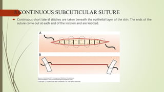
SUTURE MATERIALS AND
SUTURING TECHNIQUES
PRESENTED BY- Dr. PARTHA PRATIM DEBNATH
INTRODUCTION
 Suture means to ‘sew’ or ‘seam’. In surgery suture is the act of sewing or bringing tissue
together and holding them in apposition until healing has taken place.
 Suture material is an artificial fibre used to keep wound together until they hold sufficiently
well by themselves by natural fibre (collagen) which is synthesized and woven into a
stronger scar.
GOALS OF SUTURING
Suturing is performed to -
 Provide adequate tension
 Maintain hemostasis
 Provide support for tissue margins
 Reduce post-op pain
 Prevent bone exposure
 Permit proper flap position
 The basic purpose of a suture is to hold severed tissues in close approximation until the
healing process provides the wound with sufficient strength to withstand stress without the
need for mechanical support.
 Since wounds do not gain strength until 4-6 days after injury, the tissues are approximated
till then by sutures.
REQUISITES OF AN IDEAL SUTURE
 Tensile strength: adequate material strength will prevent suture breakdown & use
of proper knots for the material used will prevent untying or knot slippage.
 Tissue biocompatibility: sutures made from organic material will evoke a higher
tissue response than synthetic sutures.
 Low capillarity: multifilament type soak up tissue fluid by capillary action providing
a rich medium for microbes increasing chances of inflammation & infection.
 Good handling & knotting properties: ease of tying & a thread type that permits
minimal knot slippage also influence thread selection. • Sterilization without
deterioration of properties: most sutures available in packages are sterilized by dry
heat & ethylene oxide gas.
 Non allergic, non electrolytic and non carcinogènic
 Low cost
 It should not fray, should slide through tissues readily & knot should not slip after
tying
 It should be readily visualized , should not shrink & should not be extruded from
the wound.
 On break down ,it should not release toxic agents.
CLASSIFICATION OF SUTURE MATERIALS
 According to source: 1. Natural
2. Synthetic
3. Metallic
 According to structure: 1. Monofilament
2. Multifilament
 According to fate: 1. Absorbable(undergo degradation and lose T.S. < 60 days)
2. Non absorbable ( maintain T.S > 60days)
 According to coating: 1. Coated
2. Uncoated
NATURAL
Absorbable
 Catgut
 Chromic catgut
 Collagen
 Fascia lata
 Beef tendon
 Cargile membrane
Non Absorbable
 Silk
 Silk worm gut
 Linen
 Cotton
 Ramie
 Horse hair
SYNTHETIC
Absorbable
 Polyglycolic Acid
 Polyglactic Acid
 Polyglactin 910(Vicryl)
 Polydioxanone(PDS)
 Polyglecaprone 25
Non Absorbable
 Nylon/ polyamide
 PolyPropylene
 Polyesters
 Polyethelene
 Polybutester
 Polyvinylidene fluoride
Metallic
 Stainless Steel
 Tantalum
 Gold
 Silver
 Aluminium.
ABSORPTION OF SUTURE MATERIALS
 Degraded either by enzymatic process as in gut sutures, or by hydrolysis, as in
many of the synthetic materials like glycolic acid, ployglactin910 or polydioxanone.
 Non absorbable sutures are walled off or encapsulated.
 In infected tissues or in a patient who is febrile or protein deficient, suture
breakdown may be accelerated.
 If the loss of TS outpaces the healing phase, failure of the wound results.
 Absorbable sutures must be placed well into the dermis.
BIOLOGIC RESPONSE OF BODY TO SUTURE
MATERIALS
 The initial body response to sutures is almost identical in the first 4-7 days, regardless of the suture
material.
 The early response is a generalized acute aseptic inflammation, involving primarily
polymorphonuclear leukocytes.
 After few days mononuclear cells, fibroblasts & histiocytes become evident.
 Capillary formation occurs at the end of this initial phase.
SUTURE SIZES
 Largest size 1 to extremely fine 11-0. Increasing number of zeroes correlates with
decreasing suture diameter and strength.
 Thicker sutures are used for approximation of deeper layers, wounds in tension
prone areas and for ligation of blood vessels.
 Thin sutures are used for closing delicate tissues like conjunctiva and skin incisions
of the face. Size is chosen to correlate with the tensile strength of the tissue being
sutured.
 3-0 or 4-0 OMFS, muscle, deep skin
 5-0 or 6-0 facial skin closure
 9-0 or 10-0 microsurgery
CLASSIFICATION OF SURGICAL NEEDLES
1.According to eye -eye less needles
-needles with eye
2.According to shape -straight needles
-curved needles
3.According to cutting edge a)round body
b) cutting –conventional
-reverse cutting
4.According to its tip a)triangular tip b)round tip c)blunt tip
5.Others a)spatula needles b)micro point needles c)cuticular needles
PRINCIPLES OF SUTURING:
1.Needle grasped at 1/4th to half the distance from eye.
2.Needle should enter perpendicular to tissue surface.
3.Needle passed along its curve.
4.The bite should be equal on both sides of the wound margin and the point of the
entry of the needle should be closer to the wound edge than its point of exit on the
deep surface
5.The bite should be about 2-3 mm from the wound margin of the flap because after
wound closure the edge of the wound softens due to collagenolysis and the holding
power is impaired.
6. Usually the needle to be passed from mobile side to the fixed side but not
always(exception in lingual mucoperiosteum flap) and from thinner to thicker
& from deeper to superficial flap.
7.The tissues should not be closed under tension , since they will either tear
or necrose around the the suture.
8.Tie to approximate; not to blanch
9.Knot must not lie on incision line
10.The distance b/w one suture to another should be about 3-4 mm apart to
prevent strangulation of the tissue & to allow escape of the serum or
inflammatory exudate & to get more strength of the wound.
11.Sutures placed at a greater depth than distance from the incision to evert wound margins
12.Close deep wounds in layers
13.Avoid retrieving needle by tip
14.Adequate tissue bite to prevent tearing.
15.Occasionally extra tissue may be present on one side of incision and cause DOG EAR to be
formed in the final phase of wound closure.
SUTURING TECHNIQUES
1.SIMPLE INTERRUPTED SUTURE
 Most commonly used. Inserted singly through side of the wound and tied with a
surgeon’s knot.
Advantages
 Strong and can be used in areas of stress
 Placed 4-8 mm apart to close large wounds, so that tension is shared
 Each is independent and loosening one will not produce loosening of the other
 Degree of eversion produced
 In infection or hematoma, removal of few sutures
 Free of interferences b/w each stitch and easy to clean
2. SIMPLE CONTINUOUS / RUNNING
 A simple interrupted suture placed and needle reinserted in a continuous fashion such that the
suturepasses perpendicular to the incision line below and obliquely above. Ended by passing a
knot over the untightened end of the suture.
 Advantages
 Rapid technique and distributes tension uniformly
 More water tight closure (Shoen, 1975)
 Only 2 knots with associated tags
 Disadvantages
 If cut at one point, suture slackens along the whole length of the wound which will
then gape open.
3.CONTINUOUS LOCKING/BLANKET
 Similar to continuous but locking provided by withdrawing the suture through its
own loop.
 Indicated in long edentulous areas, tuberosities or retromolar area.
 Advantages
 Will avoid multiple knots
 Distributes tension uniformly
 Water tight closure
 Prevents excessive tightening.
 Disadvantage :
Prevents adjustment of tension over suture line as tissue swelling occurs.
4.VERTICAL MATTRESS
 Specially designed for use in skin.
 It passes at 2 levels, one deep to provide support and adduction of wound surfaces
at a depth and one superficial to draw the edges together and evert them.
 Used for closing deep wounds
 This approximates subcutaneous and skin edge.
 Needle passed from one edge to the other and again from latter edge to the fist
and knot tied. When needle is brought back from second flap to the first, depth of
penetration is more superficial.
Advantages :
 for better adaptation and maximum tissue approximation
 To get eversion of wound margins slightly
 Where healing is expected to be delayed for any reason, it is better to give wound added support
by vertical mattress.
 Used to control soft tissue hemorrhage.
 Runs parallel to the blood supply of the edge of the flap and therefore not interfering with healing.
Uses: abdominal surgeries & closure of skin wounds.
5.HORIZONTAL MATTRESS
 It everts mucosal or skin margins, bringing greater areas of raw tissue into
contact.So used for closing bony deficiencies such as oro-antral fistula or cystic
cavities.
 Disadvantage: constricts the blood supply to edges of incision.
 Needle passed from one edge to the other and again from the latter to the first and a knot is tied.
 Distance of needle penetration and depth of penetration is same for each entry point, but
horizontal distance of the points of penetration on the same side of the flap differs.
Advantages:
 Will evert mucosal or skin margins, bringing greater areas of raw tissue into contact. -So used for
closing bony deficiencies such as oroantral fistula or cystic cavities, extraction socket wounds.
 Prevents the flap from being inverted into the cavity.
 To control post-operative hemorrhage from gingiva around the tooth socket to tense the
mucoperiosteum over the underlying bone.
Disadvantages:
 More trouble to insert
 Constricts the blood supply to the incision if improperly used, cause wound necrosis and
dehiscence.
6. FIGURE OF 8 SUTURE
 Used for extraction socket closure and for adaption of gingival papilla around the tooth Suturing
begun on buccal surface 3-4mm from the tip of the papilla so as to prevent tearing of papilla.
 Needle first inserted into the outer surface of the buccal flap and then the lingual flap. Needle
again inserted in same fashion at a horizontal distance and then both ends tied.
7. SUBCUTICULAR SUTURE
 Used to close deep wounds in layers. Knots will be inverted or buried, so that the
knot does not lie between the skin margin and cause inflammation or infection.
 To bury the knot, first pass of the needle should be from within the wound and
through the lower portion of the dermal layer. Needle then passed through the
dermal layer and emerge through subcutaneous tissue and knot tied.
8.CONTINUOUS SUBCUTICULAR SUTURE
 Continuous short lateral stitches are taken beneath the epithelial layer of the skin. The ends of the
suture come out at each end of the incision and are knotted.
Advantages
 Excellent cosmetic result
 Useful in wounds with strong skin tension, especially for patients prone to keloid formation.
 Anchor suture in wound and, from apex, take bites below the dermal-epidermal layer
 Start next stitch directly opposite the one that precedes it
KNOT TYING
Principles of knot tying
 Use the simplest knot that will prevent slippage.
 Tying the knot as small as possible and cutting the ends of the suture as short as
reasonable to minimize foreign body reaction.
 Avoid friction or sawing
 Avoid damage to suture material
 Avoid excessive tension
 Tying sutures too tightly strangulates the tissue
 Maintenance of traction at one end of the suture after the first loop is thrown, to avoid loosening
of the knot.
 Placing the final throw as horizontally as possible to keep knot flat
 Limiting extra throws to the knot, as they do not add strength to a properly tied knot
SQUARE KNOT
 Formed by wrapping the suture around the needle holder once in opposite
directions between the ties.
 Atleast 3 ties are recommended.
 Best for gut, silk, cotton and SS.
SURGEON’S KNOT
Formed by 2 throws on the first tie and one throw in the
opposite direction in the second tie.
Recommended for tying polyester suture materials such
as Vicryl and Mersiline.
GRANNY’S KNOT
A tie in one direction followed by a tie in the same
direction and a third tie in the opposite direction to
square the knot and hold it permanently.
THANK YOU

Seminar on suture

  • 1.
    SUTURE MATERIALS AND SUTURINGTECHNIQUES PRESENTED BY- Dr. PARTHA PRATIM DEBNATH
  • 2.
    INTRODUCTION  Suture meansto ‘sew’ or ‘seam’. In surgery suture is the act of sewing or bringing tissue together and holding them in apposition until healing has taken place.  Suture material is an artificial fibre used to keep wound together until they hold sufficiently well by themselves by natural fibre (collagen) which is synthesized and woven into a stronger scar.
  • 3.
    GOALS OF SUTURING Suturingis performed to -  Provide adequate tension  Maintain hemostasis  Provide support for tissue margins  Reduce post-op pain  Prevent bone exposure  Permit proper flap position
  • 4.
     The basicpurpose of a suture is to hold severed tissues in close approximation until the healing process provides the wound with sufficient strength to withstand stress without the need for mechanical support.  Since wounds do not gain strength until 4-6 days after injury, the tissues are approximated till then by sutures.
  • 5.
    REQUISITES OF ANIDEAL SUTURE  Tensile strength: adequate material strength will prevent suture breakdown & use of proper knots for the material used will prevent untying or knot slippage.  Tissue biocompatibility: sutures made from organic material will evoke a higher tissue response than synthetic sutures.  Low capillarity: multifilament type soak up tissue fluid by capillary action providing a rich medium for microbes increasing chances of inflammation & infection.  Good handling & knotting properties: ease of tying & a thread type that permits minimal knot slippage also influence thread selection. • Sterilization without deterioration of properties: most sutures available in packages are sterilized by dry heat & ethylene oxide gas.
  • 6.
     Non allergic,non electrolytic and non carcinogènic  Low cost  It should not fray, should slide through tissues readily & knot should not slip after tying  It should be readily visualized , should not shrink & should not be extruded from the wound.  On break down ,it should not release toxic agents.
  • 7.
    CLASSIFICATION OF SUTUREMATERIALS  According to source: 1. Natural 2. Synthetic 3. Metallic  According to structure: 1. Monofilament 2. Multifilament
  • 8.
     According tofate: 1. Absorbable(undergo degradation and lose T.S. < 60 days) 2. Non absorbable ( maintain T.S > 60days)  According to coating: 1. Coated 2. Uncoated
  • 9.
    NATURAL Absorbable  Catgut  Chromiccatgut  Collagen  Fascia lata  Beef tendon  Cargile membrane Non Absorbable  Silk  Silk worm gut  Linen  Cotton  Ramie  Horse hair
  • 11.
    SYNTHETIC Absorbable  Polyglycolic Acid Polyglactic Acid  Polyglactin 910(Vicryl)  Polydioxanone(PDS)  Polyglecaprone 25 Non Absorbable  Nylon/ polyamide  PolyPropylene  Polyesters  Polyethelene  Polybutester  Polyvinylidene fluoride
  • 13.
    Metallic  Stainless Steel Tantalum  Gold  Silver  Aluminium.
  • 14.
    ABSORPTION OF SUTUREMATERIALS  Degraded either by enzymatic process as in gut sutures, or by hydrolysis, as in many of the synthetic materials like glycolic acid, ployglactin910 or polydioxanone.  Non absorbable sutures are walled off or encapsulated.  In infected tissues or in a patient who is febrile or protein deficient, suture breakdown may be accelerated.  If the loss of TS outpaces the healing phase, failure of the wound results.  Absorbable sutures must be placed well into the dermis.
  • 15.
    BIOLOGIC RESPONSE OFBODY TO SUTURE MATERIALS  The initial body response to sutures is almost identical in the first 4-7 days, regardless of the suture material.  The early response is a generalized acute aseptic inflammation, involving primarily polymorphonuclear leukocytes.  After few days mononuclear cells, fibroblasts & histiocytes become evident.  Capillary formation occurs at the end of this initial phase.
  • 16.
    SUTURE SIZES  Largestsize 1 to extremely fine 11-0. Increasing number of zeroes correlates with decreasing suture diameter and strength.  Thicker sutures are used for approximation of deeper layers, wounds in tension prone areas and for ligation of blood vessels.  Thin sutures are used for closing delicate tissues like conjunctiva and skin incisions of the face. Size is chosen to correlate with the tensile strength of the tissue being sutured.  3-0 or 4-0 OMFS, muscle, deep skin  5-0 or 6-0 facial skin closure  9-0 or 10-0 microsurgery
  • 17.
    CLASSIFICATION OF SURGICALNEEDLES 1.According to eye -eye less needles -needles with eye 2.According to shape -straight needles -curved needles 3.According to cutting edge a)round body b) cutting –conventional -reverse cutting 4.According to its tip a)triangular tip b)round tip c)blunt tip 5.Others a)spatula needles b)micro point needles c)cuticular needles
  • 20.
    PRINCIPLES OF SUTURING: 1.Needlegrasped at 1/4th to half the distance from eye. 2.Needle should enter perpendicular to tissue surface. 3.Needle passed along its curve. 4.The bite should be equal on both sides of the wound margin and the point of the entry of the needle should be closer to the wound edge than its point of exit on the deep surface 5.The bite should be about 2-3 mm from the wound margin of the flap because after wound closure the edge of the wound softens due to collagenolysis and the holding power is impaired.
  • 23.
    6. Usually theneedle to be passed from mobile side to the fixed side but not always(exception in lingual mucoperiosteum flap) and from thinner to thicker & from deeper to superficial flap. 7.The tissues should not be closed under tension , since they will either tear or necrose around the the suture. 8.Tie to approximate; not to blanch 9.Knot must not lie on incision line 10.The distance b/w one suture to another should be about 3-4 mm apart to prevent strangulation of the tissue & to allow escape of the serum or inflammatory exudate & to get more strength of the wound.
  • 24.
    11.Sutures placed ata greater depth than distance from the incision to evert wound margins 12.Close deep wounds in layers 13.Avoid retrieving needle by tip 14.Adequate tissue bite to prevent tearing. 15.Occasionally extra tissue may be present on one side of incision and cause DOG EAR to be formed in the final phase of wound closure.
  • 25.
  • 26.
    1.SIMPLE INTERRUPTED SUTURE Most commonly used. Inserted singly through side of the wound and tied with a surgeon’s knot.
  • 27.
    Advantages  Strong andcan be used in areas of stress  Placed 4-8 mm apart to close large wounds, so that tension is shared  Each is independent and loosening one will not produce loosening of the other  Degree of eversion produced  In infection or hematoma, removal of few sutures  Free of interferences b/w each stitch and easy to clean
  • 28.
    2. SIMPLE CONTINUOUS/ RUNNING  A simple interrupted suture placed and needle reinserted in a continuous fashion such that the suturepasses perpendicular to the incision line below and obliquely above. Ended by passing a knot over the untightened end of the suture.
  • 29.
     Advantages  Rapidtechnique and distributes tension uniformly  More water tight closure (Shoen, 1975)  Only 2 knots with associated tags  Disadvantages  If cut at one point, suture slackens along the whole length of the wound which will then gape open.
  • 30.
    3.CONTINUOUS LOCKING/BLANKET  Similarto continuous but locking provided by withdrawing the suture through its own loop.  Indicated in long edentulous areas, tuberosities or retromolar area.
  • 31.
     Advantages  Willavoid multiple knots  Distributes tension uniformly  Water tight closure  Prevents excessive tightening.  Disadvantage : Prevents adjustment of tension over suture line as tissue swelling occurs.
  • 32.
    4.VERTICAL MATTRESS  Speciallydesigned for use in skin.  It passes at 2 levels, one deep to provide support and adduction of wound surfaces at a depth and one superficial to draw the edges together and evert them.
  • 33.
     Used forclosing deep wounds  This approximates subcutaneous and skin edge.  Needle passed from one edge to the other and again from latter edge to the fist and knot tied. When needle is brought back from second flap to the first, depth of penetration is more superficial.
  • 34.
    Advantages :  forbetter adaptation and maximum tissue approximation  To get eversion of wound margins slightly  Where healing is expected to be delayed for any reason, it is better to give wound added support by vertical mattress.  Used to control soft tissue hemorrhage.  Runs parallel to the blood supply of the edge of the flap and therefore not interfering with healing. Uses: abdominal surgeries & closure of skin wounds.
  • 35.
    5.HORIZONTAL MATTRESS  Iteverts mucosal or skin margins, bringing greater areas of raw tissue into contact.So used for closing bony deficiencies such as oro-antral fistula or cystic cavities.  Disadvantage: constricts the blood supply to edges of incision.
  • 36.
     Needle passedfrom one edge to the other and again from the latter to the first and a knot is tied.  Distance of needle penetration and depth of penetration is same for each entry point, but horizontal distance of the points of penetration on the same side of the flap differs.
  • 37.
    Advantages:  Will evertmucosal or skin margins, bringing greater areas of raw tissue into contact. -So used for closing bony deficiencies such as oroantral fistula or cystic cavities, extraction socket wounds.  Prevents the flap from being inverted into the cavity.  To control post-operative hemorrhage from gingiva around the tooth socket to tense the mucoperiosteum over the underlying bone. Disadvantages:  More trouble to insert  Constricts the blood supply to the incision if improperly used, cause wound necrosis and dehiscence.
  • 38.
    6. FIGURE OF8 SUTURE  Used for extraction socket closure and for adaption of gingival papilla around the tooth Suturing begun on buccal surface 3-4mm from the tip of the papilla so as to prevent tearing of papilla.  Needle first inserted into the outer surface of the buccal flap and then the lingual flap. Needle again inserted in same fashion at a horizontal distance and then both ends tied.
  • 39.
    7. SUBCUTICULAR SUTURE Used to close deep wounds in layers. Knots will be inverted or buried, so that the knot does not lie between the skin margin and cause inflammation or infection.  To bury the knot, first pass of the needle should be from within the wound and through the lower portion of the dermal layer. Needle then passed through the dermal layer and emerge through subcutaneous tissue and knot tied.
  • 40.
    8.CONTINUOUS SUBCUTICULAR SUTURE Continuous short lateral stitches are taken beneath the epithelial layer of the skin. The ends of the suture come out at each end of the incision and are knotted.
  • 41.
    Advantages  Excellent cosmeticresult  Useful in wounds with strong skin tension, especially for patients prone to keloid formation.  Anchor suture in wound and, from apex, take bites below the dermal-epidermal layer  Start next stitch directly opposite the one that precedes it
  • 42.
    KNOT TYING Principles ofknot tying  Use the simplest knot that will prevent slippage.  Tying the knot as small as possible and cutting the ends of the suture as short as reasonable to minimize foreign body reaction.  Avoid friction or sawing  Avoid damage to suture material  Avoid excessive tension  Tying sutures too tightly strangulates the tissue
  • 43.
     Maintenance oftraction at one end of the suture after the first loop is thrown, to avoid loosening of the knot.  Placing the final throw as horizontally as possible to keep knot flat  Limiting extra throws to the knot, as they do not add strength to a properly tied knot
  • 44.
    SQUARE KNOT  Formedby wrapping the suture around the needle holder once in opposite directions between the ties.  Atleast 3 ties are recommended.  Best for gut, silk, cotton and SS.
  • 45.
    SURGEON’S KNOT Formed by2 throws on the first tie and one throw in the opposite direction in the second tie. Recommended for tying polyester suture materials such as Vicryl and Mersiline.
  • 46.
    GRANNY’S KNOT A tiein one direction followed by a tie in the same direction and a third tie in the opposite direction to square the knot and hold it permanently.
  • 47.