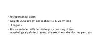
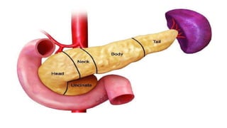
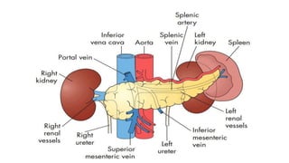
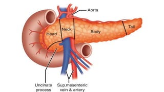
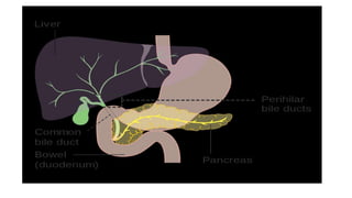
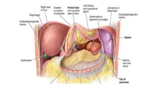
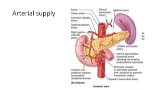
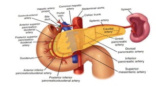
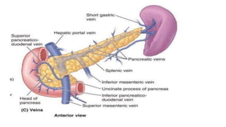
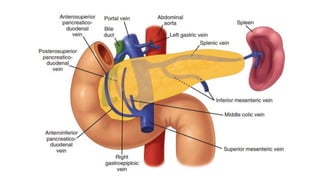
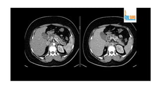
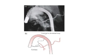
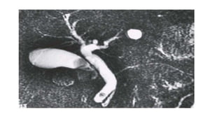
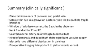
The pancreas has four regions - the head, neck, body, and tail. The head is nestled near the duodenum and contains the pancreatic duct which drains into the common bile duct. The neck lies at the level of L1-L2 near the portal vein. The body and tail are located near the spleen. Arterial blood supply comes from branches of the celiac and superior mesenteric arteries, while venous drainage involves the portal vein. The exocrine pancreas secretes enzymes to aid digestion, and the endocrine islets of Langerhans produce hormones like insulin and glucagon to regulate metabolism. Imaging techniques like ultrasound and CT are important for evaluating pancreatic anatomy and function.
3. • Retroperitoneal organ
• Weights 75 to 100 gm and is about 15 t0 20 cm long
• 4 regions
• It is an endodermally derived organ, consisting of two
morphologically distinct tissues, the exocrine and endocrine pancreas
6. Head of pancreas
• The head of the pancreas is nestled in the C-loop of the duodenum
and is posterior to the transverse mesocolon.
• Just posterior to the head of the pancreas lie the vena cava, the right
renal artery and both renal veins
• The common bile duct runs in a deep groove on the posterior aspect
of the pancreatic head
7.
8.
9.
10. Neck of pancreas
• The neck of the pancreas lies directly anterior to the portal vein.
• At the inferior border of the neck of the pancreas, the superior
mesenteric vein joins the splenic vein
• The neck of the pancreas is anterior to the vertebral body of L1 and
L2
• The neck divides the pancreas into approximately two equal halves.
11. Body and tail
• The body and tail of the pancreas lie just anterior to the splenic artery and
vein.
• The anterior surface of the body of the pancreas is covered by peritoneum.
• Once the gastrocolic omentum is divided, the body and tail of the pancreas
can be seen along the floor of the lesser sac, just posterior to the stomach
• The base of the transverse mesocolon attaches to the inferior margin of the
body and tail of the pancreas
• the tail is nestled in the hilum of the spleen near the splenic flexure of the
left colon
12.
13. Pancreatic Duct Anatomy
• The duct of the ventral anlage becomes the duct of Wirsung, and the
duct from the dorsal anlage becomes the duct of Santorini.
• The ducts from each anlage usually fuse together in the pancreatic
head
• most of the pancreas drains through the duct of Wirsung, or main
pancreatic duct, into the common channel formed from the bile duct
and pancreatic duct.
14. • The length of the common channel is variable.
• In approximately 30% of patients, the duct of Santorini ends as a blind
accessory duct and does not empty into the duodenum
• main pancreatic duct is usually only 2 to 3 mm in diameter and runs
midway between the superior and inferior borders of the pancreas,
• Main pancreatic duct closer to the posterior than to the anterior
surface
15. • The muscle fibers around the ampulla form the sphincter of Oddi,
which controls the flow of pancreatic and biliary secretions into the
duodenum.
• Contraction and relaxation of the sphincter is regulated by complex
neural and hormonal factors.
18. • The blood supply to the pancreas comes from multiple branches from
the celiac and superior mesenteric arteries
• The common hepatic artery gives rise to the gastroduodenal artery
• The gastroduodenal artery then travels inferiorly anterior to the neck
of the pancreas and posterior to the duodenal bulb.
• At the inferior border of the duodenum, the gastroduodenal artery
then gives rise to the right gastroepiploic artery then continues on as
the anterior superior pancreaticoduodenal artery
19. • superior mesenteric artery gives off inferior pancreaticoduodenal
artery .
• Variations in the arterial anatomy occur in one out of five patients.
• It is important to look for this variation on preoperative computed
tomographic (CT) scans and in the operating room
• The body and tail of the pancreas are supplied by multiple branches
of the splenic artery.
• The inferior pancreatic artery usually arises from the superior
mesenteric artery
20.
21.
22. Venous drainage
• The venous drainage of the pancreas follows a pattern similar to that
of the arterial supply (Fig. 33-5). The veins are usually superficial to
the arteries within the parenchyma of the pancreas.
• There is an anterior and posterior venous arcade within the head of
the pancreas.
• The superior veins drain directly into the portal vein just above the
neck of the pancreas.
23.
24. • The posterior inferior arcade drains directly into the inferior
mesenteric vein.
• These venous tributaries must be divided during a Whipple procedure
• The anterior inferior pancreaticoduodenal vein joins the right
gastroepiploic vein and the middle colic vein.
25. • There also are numerous small venous branches coming from the
pancreatic parenchyma directly into the lateral and posterior aspect
of the portal vein.
• Venous return from the body and tail of the pancreas drains into the
splenic vein
28. • The profuse network of lymphatic drainage provides egress to tumor
cells arising from the pancreas.
• The pancreatic lymphatics also communicate with lymph nodes in the
transverse mesocolon and mesentery of the proximal jejunum.
29. Neuroanatomy
• The pancreas is innervated by the sympathetic and parasympathetic
nervous systems.
• The parasympathetic system stimulates endocrine and exocrine
secretion and the sympathetic system inhibits secretion.
• The pancreas also has a rich supply of afferent sensory fibers, which
are responsible for the intense pain
30.
31. Physiology
• The exocrine pancreas accounts for about 85% of the pancreatic mass
• 10% of the gland is accounted for by extracellular matrix, and 4% by
blood vessels and the major ducts
• only 2% of the gland is comprised of endocrine tissue.
• regulates the type of digestion, its rate, and the processing and
distribution of absorbed nutrients.
• only approximately 20% of the normal pancreas is required to prevent
insufficiency
32. Exocrine pancreas
• patients can live without a pancreas when insulin and digestive enzyme
replacement are administered.
• The pancreas secretes approximately 500 to 800 mL per day of colourless ,
odourless , alkaline, isosmotic pancreatic juice.
• Pancreatic juice is a combination of acinar cell and duct cell secretions.
33. • The centroacinar and intercalated duct cells secrete the water and
electrolytes present in the pancreatic juice.
• About 40 acinar cells are arranged into a spherical unit called an
acinus.
• Centroacinar cells are located near the center of the acinus and are
responsible for fluid and electrolyte secretion.
• These cells contain the enzyme carbonic anhydrase, which is needed
for bicarbonate secretion.
34.
35.
36. • Secretin is the major stimulant for bicarbonate secretion
• CCK also stimulates bicarbonate secretion, but to a much lesser
extent than secretin.
• CCK potentiates secretin-stimulated bicarbonate secretion.
• Gastrin and acetylcholine, both stimulants of gastric acid secretion,
are also weak stimulants of pancreatic bicarbonate secretion.
• The endocrine pancreas also influences the adjacent exocrine
pancreatic secretions.
37.
38. Endocrine pancreas
• There are nearly 1 million islets of Langerhans in the normal adult
pancreas
• Most islets contain 3000 to 4000 cells of five major types
39.
40. Investigation
• Pancreatic function tests
• Estimation of pancreatic enzymes in body fluids
• Imaging
• Ultrasonography
• Computed tomography
• Magnetic resonance imaging
• Endoscopic retrograde cholangiopancreatography
• Endoscopic ultrasound
41.
42.
43.
44.
45.
46. Summary (clinically significant )
• Plane between neck of pancreas and portal vein
• Splenic vein run in a groove on posterior side fed by multiple fragile
branches
• Window of wisnlsow connect the 2 sac in the abdomen
• Neck found at the L1 nd L2
• Gastroduodenal artery pass through duodenal bulb
• Head of pancreas and duodenum share significant vascular supply
• Islet cells have different distribution in pancreas
• Preoperative imaging is important to pick anatomic variant