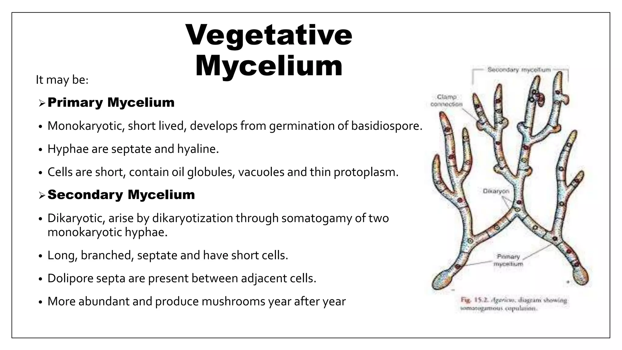
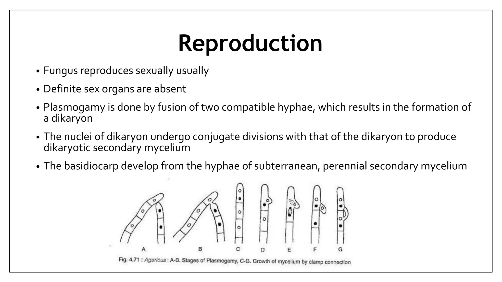
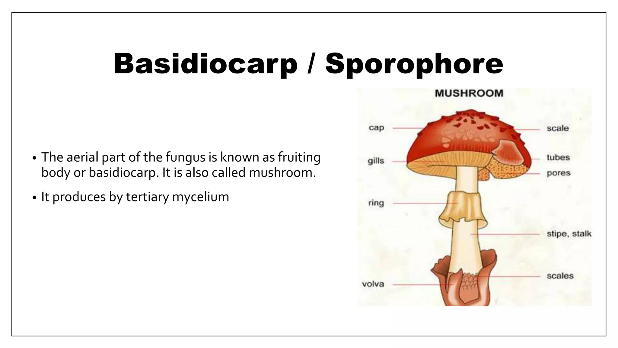
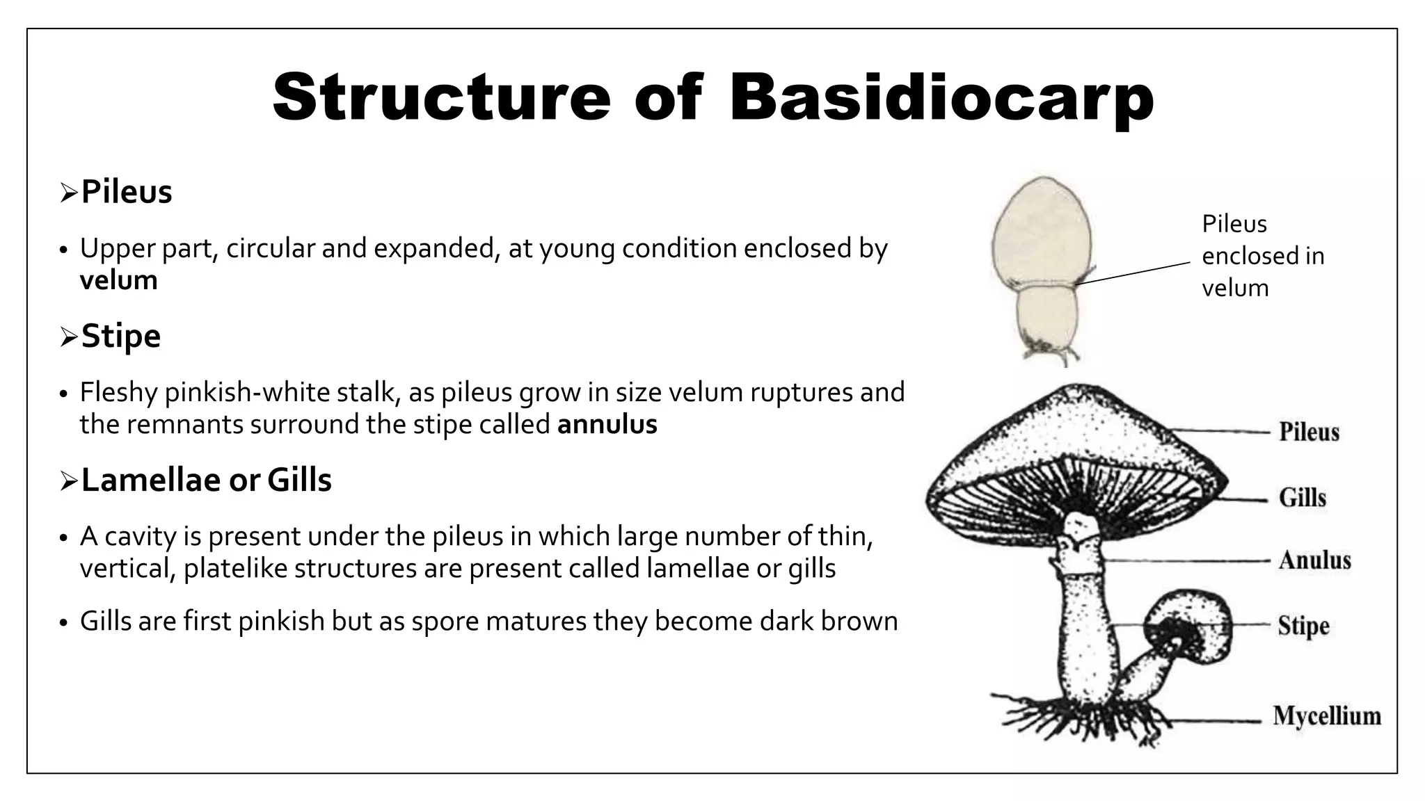
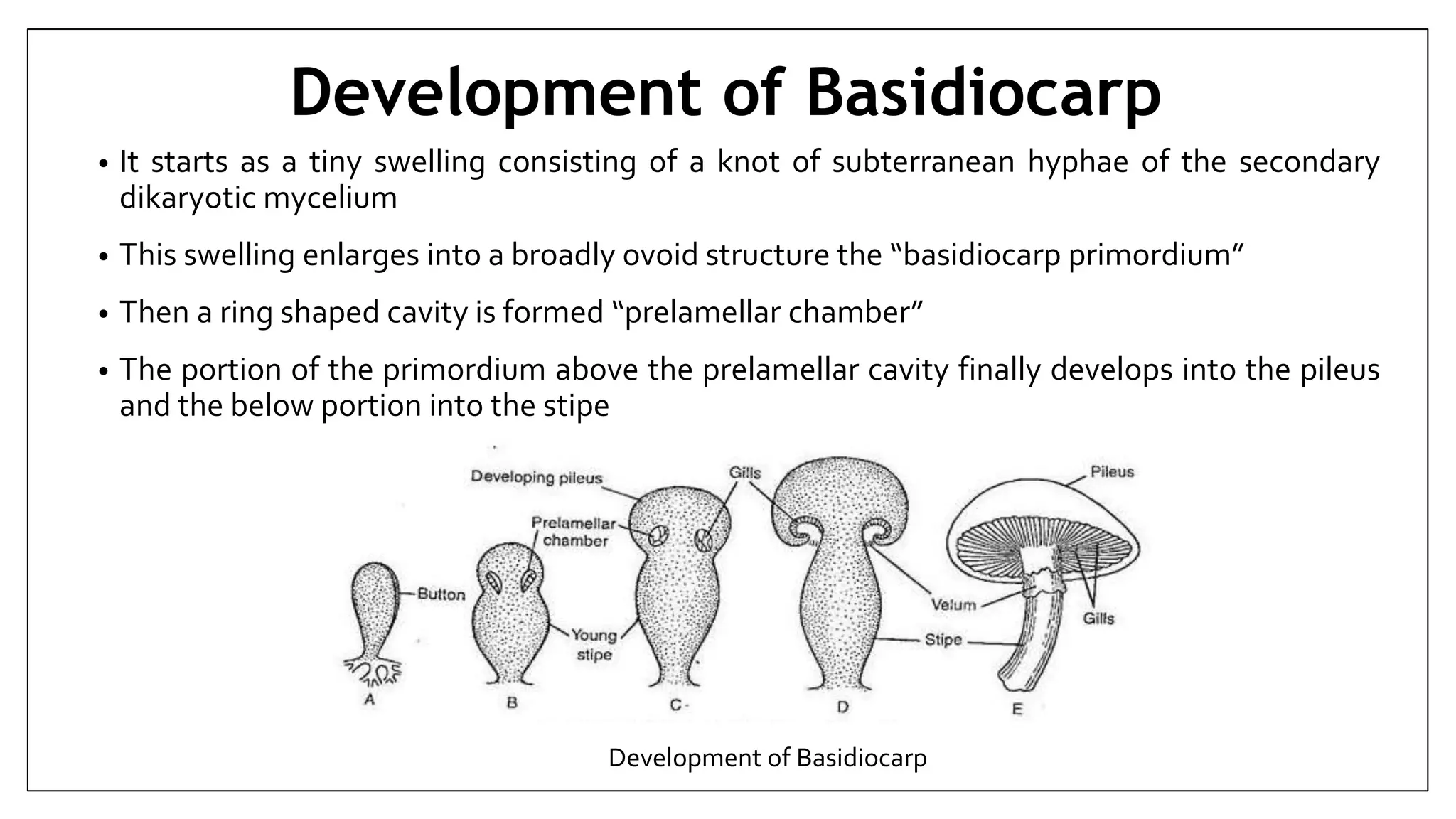
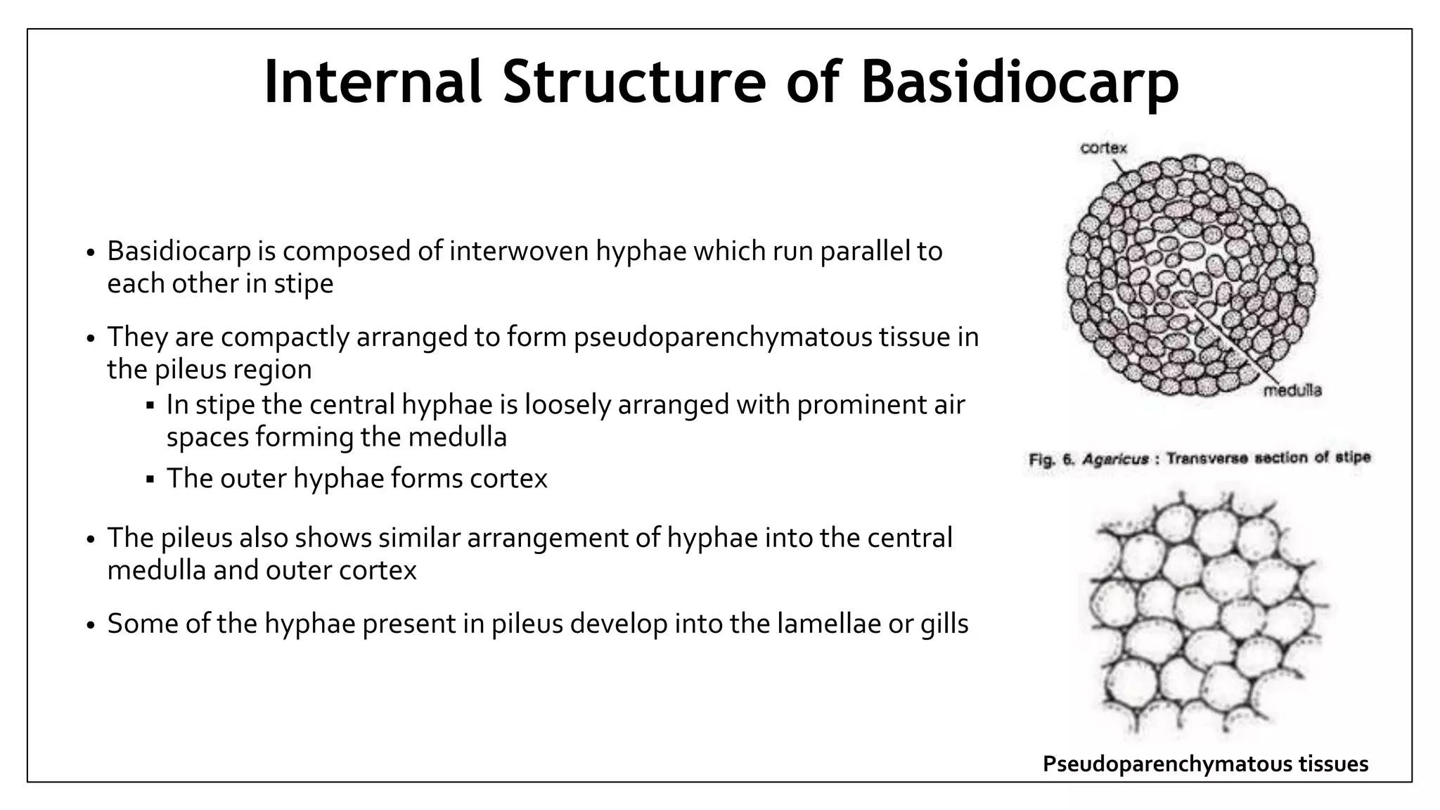
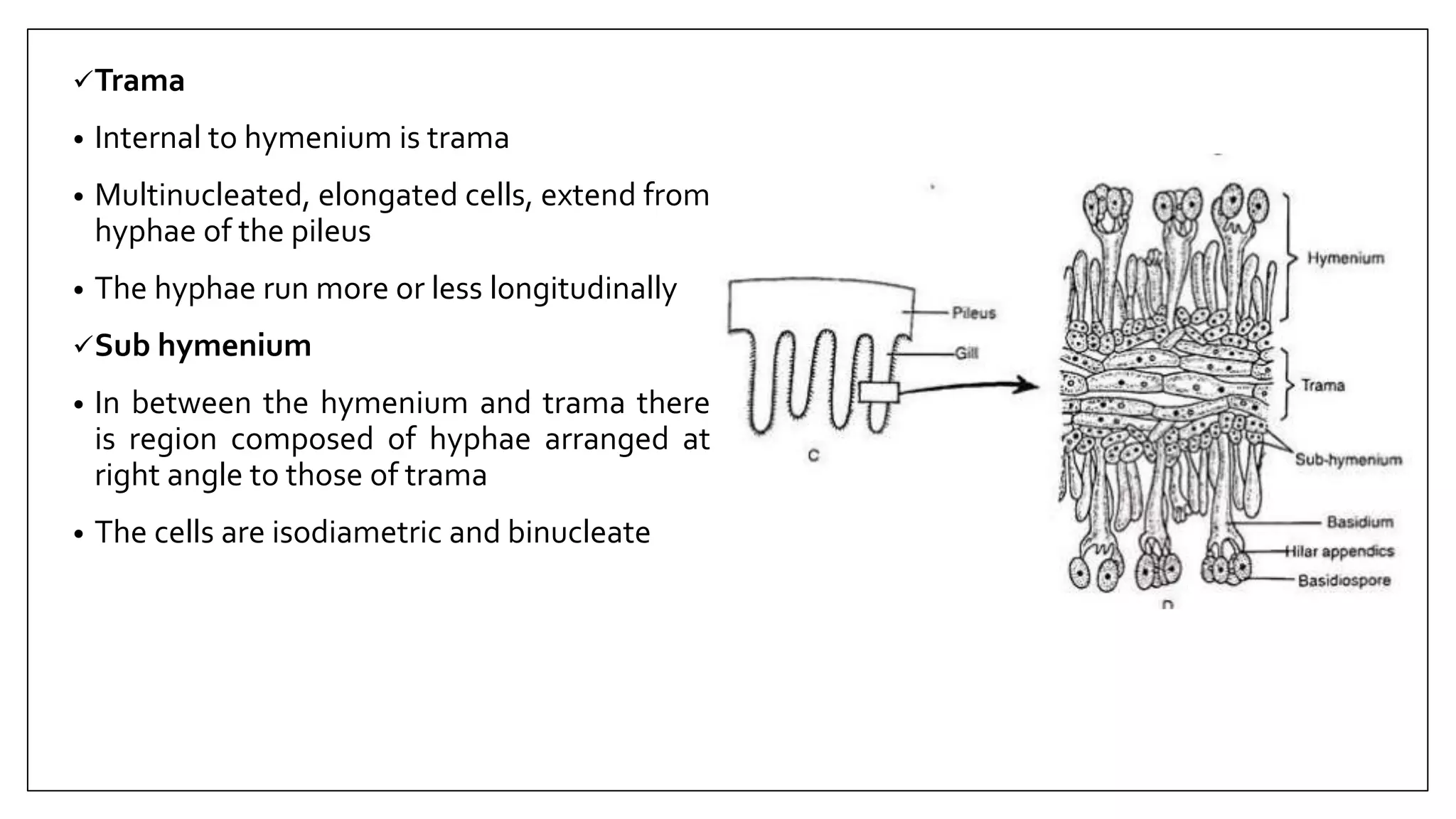
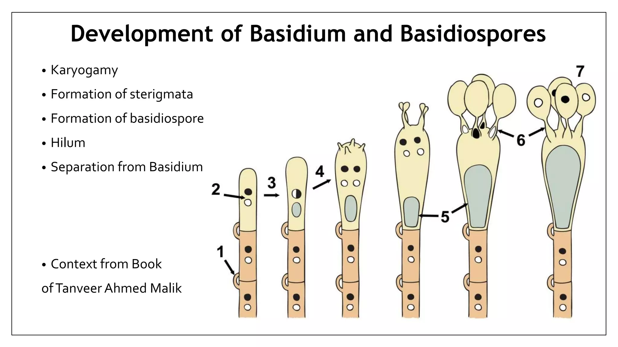
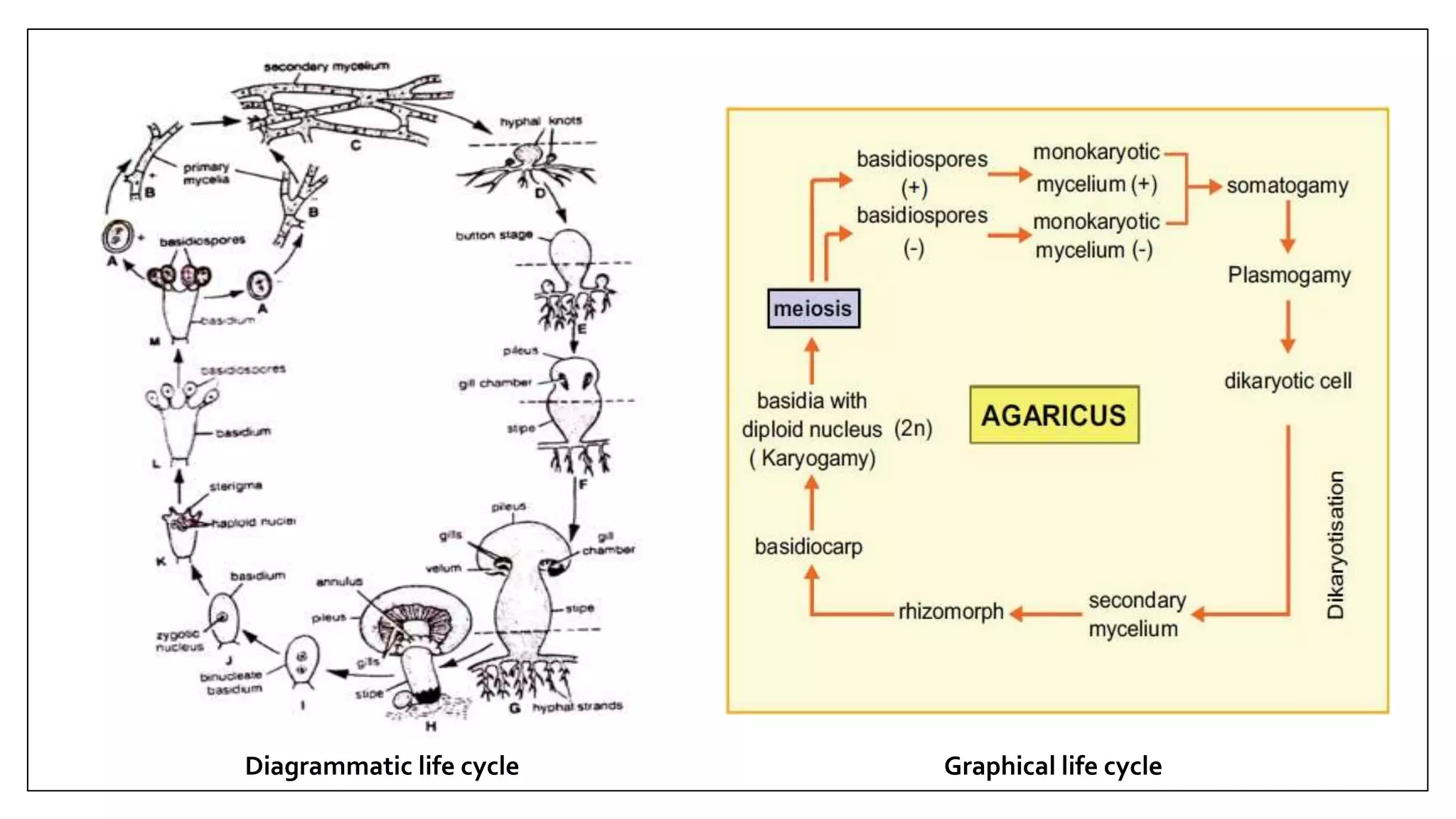
STRUCTURE AND
REPRODUCTION IN AGARICUS
Presented by: Ali Abbas Aslam
Contact me onWhatsApp for ppt file
0309-8400094
Taxonomic Position
• Kingdom : Fungi
• Division : Basidiomycota
• Class : Agaricomycetes
• Order : Agaricales
• Family : Agaricaceae
• Genus : Agaricus
• Specie : A.campestris / A.californicus
Structure
Plant Body consists of two major parts:
1. Vegetative mycelium
2. Basidiocarp
Mycelium Basidiocarp
Vegetative
MyceliumIt may be:
Primary Mycelium
• Monokaryotic, short lived, develops from germination of basidiospore.
• Hyphae are septate and hyaline.
• Cells are short, contain oil globules, vacuoles and thin protoplasm.
Secondary Mycelium
• Dikaryotic, arise by dikaryotization through somatogamy of two
monokaryotic hyphae.
• Long, branched, septate and have short cells.
• Dolipore septa are present between adjacent cells.
• More abundant and produce mushrooms year after year
Vegetative Mycelium
Tertiary Mycelium
• Mycelium producing basidiocarps is sometimes referred to as tertiary mycelium.
• It forms complex tissue which produces basidia.
Reproduction
• Fungus reproduces sexually usually
• Definite sex organs are absent
• Plasmogamy is done by fusion of two compatible hyphae, which results in the formation of
a dikaryon
• The nuclei of dikaryon undergo conjugate divisions with that of the dikaryon to produce
dikaryotic secondary mycelium
• The basidiocarp develop from the hyphae of subterranean, perennial secondary mycelium
Basidiocarp / Sporophore
• The aerial part of the fungus is known as fruiting
body or basidiocarp. It is also called mushroom.
• It produces by tertiary mycelium
Structure of Basidiocarp
Pileus
• Upper part, circular and expanded, at young condition enclosed by
velum
Stipe
• Fleshy pinkish-white stalk, as pileus grow in size velum ruptures and
the remnants surround the stipe called annulus
Lamellae or Gills
• A cavity is present under the pileus in which large number of thin,
vertical, platelike structures are present called lamellae or gills
• Gills are first pinkish but as spore matures they become dark brown
Pileus
enclosed in
velum
Development of Basidiocarp
• It starts as a tiny swelling consisting of a knot of subterranean hyphae of the secondary
dikaryotic mycelium
• This swelling enlarges into a broadly ovoid structure the “basidiocarp primordium”
• Then a ring shaped cavity is formed “prelamellar chamber”
• The portion of the primordium above the prelamellar cavity finally develops into the pileus
and the below portion into the stipe
Development of Basidiocarp
Internal Structure of Basidiocarp
• Basidiocarp is composed of interwoven hyphae which run parallel to
each other in stipe
• They are compactly arranged to form pseudoparenchymatous tissue in
the pileus region
 In stipe the central hyphae is loosely arranged with prominent air
spaces forming the medulla
 The outer hyphae forms cortex
• The pileus also shows similar arrangement of hyphae into the central
medulla and outer cortex
• Some of the hyphae present in pileus develop into the lamellae or gills
Pseudoparenchymatous tissues
Structure of lamellae
• Each lamella consists of three parts:
Hymenium
Sub hymenium
Trama
Hymenium
• Fertile region, club shaped, binucleate forming palisade like layer on both sides of lamella,
they developed to form basidia
• Some are sterile and are called paraphysis
• Rest of them are elongated cells. They protrude beyond the paraphysis and are called
cystidia
Lamella
Trama
• Internal to hymenium is trama
• Multinucleated, elongated cells, extend from
hyphae of the pileus
• The hyphae run more or less longitudinally
Sub hymenium
• In between the hymenium and trama there
is region composed of hyphae arranged at
right angle to those of trama
• The cells are isodiametric and binucleate
• Karyogamy
• Formation of sterigmata
• Formation of basidiospore
• Hilum
• Separation from Basidium
• Context from Book
ofTanveer Ahmed Malik
Development of Basidium and Basidiospores
• The mycelium of Agaricus has a tendency to grow in all directions from a central point to
form a large invisible circular colony
• When the mycelium becomes mature at tips, sporophores are produced. These sporophores
appear in a circle.These circles of mushrooms are commonly called “fairy rings”
• A.campestris produces dark green rings in grass
• Damages the lawns, produce a thick fungal mat that prevents water from reaching the grass
roots below.
• Also deplete soil nutrients
Fairy Rings
Diagrammatic life cycle Graphical life cycle

Structure and reproduction in agaricus

  • 1.
    STRUCTURE AND REPRODUCTION INAGARICUS Presented by: Ali Abbas Aslam Contact me onWhatsApp for ppt file 0309-8400094
  • 2.
    Taxonomic Position • Kingdom: Fungi • Division : Basidiomycota • Class : Agaricomycetes • Order : Agaricales • Family : Agaricaceae • Genus : Agaricus • Specie : A.campestris / A.californicus
  • 3.
    Structure Plant Body consistsof two major parts: 1. Vegetative mycelium 2. Basidiocarp Mycelium Basidiocarp
  • 4.
    Vegetative MyceliumIt may be: PrimaryMycelium • Monokaryotic, short lived, develops from germination of basidiospore. • Hyphae are septate and hyaline. • Cells are short, contain oil globules, vacuoles and thin protoplasm. Secondary Mycelium • Dikaryotic, arise by dikaryotization through somatogamy of two monokaryotic hyphae. • Long, branched, septate and have short cells. • Dolipore septa are present between adjacent cells. • More abundant and produce mushrooms year after year
  • 5.
    Vegetative Mycelium Tertiary Mycelium •Mycelium producing basidiocarps is sometimes referred to as tertiary mycelium. • It forms complex tissue which produces basidia.
  • 6.
    Reproduction • Fungus reproducessexually usually • Definite sex organs are absent • Plasmogamy is done by fusion of two compatible hyphae, which results in the formation of a dikaryon • The nuclei of dikaryon undergo conjugate divisions with that of the dikaryon to produce dikaryotic secondary mycelium • The basidiocarp develop from the hyphae of subterranean, perennial secondary mycelium
  • 7.
    Basidiocarp / Sporophore •The aerial part of the fungus is known as fruiting body or basidiocarp. It is also called mushroom. • It produces by tertiary mycelium
  • 8.
    Structure of Basidiocarp Pileus •Upper part, circular and expanded, at young condition enclosed by velum Stipe • Fleshy pinkish-white stalk, as pileus grow in size velum ruptures and the remnants surround the stipe called annulus Lamellae or Gills • A cavity is present under the pileus in which large number of thin, vertical, platelike structures are present called lamellae or gills • Gills are first pinkish but as spore matures they become dark brown Pileus enclosed in velum
  • 9.
    Development of Basidiocarp •It starts as a tiny swelling consisting of a knot of subterranean hyphae of the secondary dikaryotic mycelium • This swelling enlarges into a broadly ovoid structure the “basidiocarp primordium” • Then a ring shaped cavity is formed “prelamellar chamber” • The portion of the primordium above the prelamellar cavity finally develops into the pileus and the below portion into the stipe Development of Basidiocarp
  • 10.
    Internal Structure ofBasidiocarp • Basidiocarp is composed of interwoven hyphae which run parallel to each other in stipe • They are compactly arranged to form pseudoparenchymatous tissue in the pileus region  In stipe the central hyphae is loosely arranged with prominent air spaces forming the medulla  The outer hyphae forms cortex • The pileus also shows similar arrangement of hyphae into the central medulla and outer cortex • Some of the hyphae present in pileus develop into the lamellae or gills Pseudoparenchymatous tissues
  • 11.
    Structure of lamellae •Each lamella consists of three parts: Hymenium Sub hymenium Trama Hymenium • Fertile region, club shaped, binucleate forming palisade like layer on both sides of lamella, they developed to form basidia • Some are sterile and are called paraphysis • Rest of them are elongated cells. They protrude beyond the paraphysis and are called cystidia Lamella
  • 12.
    Trama • Internal tohymenium is trama • Multinucleated, elongated cells, extend from hyphae of the pileus • The hyphae run more or less longitudinally Sub hymenium • In between the hymenium and trama there is region composed of hyphae arranged at right angle to those of trama • The cells are isodiametric and binucleate
  • 13.
    • Karyogamy • Formationof sterigmata • Formation of basidiospore • Hilum • Separation from Basidium • Context from Book ofTanveer Ahmed Malik Development of Basidium and Basidiospores
  • 14.
    • The myceliumof Agaricus has a tendency to grow in all directions from a central point to form a large invisible circular colony • When the mycelium becomes mature at tips, sporophores are produced. These sporophores appear in a circle.These circles of mushrooms are commonly called “fairy rings” • A.campestris produces dark green rings in grass • Damages the lawns, produce a thick fungal mat that prevents water from reaching the grass roots below. • Also deplete soil nutrients Fairy Rings
  • 15.
    Diagrammatic life cycleGraphical life cycle