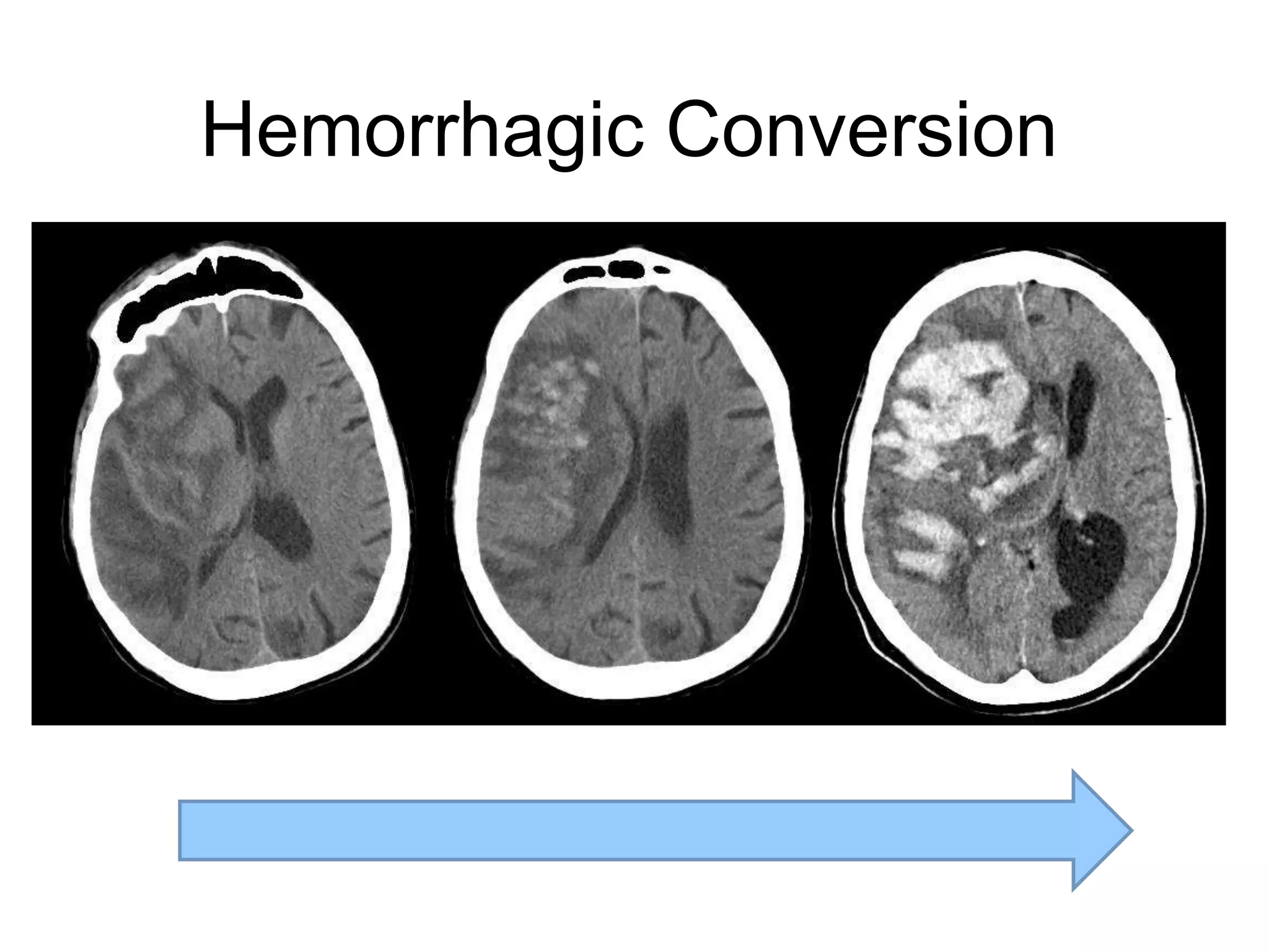
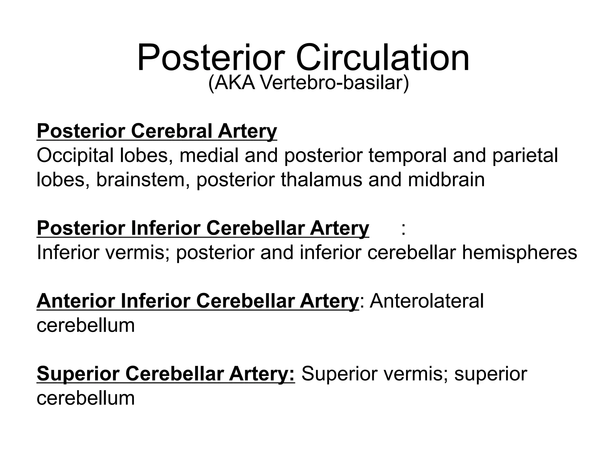
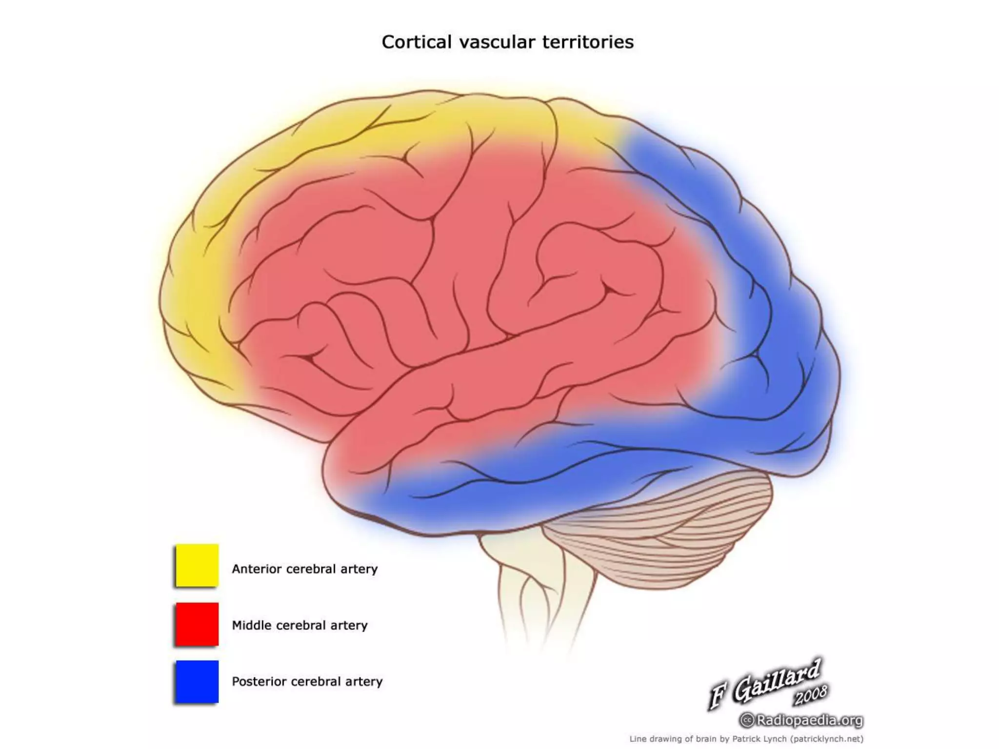
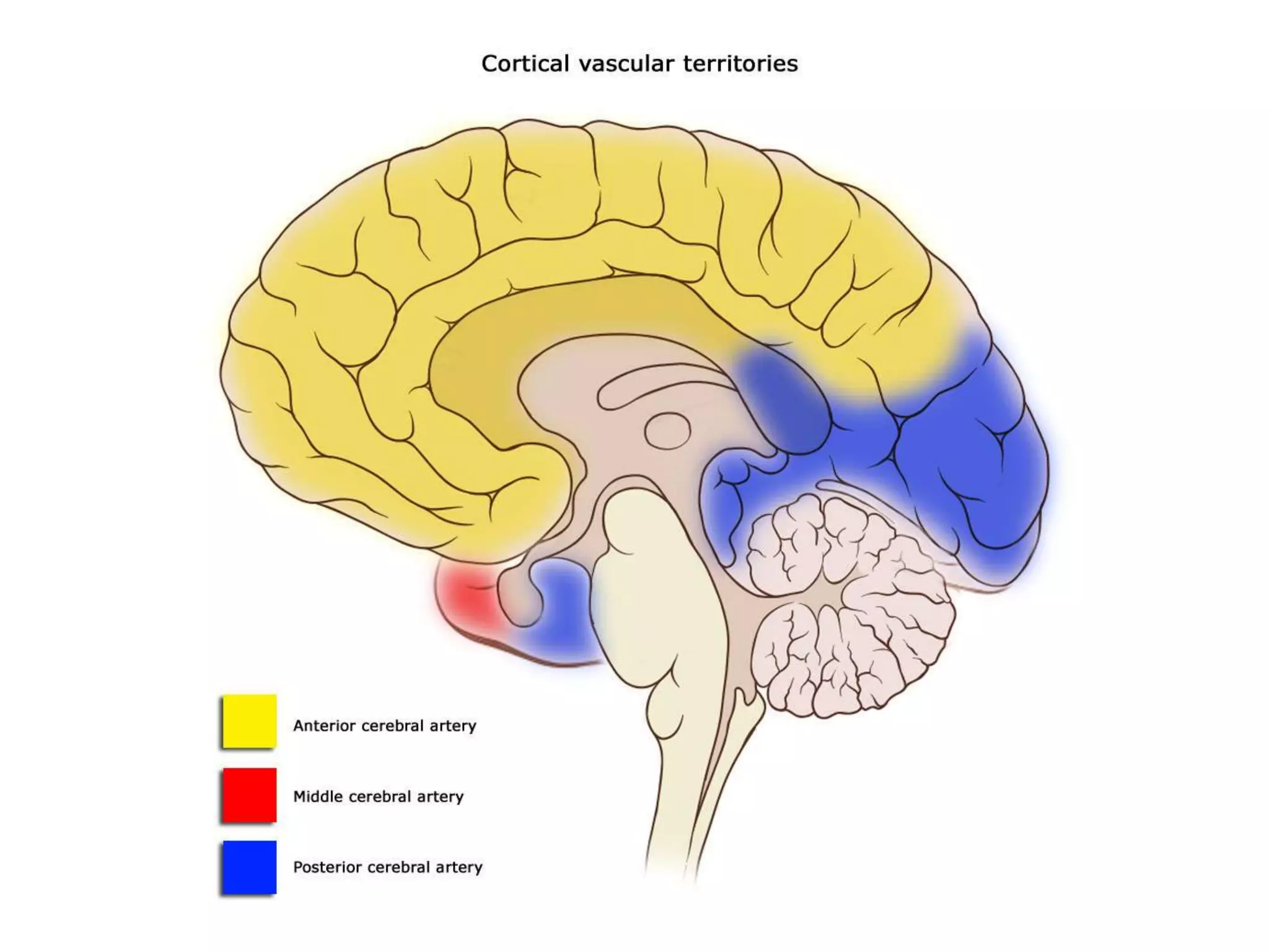
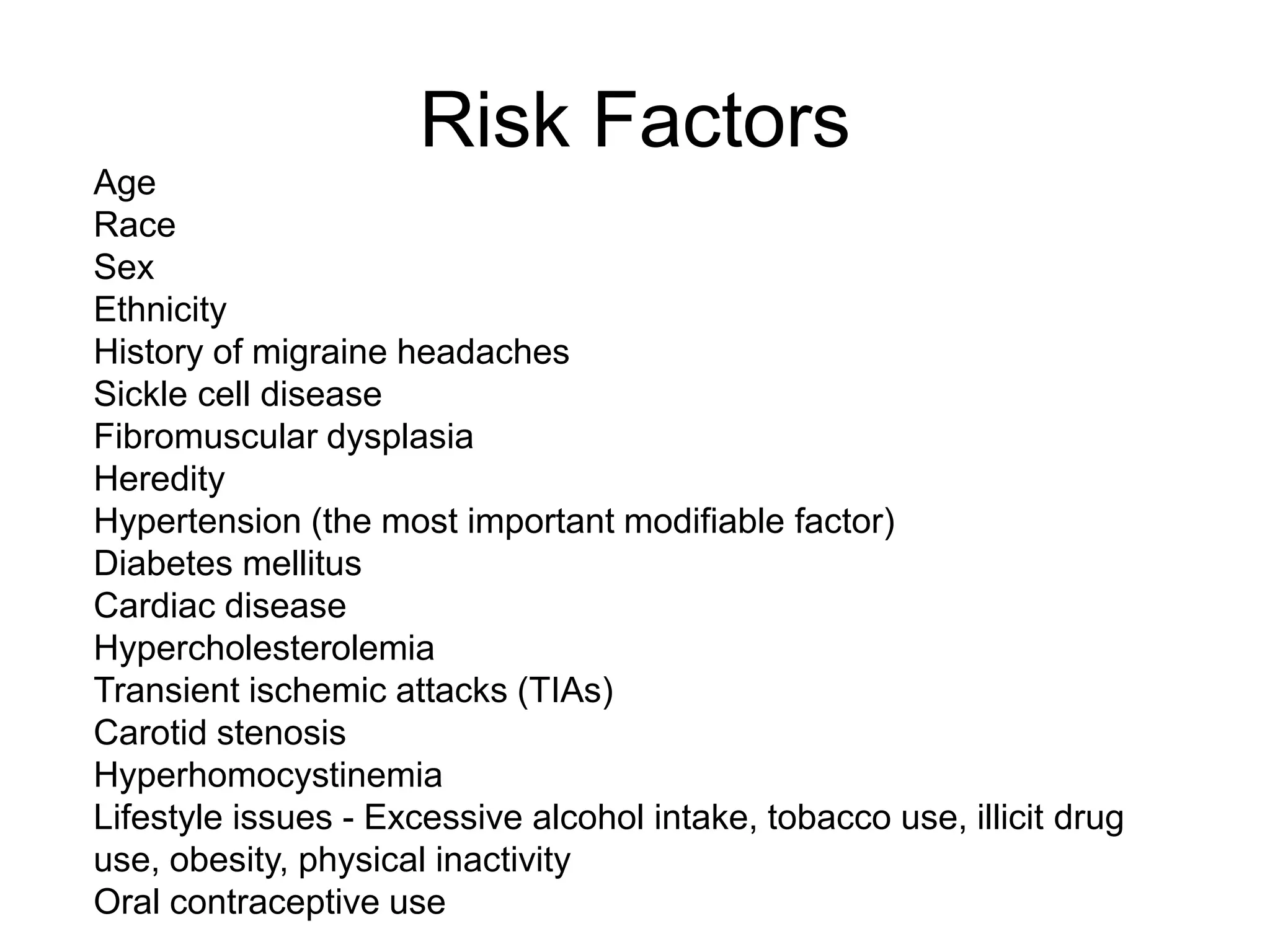
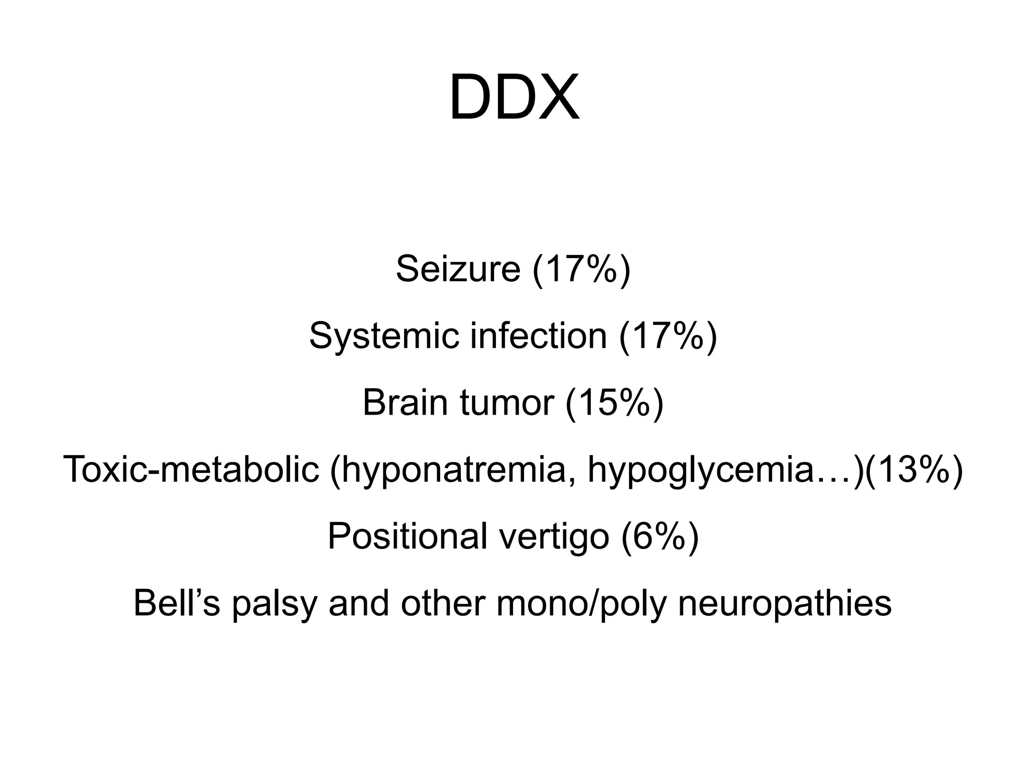
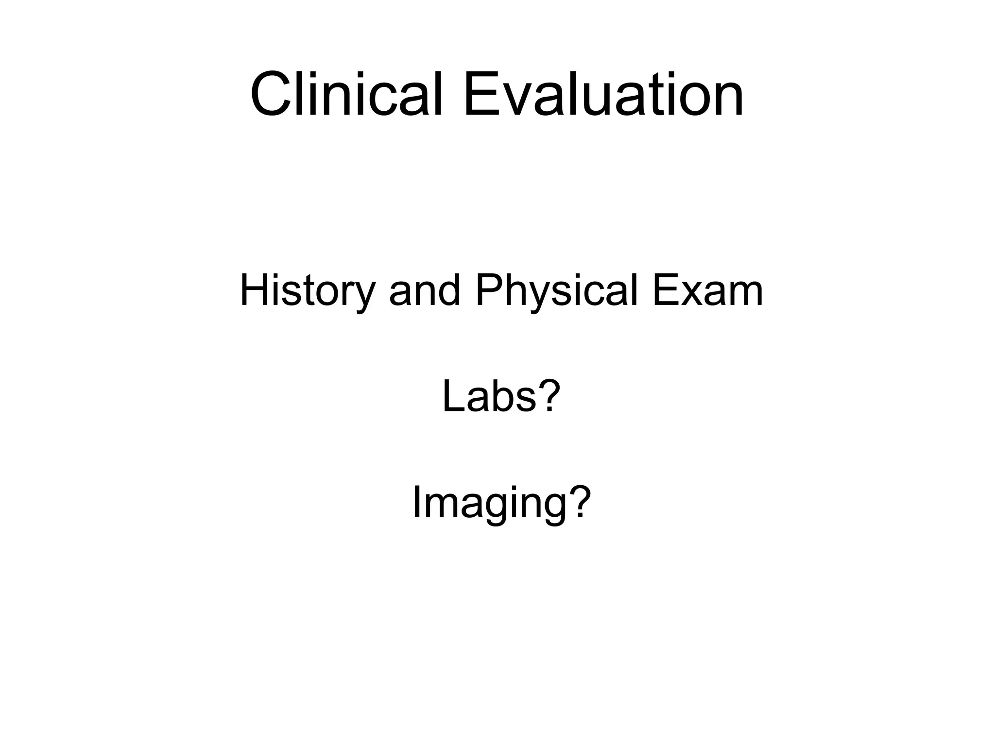
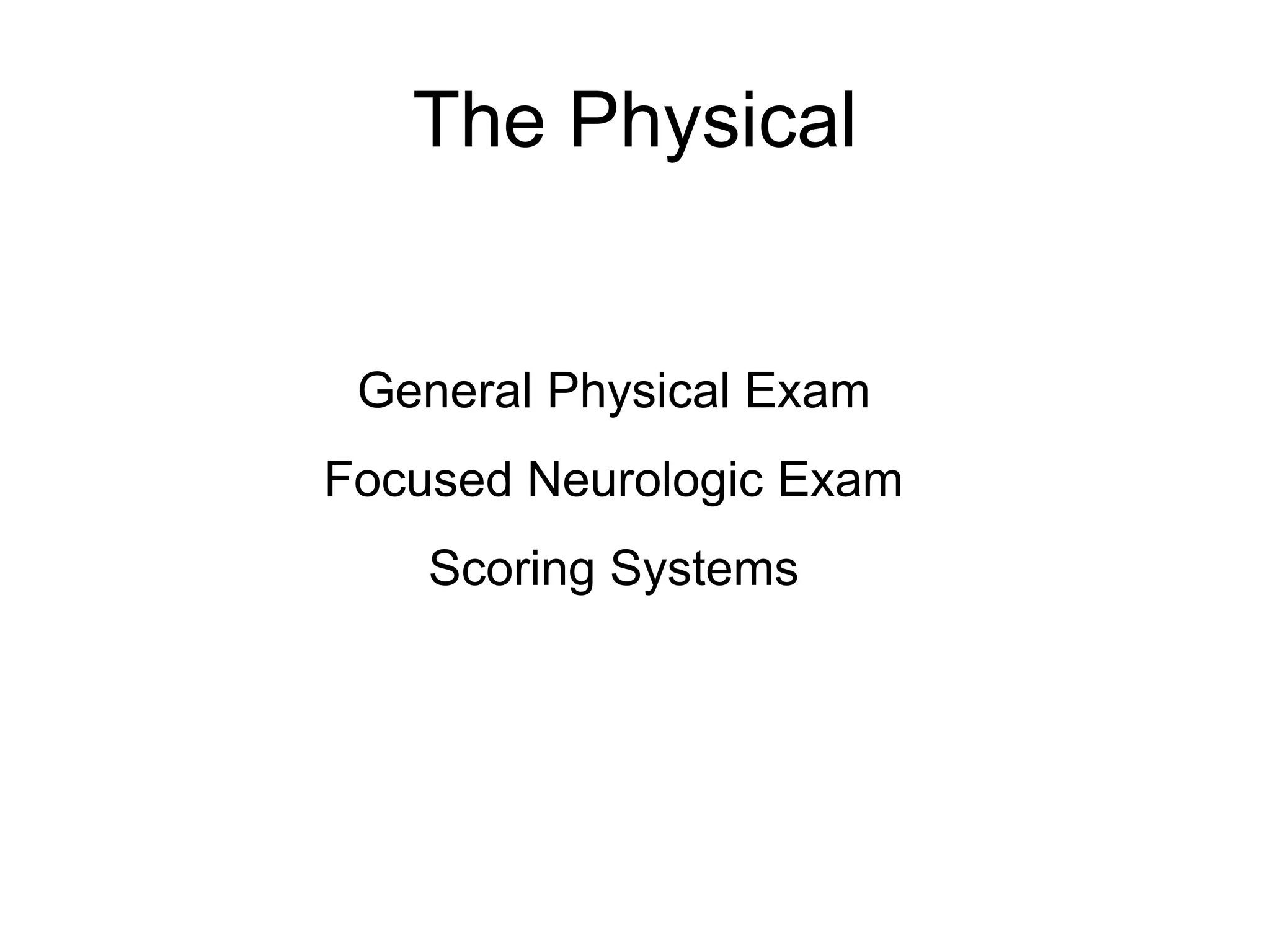
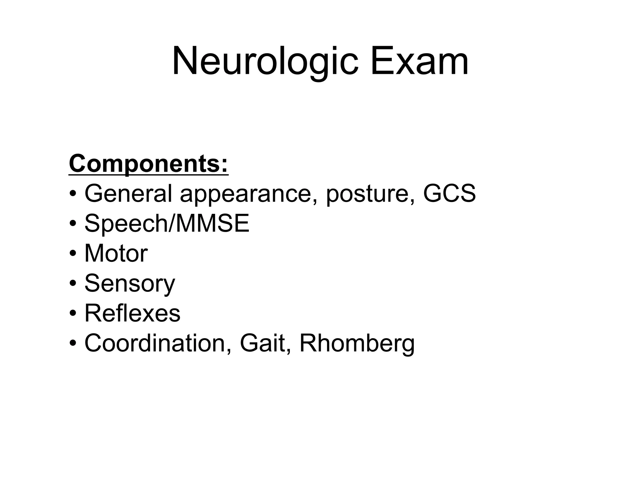
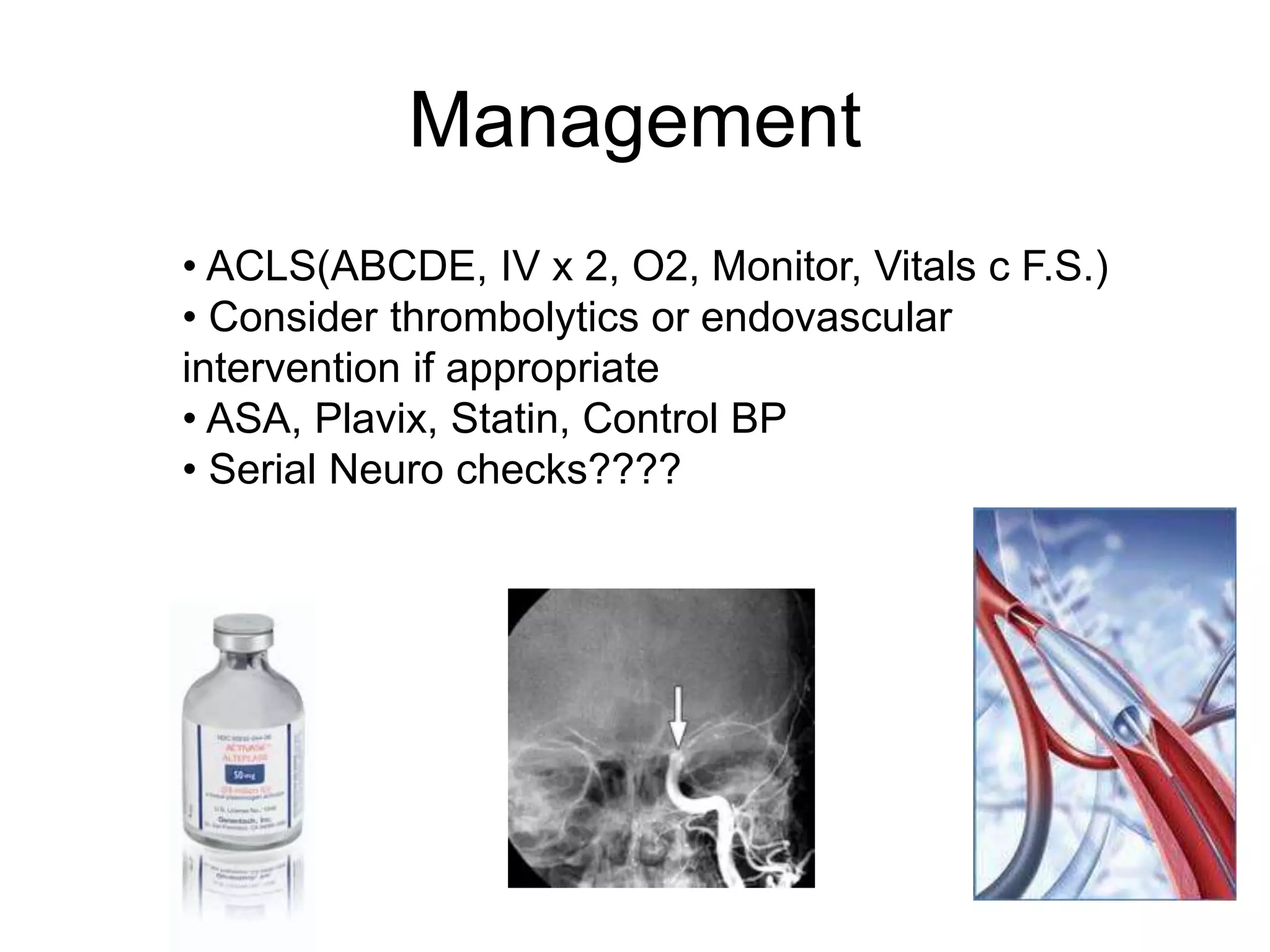
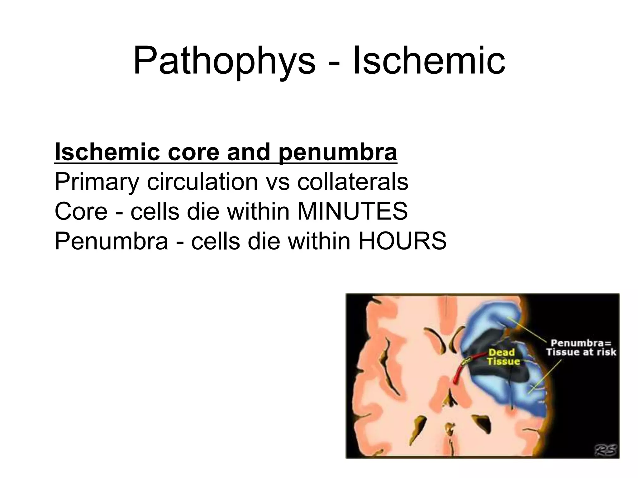
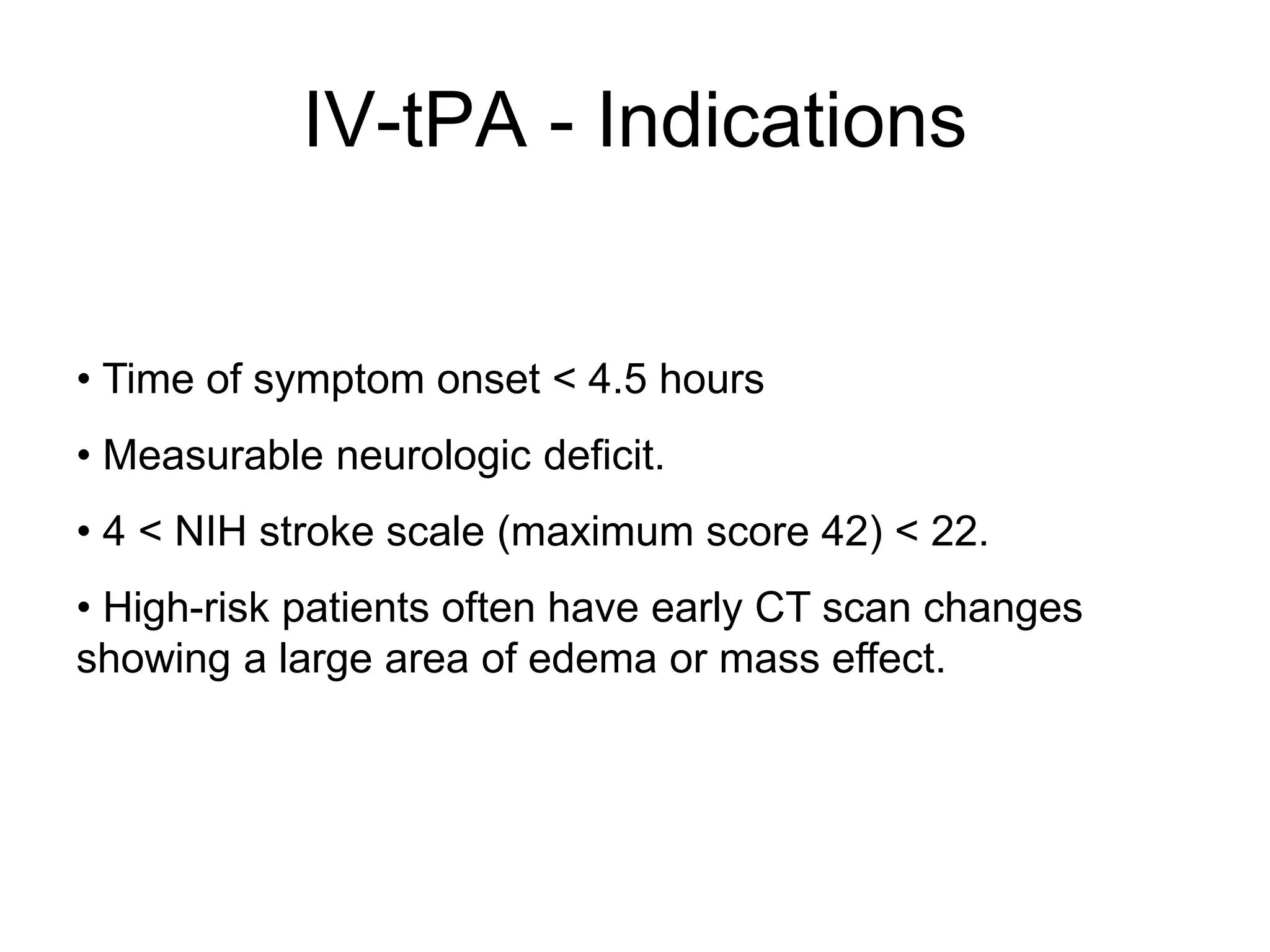
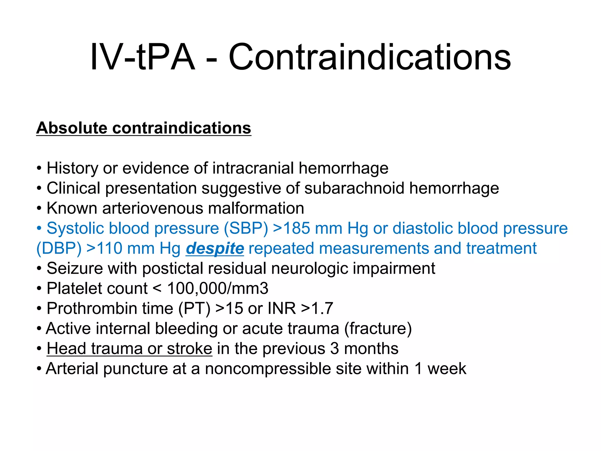
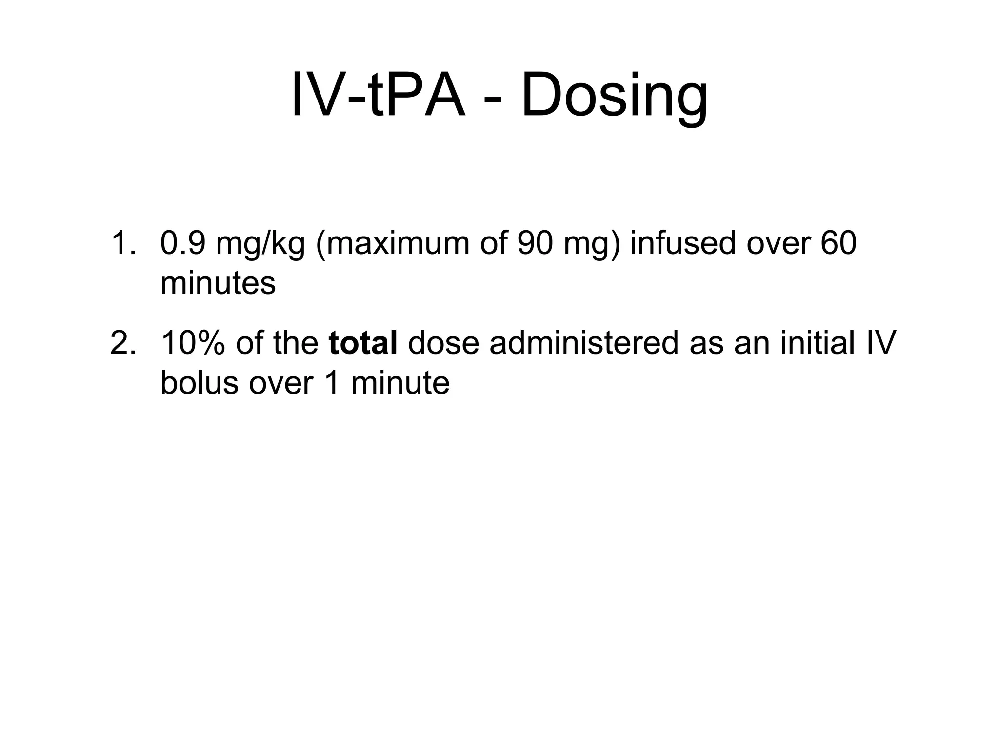
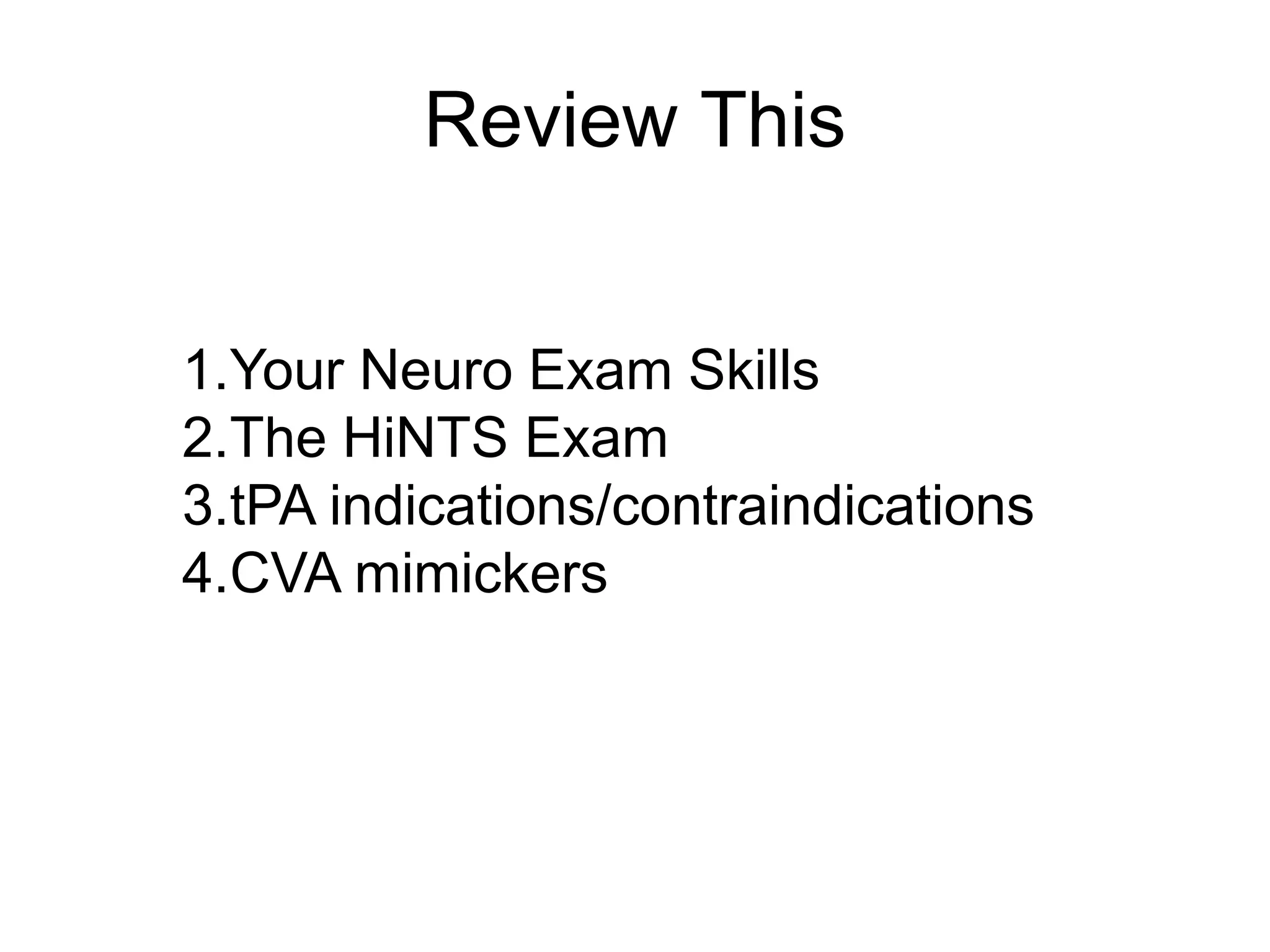
David Marcus, MD
@EMIMDoc - EMIMDoc.org
Assistant Program Director – LIJ EM/IM
Co-Director of Student Education - LIJ
Stroke and the Neurologic Exam
Doctor – What’s Wrong With Me?!?!?!
A 78 year old woman is brought in by her daughter after waking up
this morning unable to get out of bed. She is alert, appears tired,
follows commands slowly and cannot move the left side of her body.
Your patient, a 66 year old man with diabetes and HTN presents with
1 full day of severe dizziness. He says the room is spinning and he
cannot walk unassisted.
The 48 year old woman you are examining in room in intake is
complaining of a sudden onset of severe headache about 1 hour
ago. This is the worst headache of her life. It is associated with
drooping of her left eyelid and facial asymmetry. She has a history
of HTN, occasional headaches and asthma.
Intro to Stroke: epidemiology, definitions, pathophys
Categories
Risk Factors
Evaluation of Suspected Stroke
Management
Goals
Epidemiology
Epidemiology
Black > White > Hispanic
Men > Women
“Sudden loss of circulation to an
area of the brain, resulting in a
corresponding loss of
neurologic function.”
Definition
Cerebrovascular Accident (CVA)
Stroke syndrome
Brain Attack
AKA
TIA
“…a transient episode of neurological
dysfunction caused by focal brain, spinal cord,
or retinal ischemia, without acute infarction.”
(AHA)
10% - CVA in 90 days
Ischemic:Hemorrhagic/ICH
Categories
Hemorrhagic Conversion
Vascular occlusion >>> Ischemia >>> cell hypoxia
and depletion of ATP>>>
Cell membrane failure >>> Cytotoxic Edema
4-6 hours: breakdown of BBB >>> Vasogenic edema
Pathophys - Ischemic
Pathophys - Hemorrhagic
Direct damage due to hematoma
Increasing mass effect and ICP
Inflammatory changes
Anatomy - Circle of Willis
Anterior Cerebral Artery
Medial frontal and parietal lobe, caudate head, globus
pallidus, anterior limb of internal capsule
Middle Cerebral Artery
Lateral frontal and parietal lobes lateral and anterior
temporal lobe, globus pallidus and putamen, internal
capsule
Anterior Choroidal Artery
Optic tracts, medial temporal lobe, ventrolateral thalamus,
corona radiata, posterior limb of the internal capsule
Anterior Circulation (Carotid)
Posterior Circulation
(AKA Vertebro-basilar)
Posterior Cerebral Artery
Occipital lobes, medial and posterior temporal and parietal
lobes, brainstem, posterior thalamus and midbrain
Posterior Inferior Cerebellar Artery :
Inferior vermis; posterior and inferior cerebellar hemispheres
Anterior Inferior Cerebellar Artery: Anterolateral
cerebellum
Superior Cerebellar Artery: Superior vermis; superior
cerebellum
Age
Race
Sex
Ethnicity
History of migraine headaches
Sickle cell disease
Fibromuscular dysplasia
Heredity
Hypertension (the most important modifiable factor)
Diabetes mellitus
Cardiac disease
Hypercholesterolemia
Transient ischemic attacks (TIAs)
Carotid stenosis
Hyperhomocystinemia
Lifestyle issues - Excessive alcohol intake, tobacco use, illicit drug
use, obesity, physical inactivity
Oral contraceptive use
Risk Factors
Remember Us?
A 78 year old woman is brought in by her daughter after waking up
this morning unable to get out of bed. She is alert, appears tired,
follows commands slowly and cannot move the left side of her body.
Your patient, a 66 year old man with diabetes and HTN presents with
1 full day of severe dizziness. He says the room is spinning and he
cannot walk unassisted.
The 48 year old woman you are examining in room in intake is
complaining of a sudden onset of severe headache about 1 hour
ago. This is the worst headache of her life. It is associated with
drooping of her left eyelid and facial asymmetry. She has a history
of HTN, occasional headaches and asthma.
DDX
Seizure (17%)
Systemic infection (17%)
Brain tumor (15%)
Toxic-metabolic (hyponatremia, hypoglycemia…)(13%)
Positional vertigo (6%)
Bell’s palsy and other mono/poly neuropathies
History and Physical Exam
Labs?
Imaging?
Clinical Evaluation
The History
In addition to all the usual, focus on:
•Onset: When was patient last seen normal?
• Fluctuating symptoms
• Previous episodes
• Medications, anticoagulation?
The Physical
General Physical Exam
Focused Neurologic Exam
Scoring Systems
Components:
• General appearance, posture, GCS
• Speech/MMSE
• Motor
• Sensory
• Reflexes
• Coordination, Gait, Rhomberg
Neurologic Exam
Well organized exam and good instructions -
http://cloud.med.nyu.edu/modules/pub/neurosurgery/
Good videos, especially of abnormals -
http://library.med.utah.edu/neurologicexam/html/home_ex
am.html
Neuro Exam - Aides
• Assess level of responsiveness (AVPU/GCS)
• Focus on signs of persistent lateralizing asymmetry
• Reflex abnormalities may localize to brainstem
• Prognosis of decorticate better than decerebrate
• May assess the following, even in unresponsive:
•Corneal reflex (CNV)
•Doll’s eye (Brainstem, EOM)
•Calorics (EOM)
•Pupillary response
•Introducing objects into field of view
•Facial grimacing (CNVII)
•Gag reflex (CN IX, X)
The Altered Patient
Scoring Systems
Scoring Systems
Differentiating between central and peripheral vertigo
• ACLS(ABCDE, IV x 2, O2, Monitor, Vitals c F.S.)
• Consider thrombolytics or endovascular
intervention if appropriate
• ASA, Plavix, Statin, Control BP
• Serial Neuro checks????
Management
Pathophys - Ischemic
Ischemic core and penumbra
Primary circulation vs collaterals
Core - cells die within MINUTES
Penumbra - cells die within HOURS
IV-tPA - Indications
• Time of symptom onset < 4.5 hours
• Measurable neurologic deficit.
• 4 < NIH stroke scale (maximum score 42) < 22.
• High-risk patients often have early CT scan changes
showing a large area of edema or mass effect.
IV-tPA - Contraindications
Absolute contraindications
• History or evidence of intracranial hemorrhage
• Clinical presentation suggestive of subarachnoid hemorrhage
• Known arteriovenous malformation
• Systolic blood pressure (SBP) >185 mm Hg or diastolic blood pressure
(DBP) >110 mm Hg despite repeated measurements and treatment
• Seizure with postictal residual neurologic impairment
• Platelet count < 100,000/mm3
• Prothrombin time (PT) >15 or INR >1.7
• Active internal bleeding or acute trauma (fracture)
• Head trauma or stroke in the previous 3 months
• Arterial puncture at a noncompressible site within 1 week
IV-tPA - Dosing
1. 0.9 mg/kg (maximum of 90 mg) infused over 60
minutes
2. 10% of the total dose administered as an initial IV
bolus over 1 minute
• 5% of ischemic strokes undergo hemorrhagic conversion
• In the US, 20% of individuals die within one year after a
first-time stroke
• In stroke survivors from the Framingham Heart Study:
• 31% needed help caring for themselves
• 20% needed help when walking
• 71% had impaired vocational capacity
Prognosis
Review This
1.Your Neuro Exam Skills
2.The HiNTS Exam
3.tPA indications/contraindications
4.CVA mimickers

Stroke Overview - EM Orientation

  • 1.
    David Marcus, MD @EMIMDoc- EMIMDoc.org Assistant Program Director – LIJ EM/IM Co-Director of Student Education - LIJ Stroke and the Neurologic Exam
  • 2.
    Doctor – What’sWrong With Me?!?!?!
  • 3.
    Doctor – What’sWrong With Me?!?!?! A 78 year old woman is brought in by her daughter after waking up this morning unable to get out of bed. She is alert, appears tired, follows commands slowly and cannot move the left side of her body. Your patient, a 66 year old man with diabetes and HTN presents with 1 full day of severe dizziness. He says the room is spinning and he cannot walk unassisted. The 48 year old woman you are examining in room in intake is complaining of a sudden onset of severe headache about 1 hour ago. This is the worst headache of her life. It is associated with drooping of her left eyelid and facial asymmetry. She has a history of HTN, occasional headaches and asthma.
  • 4.
    Intro to Stroke:epidemiology, definitions, pathophys Categories Risk Factors Evaluation of Suspected Stroke Management Goals
  • 5.
  • 6.
    Epidemiology Black > White> Hispanic Men > Women
  • 7.
    “Sudden loss ofcirculation to an area of the brain, resulting in a corresponding loss of neurologic function.” Definition
  • 8.
    Cerebrovascular Accident (CVA) Strokesyndrome Brain Attack AKA
  • 9.
    TIA “…a transient episodeof neurological dysfunction caused by focal brain, spinal cord, or retinal ischemia, without acute infarction.” (AHA) 10% - CVA in 90 days
  • 10.
  • 12.
  • 13.
    Vascular occlusion >>>Ischemia >>> cell hypoxia and depletion of ATP>>> Cell membrane failure >>> Cytotoxic Edema 4-6 hours: breakdown of BBB >>> Vasogenic edema Pathophys - Ischemic
  • 14.
    Pathophys - Hemorrhagic Directdamage due to hematoma Increasing mass effect and ICP Inflammatory changes
  • 16.
  • 17.
    Anterior Cerebral Artery Medialfrontal and parietal lobe, caudate head, globus pallidus, anterior limb of internal capsule Middle Cerebral Artery Lateral frontal and parietal lobes lateral and anterior temporal lobe, globus pallidus and putamen, internal capsule Anterior Choroidal Artery Optic tracts, medial temporal lobe, ventrolateral thalamus, corona radiata, posterior limb of the internal capsule Anterior Circulation (Carotid)
  • 18.
    Posterior Circulation (AKA Vertebro-basilar) PosteriorCerebral Artery Occipital lobes, medial and posterior temporal and parietal lobes, brainstem, posterior thalamus and midbrain Posterior Inferior Cerebellar Artery : Inferior vermis; posterior and inferior cerebellar hemispheres Anterior Inferior Cerebellar Artery: Anterolateral cerebellum Superior Cerebellar Artery: Superior vermis; superior cerebellum
  • 21.
    Age Race Sex Ethnicity History of migraineheadaches Sickle cell disease Fibromuscular dysplasia Heredity Hypertension (the most important modifiable factor) Diabetes mellitus Cardiac disease Hypercholesterolemia Transient ischemic attacks (TIAs) Carotid stenosis Hyperhomocystinemia Lifestyle issues - Excessive alcohol intake, tobacco use, illicit drug use, obesity, physical inactivity Oral contraceptive use Risk Factors
  • 22.
    Remember Us? A 78year old woman is brought in by her daughter after waking up this morning unable to get out of bed. She is alert, appears tired, follows commands slowly and cannot move the left side of her body. Your patient, a 66 year old man with diabetes and HTN presents with 1 full day of severe dizziness. He says the room is spinning and he cannot walk unassisted. The 48 year old woman you are examining in room in intake is complaining of a sudden onset of severe headache about 1 hour ago. This is the worst headache of her life. It is associated with drooping of her left eyelid and facial asymmetry. She has a history of HTN, occasional headaches and asthma.
  • 23.
    DDX Seizure (17%) Systemic infection(17%) Brain tumor (15%) Toxic-metabolic (hyponatremia, hypoglycemia…)(13%) Positional vertigo (6%) Bell’s palsy and other mono/poly neuropathies
  • 24.
    History and PhysicalExam Labs? Imaging? Clinical Evaluation
  • 25.
    The History In additionto all the usual, focus on: •Onset: When was patient last seen normal? • Fluctuating symptoms • Previous episodes • Medications, anticoagulation?
  • 26.
    The Physical General PhysicalExam Focused Neurologic Exam Scoring Systems
  • 27.
    Components: • General appearance,posture, GCS • Speech/MMSE • Motor • Sensory • Reflexes • Coordination, Gait, Rhomberg Neurologic Exam
  • 28.
    Well organized examand good instructions - http://cloud.med.nyu.edu/modules/pub/neurosurgery/ Good videos, especially of abnormals - http://library.med.utah.edu/neurologicexam/html/home_ex am.html Neuro Exam - Aides
  • 29.
    • Assess levelof responsiveness (AVPU/GCS) • Focus on signs of persistent lateralizing asymmetry • Reflex abnormalities may localize to brainstem • Prognosis of decorticate better than decerebrate • May assess the following, even in unresponsive: •Corneal reflex (CNV) •Doll’s eye (Brainstem, EOM) •Calorics (EOM) •Pupillary response •Introducing objects into field of view •Facial grimacing (CNVII) •Gag reflex (CN IX, X) The Altered Patient
  • 30.
  • 31.
    Scoring Systems Differentiating betweencentral and peripheral vertigo
  • 32.
    • ACLS(ABCDE, IVx 2, O2, Monitor, Vitals c F.S.) • Consider thrombolytics or endovascular intervention if appropriate • ASA, Plavix, Statin, Control BP • Serial Neuro checks???? Management
  • 33.
    Pathophys - Ischemic Ischemiccore and penumbra Primary circulation vs collaterals Core - cells die within MINUTES Penumbra - cells die within HOURS
  • 34.
    IV-tPA - Indications •Time of symptom onset < 4.5 hours • Measurable neurologic deficit. • 4 < NIH stroke scale (maximum score 42) < 22. • High-risk patients often have early CT scan changes showing a large area of edema or mass effect.
  • 35.
    IV-tPA - Contraindications Absolutecontraindications • History or evidence of intracranial hemorrhage • Clinical presentation suggestive of subarachnoid hemorrhage • Known arteriovenous malformation • Systolic blood pressure (SBP) >185 mm Hg or diastolic blood pressure (DBP) >110 mm Hg despite repeated measurements and treatment • Seizure with postictal residual neurologic impairment • Platelet count < 100,000/mm3 • Prothrombin time (PT) >15 or INR >1.7 • Active internal bleeding or acute trauma (fracture) • Head trauma or stroke in the previous 3 months • Arterial puncture at a noncompressible site within 1 week
  • 36.
    IV-tPA - Dosing 1.0.9 mg/kg (maximum of 90 mg) infused over 60 minutes 2. 10% of the total dose administered as an initial IV bolus over 1 minute
  • 37.
    • 5% ofischemic strokes undergo hemorrhagic conversion • In the US, 20% of individuals die within one year after a first-time stroke • In stroke survivors from the Framingham Heart Study: • 31% needed help caring for themselves • 20% needed help when walking • 71% had impaired vocational capacity Prognosis
  • 38.
    Review This 1.Your NeuroExam Skills 2.The HiNTS Exam 3.tPA indications/contraindications 4.CVA mimickers

Editor's Notes

  • #9 Stroke is a nonspecific term
  • #10 Transient Ischemic Attack The clinical symptoms of TIA typically last less than an hour, but prolonged episodes can occur. While the classical definition of TIA included symptoms lasting as long as 24 hours, advances in neuroimaging have suggested that many such cases represent minor strokes with resolved symptoms rather than true TIAs. The AHA/ASA-endorsed definition of TIA is as follows: Transient ischemic attack (TIA) is a transient episode of neurological dysfunction caused by focal brain, spinal cord, or retinal ischemia, without acute infarction
  • #11 (Prior literature indicated that only 8-18% of strokes are hemorrhagic, but a retrospective review from a stroke center found that 40.9% of 757 strokes included in the study were hemorrhagic.[1] ) Cardioembolic: may account for up to 20% of acute strokes and have been reported to have the highest 1-month mortality.
  • #12 Hemorrhagic, Ischemic
  • #13 Hemorrhagic conversion
  • #14 Regions of the brain with CBF lower than 10 mL/100g of tissue/min are referred to collectively as the core, and these cells are presumed to die within minutes of stroke onset. Zones of decreased or marginal perfusion (CBF < 25 mL/100g of tissue/min) are collectively called the ischemic penumbra. Tissue in the penumbra can remain viable for several hours because of marginal tissue perfusion
  • #16 Thomas Willis - described circle of Willis in 1664, Mellitus, of DM is attributed to him as well. Also was first to describe the cranial nerves, in the current order.
  • #35 Patients with a score above 22 are considered high risk for hemorrhagic conversion due to the probability of a large infarcted area. Patients with a score less than 4 have only minor neurologic deficits, for which thrombolytic therapy is not indicated.