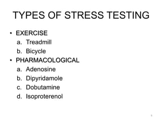
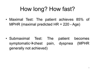
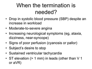
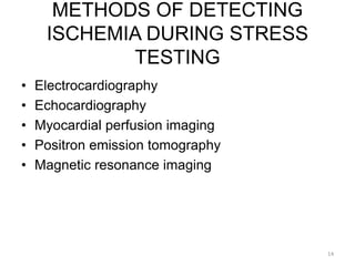
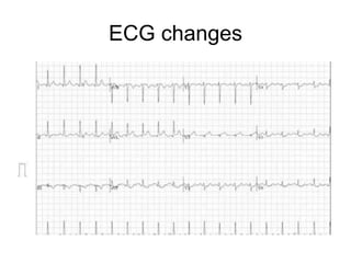
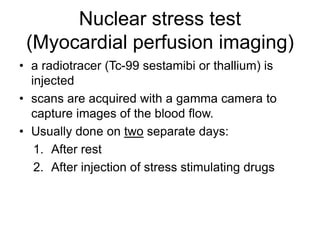
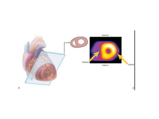
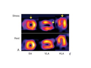
Cardiac stress tests
Porya Hassan Abadi
Introduction
• Diagnostic test for coronary artery disease
• Main aim is to assess the coronary flow system
• Measures ability of heart to respond to stress
• Stress is induced by drugs or exercise under
clinical supervision
Clinical case
• 50 y/o female with diabetes, HTN, smoker
• Nonspecific substernal chest pain
• Slight dyspnoea
• ECG nonspecific
• Normal cardiac markers
Indications
• Diagnosis of coronary disease in patients with:
– Stable chest pain
– Unstable chest pain stabilised with therapy
– Previous MI
– Post revascularisation (CABG)
Contra-indications
• Acute myocardial infarction (within 2 d)
• Unstable angina not previously stabilized by
medical therapy
• Uncontrolled cardiac arrhythmias causing
symptoms or hemodynamic compromise
• Symptomatic severe aortic stenosis
• Uncontrolled symptomatic heart failure
• Acute pulmonary embolus or pulmonary
infarction
• Acute myocarditis or pericarditis
• Acute aortic dissection
TYPES OF STRESS TESTING
6
• EXERCISE
a. Treadmill
b. Bicycle
• PHARMACOLOGICAL
a. Adenosine
b. Dipyridamole
c. Dobutamine
d. Isoproterenol
Physical stimulation
• Test is done by exercise on treadmill or pedaling
an stationary bicycle.
• Constantly monitored (ECG,BP and heart rate)
8
TEST OF CHOICE IN THOSE
WITH:
• NORMAL BASLINE ECG
• ABLE TO EXERCISE
• NO PRIOR REVASCULARISATION
How long? How fast?
9
• Maximal Test: The patient achieves 85% of
MPHR (maximal predicted HR = 220 - Age)
• Submaximal Test: The patient becomes
symptomaticchest pain, dyspnea (MPHR
generally not achieved)
When the termination is
needed?
• Drop in systolic blood pressure (SBP) despite an
increase in workload
• Moderate-to-severe angina
• Increasing neurological symptoms (eg, ataxia,
dizziness, near-syncope)
• Signs of poor perfusion (cyanosis or pallor)
• Subject’s desire to stop
• Sustained ventricular tachycardia
• ST elevation (> 1 mm) in leads (other than V 1
or aVR)
Safety and risk of exercise test
• Nonselected patient population: Mortality <
0.01%
• Within 4 weeks of MI: Mortality = 0.03% and
Morbidity = 0.09% (reinfarction, cardiac arrest)
Pharmacological Stress Test
• Stress is induced using agents such as :
1. Vasodilators such as: Adenosine and
Dipyridamole not used asthma or COPD
2. B2 agonist such as: Dobutamine
INDICATIONS OF
PHARMACOLOGICAL STRESS
TESTING
13
• Patients inability to exercise adequately because
of physical or psychological limitations.
• The chosen test cannot be performed readily
with exercise (e.g. PET scanning).
METHODS OF DETECTING
ISCHEMIA DURING STRESS
TESTING
14
• Electrocardiography
• Echocardiography
• Myocardial perfusion imaging
• Positron emission tomography
• Magnetic resonance imaging
ECG changes
Stress echocardiography
• echocardiography is performed both before and
after the exercise used to detect:
1. Wall motion abnormalities
2. Ejection fraction
3. Valvular heart disease
Nuclear stress test
(Myocardial perfusion imaging)
• a radiotracer (Tc-99 sestamibi or thallium) is
injected
• scans are acquired with a gamma camera to
capture images of the blood flow.
• Usually done on two separate days:
1. After rest
2. After injection of stress stimulating drugs
Cardiac stress test
Cardiac stress test

Cardiac stress test

  • 1.
  • 2.
    Introduction • Diagnostic testfor coronary artery disease • Main aim is to assess the coronary flow system • Measures ability of heart to respond to stress • Stress is induced by drugs or exercise under clinical supervision
  • 3.
    Clinical case • 50y/o female with diabetes, HTN, smoker • Nonspecific substernal chest pain • Slight dyspnoea • ECG nonspecific • Normal cardiac markers
  • 4.
    Indications • Diagnosis ofcoronary disease in patients with: – Stable chest pain – Unstable chest pain stabilised with therapy – Previous MI – Post revascularisation (CABG)
  • 5.
    Contra-indications • Acute myocardialinfarction (within 2 d) • Unstable angina not previously stabilized by medical therapy • Uncontrolled cardiac arrhythmias causing symptoms or hemodynamic compromise • Symptomatic severe aortic stenosis • Uncontrolled symptomatic heart failure • Acute pulmonary embolus or pulmonary infarction • Acute myocarditis or pericarditis • Acute aortic dissection
  • 6.
    TYPES OF STRESSTESTING 6 • EXERCISE a. Treadmill b. Bicycle • PHARMACOLOGICAL a. Adenosine b. Dipyridamole c. Dobutamine d. Isoproterenol
  • 7.
    Physical stimulation • Testis done by exercise on treadmill or pedaling an stationary bicycle. • Constantly monitored (ECG,BP and heart rate)
  • 8.
    8 TEST OF CHOICEIN THOSE WITH: • NORMAL BASLINE ECG • ABLE TO EXERCISE • NO PRIOR REVASCULARISATION
  • 9.
    How long? Howfast? 9 • Maximal Test: The patient achieves 85% of MPHR (maximal predicted HR = 220 - Age) • Submaximal Test: The patient becomes symptomaticchest pain, dyspnea (MPHR generally not achieved)
  • 10.
    When the terminationis needed? • Drop in systolic blood pressure (SBP) despite an increase in workload • Moderate-to-severe angina • Increasing neurological symptoms (eg, ataxia, dizziness, near-syncope) • Signs of poor perfusion (cyanosis or pallor) • Subject’s desire to stop • Sustained ventricular tachycardia • ST elevation (> 1 mm) in leads (other than V 1 or aVR)
  • 11.
    Safety and riskof exercise test • Nonselected patient population: Mortality < 0.01% • Within 4 weeks of MI: Mortality = 0.03% and Morbidity = 0.09% (reinfarction, cardiac arrest)
  • 12.
    Pharmacological Stress Test •Stress is induced using agents such as : 1. Vasodilators such as: Adenosine and Dipyridamole not used asthma or COPD 2. B2 agonist such as: Dobutamine
  • 13.
    INDICATIONS OF PHARMACOLOGICAL STRESS TESTING 13 •Patients inability to exercise adequately because of physical or psychological limitations. • The chosen test cannot be performed readily with exercise (e.g. PET scanning).
  • 14.
    METHODS OF DETECTING ISCHEMIADURING STRESS TESTING 14 • Electrocardiography • Echocardiography • Myocardial perfusion imaging • Positron emission tomography • Magnetic resonance imaging
  • 15.
  • 16.
    Stress echocardiography • echocardiographyis performed both before and after the exercise used to detect: 1. Wall motion abnormalities 2. Ejection fraction 3. Valvular heart disease
  • 17.
    Nuclear stress test (Myocardialperfusion imaging) • a radiotracer (Tc-99 sestamibi or thallium) is injected • scans are acquired with a gamma camera to capture images of the blood flow. • Usually done on two separate days: 1. After rest 2. After injection of stress stimulating drugs