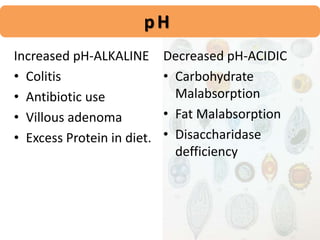
Muhammad Asif Zeb
Lecturer Hematology
IPMS-KMU
Human feces is called as STOOL.
Faeces / Feces is plural of latin term faex meaning
RESIDUE.
It is the waste residue of indigestible materials of
human digestive tract expelled through the anus
during defecation.
Meconium is newborn’s first feces.
SCATOLOGY or CAPROLOGY is the study of feces.
DEFINITION
• Undigested and Unabsorbed food
• Intestinal secretions, Mucous
• Bile pigments and Salts
• Bacteria and Inorganic material
• Epithelial cells,
COMPOSITION
• Universal Precautions
• Stool should be collected in a dry, sterilized,
wide mouthed container.
• It should be uncontaminated with Urine or any
other body secretions.
• Properly labeled and always a fresh sample
should be tested.
COLLECTION
MACROSCOPIC EXAMINATION
• Volume
• Colour
• Consistency
• Adour
• Blood, Mucous
• Parts of parasite and Adult Parasite
COLOUR OF STOOL
n
color which results from a combination of bile
and stercobilirubin.
VARIATIONS
Bright Red/Maroon Clay
Blood streak White
Yellow Pale greasy
Green
Black
COLOUR OF FECES -in Infants
As breast fed infants have no bacteria in their intestine so
stercobilinogen is not produced and the colour of faeces remain
yellow
 Some healthy children may pass frequent, loose stools containing
undigested vegetable matter called as Toddler’s diarrhoea.
In diarrhoea the movement of intestine is so rapid that bacteria
do not have time to decompose bilirubin and green faeces may be
passed
• obstruction to the flow of bile into the intestine,
gives rise to pale, tan or clay coloured faeces.
• Chlorophyll rich foods produce green faeces.
• Bleeding into upper gut gives rise to black faeces due
to altered blood
• In addition, oral iron ingestion results in black faeces
• Diarrhoea mixed with mucous and
Blood is suggestive of Typhoid,
Amoebiasis, Large bowel Carcinoma.
• Diarrhoea mixed with mucous and
Pus is suggestive of Ulcerative
Collitis, Regional Enteritis, Shigellosis,
Salmonellosis, Acute diverticullitis,
Intestinal TB.
O D O U R O F S T O O L
 Basically depends on the pH of the stool and INDOLE
and SKETOLE are the substances that produce normal
odour formed by Intestinal bacterial fermentation .
Faeces are particularly offensive in amoebic dysentery
 A foul odour is caused by degradation of undigested
protein and excessive carbohydrate intake.
NORMALCY
• Water – Upto 75%
• pH – 5.8 to 7.5
• Occult blood, RS – Negative
• Bile – Negative in Adults
Positive in Children
• Sodium – 5.8 to 9.8 mEq/24hrs
• Chlorides – 2.5 to 3.9 mEq/24hrs
• Potassium – 15.7 to 20.7 mEq/24hrs
• Lipids / Fatty acids – 0 to 6 gms/24hrs
• Nitrogen - <2.5g/24hrs
pH
Increased pH-ALKALINE
• Colitis
• Antibiotic use
• Villous adenoma
• Excess Protein in diet.
Decreased pH-ACIDIC
• Carbohydrate
Malabsorption
• Fat Malabsorption
• Disaccharidase
defficiency
•Round worm
•Hook worm
•Tape worm
•Pin worm
•Whipworm
PARASITE
MICROSCOPIC
EXAMINATION
• Microscope slides
• Cover slips
• Sodium chloride solution
• Lugol’s Iodine Solution
• Wooden applicator
• Fresh stool
• Gloves
MATERIALS
SLIDE PREPARATION
Saline Specimen Prpn.
Iodine Specimen Prpn.
• A drop of Saline or Lugol’s Iodine is placed over a
clean microscopic slide.
• About 2mg of stool sample should be taken and
mixed with wooden stick, placed over the slide.
• Coverslip is placed avoiding air bubbles.
• Examined under Microscope.
SLIDES
Preparation of Stool slide
• Use cleaned microscope
slides
• Place a drop of saline or
ioidin
• Take a small amount of
stool with a wooden stick
• Mix stool with saline
• Mistake: Too much stool!
• Place coverslip
• Avoid air bubbles!
PIN WORM EGG COLLECTION
Eggs of Pin worm – Enterobius vermicularis rarely
appear in stools. These are usually collected in
the folds of skin in perianal region.
COLLECTION
Cotton swab / Plaster patch – Anus especially in
early morning – Dipped in Saline – Observed.
EXAMINATION OF PARASITES
Warm stools are best for detecting Ova or
parasites. Do not refrigerate the specimen.
Because of cyclic life cycle of parasites,
three separate random stool specimens are
recommended for examination.
LEUKOCYTES IN STOOL
Large amounts of leukocytes is suggestive of
Chronic Ulceratice Collitis, Chronic Bacillary
Dysentry, Localised Abscess, Fistulas.
Mononuclear Leukocytes appear in Typhoid.
Polymorphonuclear Leukocytes appear in
Shigellosis, Salmonellosis, Invasice E. coli
diarrhoea, Ulceratice Collitis.
HOOKWORM
Ancylostoma duodenale.
● It is colourless with a thin shell
which
appears microscopically as a black line
around the ovum.
● Oval in shape,.
● Contains an ovum which appears
segmented (usually 4–8 cell stage).
ROUNDWORM
Ascaris lumbricoides
TAPEWORM
Taenia solium-Pork
Taenia saginata-Beef
WHIPWORM
Trichuris trichura
PINWORM
Enterobius vermicularis
ENTAMOEBA
Entamoeba histolytica
• The cysts are retractile, round structures, 12-15
μm in diameter.
• contain 1-4 nuclei.
• The nuclear membrane is thin,regular and
circular and a small central karyosome is easily
visible in iodine stained preparation. Cytoplasm in
iodine preparation is
• yellowish grey and granular.
• chromatoid bodies
GIADIASIS
CHEMICAL
EXAMINATION
OCCULT BLOOD
PRINCIPLE – BENZIDINE TEST(a sensitive test for presence of blood
based on prodctn of blue)
Perioxidase action of hemoglobin in
blood converts hydrogen peroxide to water and
nascent oxygen. This oxygen oxidizes benzidine
in acid medium to form green to blue coloured
complex.
• Make a suspension of faeces in 10 ml saline and boil to
inactivate the normally present oxidising enzymes in
faeces.
• Make 2 ml of saturated solution of benzidine in glacial
• acetic acid in other tube.
• Add 2 ml of H2O2 and check whether blue or green
colour develops. If so discard the reagents.
• Add faecal suspension drop by drop to the solution of
benzidine and H2O2 till there is change of colour.
• Appearance of deep blue colour indicates presence of
blood.
Found in Ulcers,
Diverticullitis,
Ulcerative Collitis,
Diaphragmatic Hernia,
Adenoma,
CA Colon, Gastrium
OCCULT BLOOD cont…
REDUCING SUBSTANCES
Tested for RS especially in infants with Chronic
diarrhea to rule out Lactose Intolerance.
Stool will be positive for RS in variety of conditions
especially in Rota viral Infection in Infants.
(rota virus is virus that infect bowels causing a severe inflamaatiom of stomach)

Stool examination.pptx

  • 1.
    Muhammad Asif Zeb LecturerHematology IPMS-KMU
  • 2.
    Human feces iscalled as STOOL. Faeces / Feces is plural of latin term faex meaning RESIDUE. It is the waste residue of indigestible materials of human digestive tract expelled through the anus during defecation. Meconium is newborn’s first feces. SCATOLOGY or CAPROLOGY is the study of feces. DEFINITION
  • 3.
    • Undigested andUnabsorbed food • Intestinal secretions, Mucous • Bile pigments and Salts • Bacteria and Inorganic material • Epithelial cells, COMPOSITION
  • 4.
    • Universal Precautions •Stool should be collected in a dry, sterilized, wide mouthed container. • It should be uncontaminated with Urine or any other body secretions. • Properly labeled and always a fresh sample should be tested. COLLECTION
  • 5.
    MACROSCOPIC EXAMINATION • Volume •Colour • Consistency • Adour • Blood, Mucous • Parts of parasite and Adult Parasite
  • 6.
    COLOUR OF STOOL n colorwhich results from a combination of bile and stercobilirubin. VARIATIONS Bright Red/Maroon Clay Blood streak White Yellow Pale greasy Green Black
  • 7.
    COLOUR OF FECES-in Infants As breast fed infants have no bacteria in their intestine so stercobilinogen is not produced and the colour of faeces remain yellow  Some healthy children may pass frequent, loose stools containing undigested vegetable matter called as Toddler’s diarrhoea. In diarrhoea the movement of intestine is so rapid that bacteria do not have time to decompose bilirubin and green faeces may be passed
  • 8.
    • obstruction tothe flow of bile into the intestine, gives rise to pale, tan or clay coloured faeces. • Chlorophyll rich foods produce green faeces. • Bleeding into upper gut gives rise to black faeces due to altered blood • In addition, oral iron ingestion results in black faeces
  • 9.
    • Diarrhoea mixedwith mucous and Blood is suggestive of Typhoid, Amoebiasis, Large bowel Carcinoma. • Diarrhoea mixed with mucous and Pus is suggestive of Ulcerative Collitis, Regional Enteritis, Shigellosis, Salmonellosis, Acute diverticullitis, Intestinal TB.
  • 10.
    O D OU R O F S T O O L  Basically depends on the pH of the stool and INDOLE and SKETOLE are the substances that produce normal odour formed by Intestinal bacterial fermentation . Faeces are particularly offensive in amoebic dysentery  A foul odour is caused by degradation of undigested protein and excessive carbohydrate intake.
  • 11.
    NORMALCY • Water –Upto 75% • pH – 5.8 to 7.5 • Occult blood, RS – Negative • Bile – Negative in Adults Positive in Children • Sodium – 5.8 to 9.8 mEq/24hrs • Chlorides – 2.5 to 3.9 mEq/24hrs • Potassium – 15.7 to 20.7 mEq/24hrs • Lipids / Fatty acids – 0 to 6 gms/24hrs • Nitrogen - <2.5g/24hrs
  • 12.
    pH Increased pH-ALKALINE • Colitis •Antibiotic use • Villous adenoma • Excess Protein in diet. Decreased pH-ACIDIC • Carbohydrate Malabsorption • Fat Malabsorption • Disaccharidase defficiency
  • 13.
    •Round worm •Hook worm •Tapeworm •Pin worm •Whipworm PARASITE
  • 14.
  • 15.
    • Microscope slides •Cover slips • Sodium chloride solution • Lugol’s Iodine Solution • Wooden applicator • Fresh stool • Gloves MATERIALS
  • 16.
    SLIDE PREPARATION Saline SpecimenPrpn. Iodine Specimen Prpn. • A drop of Saline or Lugol’s Iodine is placed over a clean microscopic slide. • About 2mg of stool sample should be taken and mixed with wooden stick, placed over the slide. • Coverslip is placed avoiding air bubbles. • Examined under Microscope. SLIDES
  • 17.
    Preparation of Stoolslide • Use cleaned microscope slides
  • 18.
    • Place adrop of saline or ioidin
  • 19.
    • Take asmall amount of stool with a wooden stick
  • 20.
    • Mix stoolwith saline
  • 21.
    • Mistake: Toomuch stool!
  • 22.
    • Place coverslip •Avoid air bubbles!
  • 23.
    PIN WORM EGGCOLLECTION Eggs of Pin worm – Enterobius vermicularis rarely appear in stools. These are usually collected in the folds of skin in perianal region. COLLECTION Cotton swab / Plaster patch – Anus especially in early morning – Dipped in Saline – Observed.
  • 24.
    EXAMINATION OF PARASITES Warmstools are best for detecting Ova or parasites. Do not refrigerate the specimen. Because of cyclic life cycle of parasites, three separate random stool specimens are recommended for examination.
  • 25.
    LEUKOCYTES IN STOOL Largeamounts of leukocytes is suggestive of Chronic Ulceratice Collitis, Chronic Bacillary Dysentry, Localised Abscess, Fistulas. Mononuclear Leukocytes appear in Typhoid. Polymorphonuclear Leukocytes appear in Shigellosis, Salmonellosis, Invasice E. coli diarrhoea, Ulceratice Collitis.
  • 26.
    HOOKWORM Ancylostoma duodenale. ● Itis colourless with a thin shell which appears microscopically as a black line around the ovum. ● Oval in shape,. ● Contains an ovum which appears segmented (usually 4–8 cell stage).
  • 27.
  • 28.
  • 29.
  • 30.
  • 31.
  • 32.
    • The cystsare retractile, round structures, 12-15 μm in diameter. • contain 1-4 nuclei. • The nuclear membrane is thin,regular and circular and a small central karyosome is easily visible in iodine stained preparation. Cytoplasm in iodine preparation is • yellowish grey and granular. • chromatoid bodies
  • 33.
  • 34.
  • 35.
    OCCULT BLOOD PRINCIPLE –BENZIDINE TEST(a sensitive test for presence of blood based on prodctn of blue) Perioxidase action of hemoglobin in blood converts hydrogen peroxide to water and nascent oxygen. This oxygen oxidizes benzidine in acid medium to form green to blue coloured complex.
  • 36.
    • Make asuspension of faeces in 10 ml saline and boil to inactivate the normally present oxidising enzymes in faeces. • Make 2 ml of saturated solution of benzidine in glacial • acetic acid in other tube. • Add 2 ml of H2O2 and check whether blue or green colour develops. If so discard the reagents. • Add faecal suspension drop by drop to the solution of benzidine and H2O2 till there is change of colour. • Appearance of deep blue colour indicates presence of blood.
  • 37.
    Found in Ulcers, Diverticullitis, UlcerativeCollitis, Diaphragmatic Hernia, Adenoma, CA Colon, Gastrium OCCULT BLOOD cont…
  • 38.
    REDUCING SUBSTANCES Tested forRS especially in infants with Chronic diarrhea to rule out Lactose Intolerance. Stool will be positive for RS in variety of conditions especially in Rota viral Infection in Infants. (rota virus is virus that infect bowels causing a severe inflamaatiom of stomach)

Editor's Notes

  • #5 Preservative fluid is Bayer’s solution or Methiolate-Iodine-Formalin. Only formed stools can be refrigerated at 4’C for an overnight.
  • #7 Colour – Disease.
  • #11 Indole, crystalline o,c wth unpleasent odour present in feces)))) sketole present naturaly in feces and strong odour
  • #17 Explain Concentration method/Cover slip method.
  • #32 To detect Entamoeba, the fecal specimen must be kept at body temperature until it can be examined.
  • #36 Guaic test in Hutchison’s. l((( fecal occult blood is a test to checks for hidden blood in in stool)
  • #39 Here explain wat is RS, its mechanisms……..