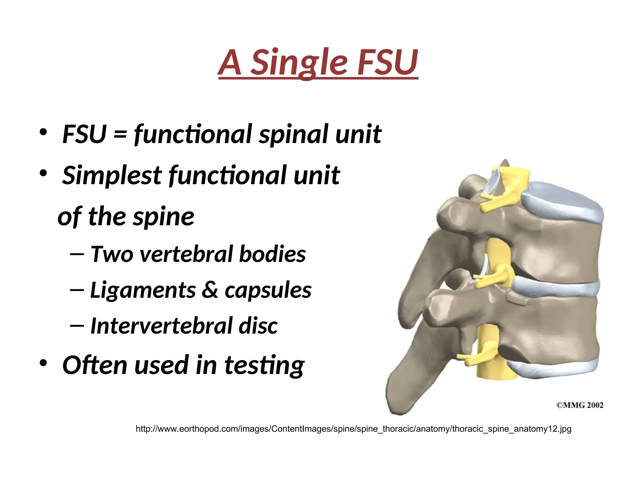
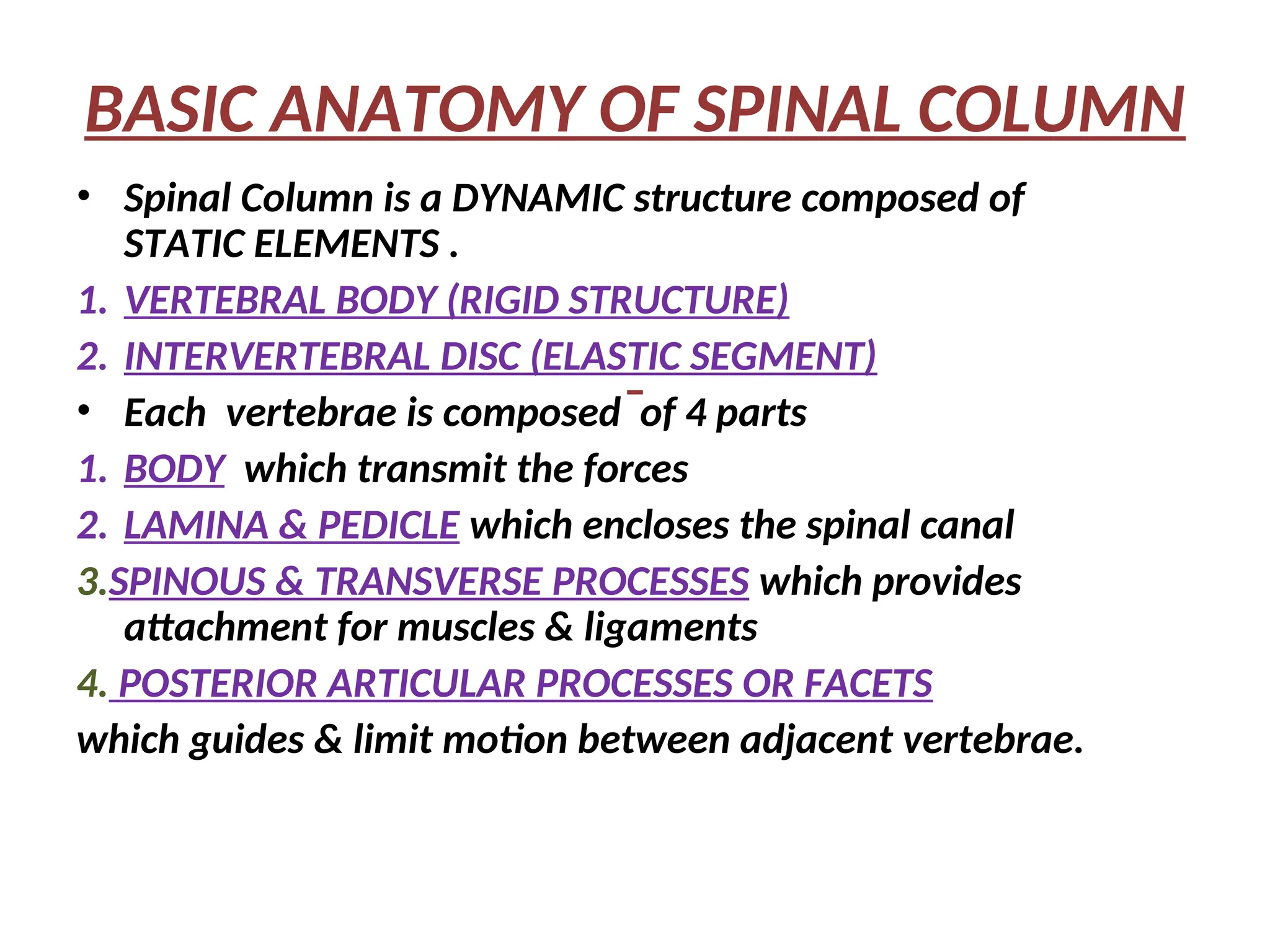
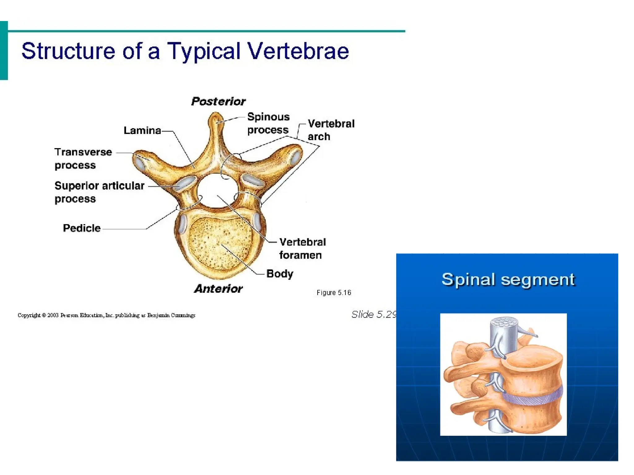
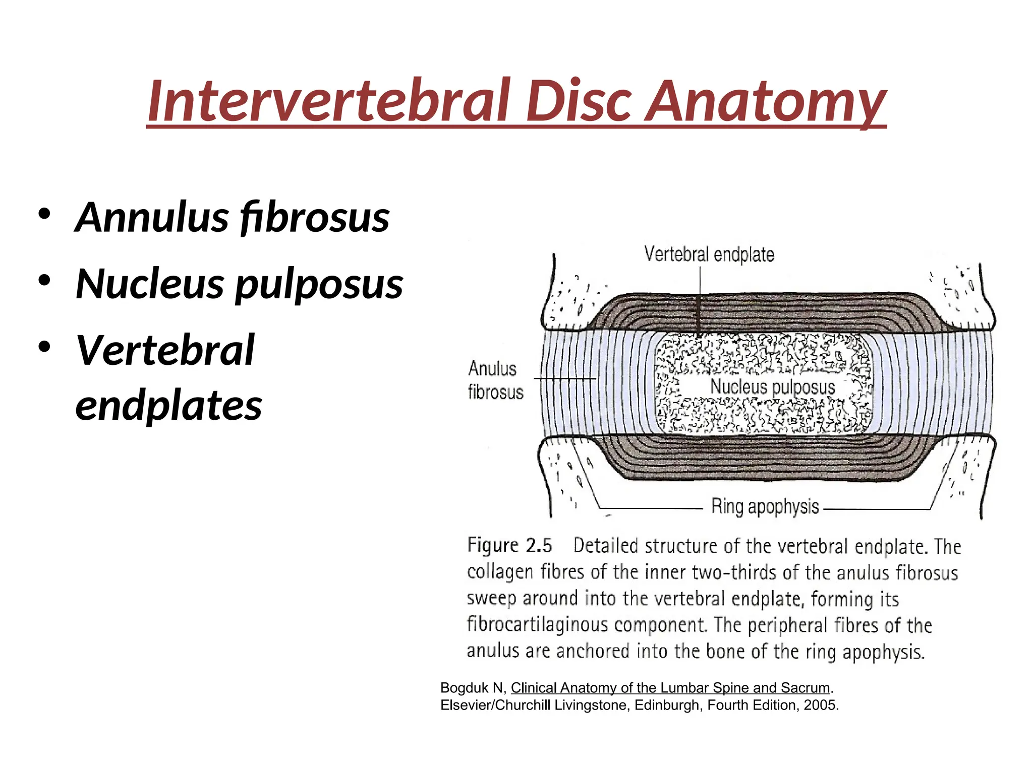
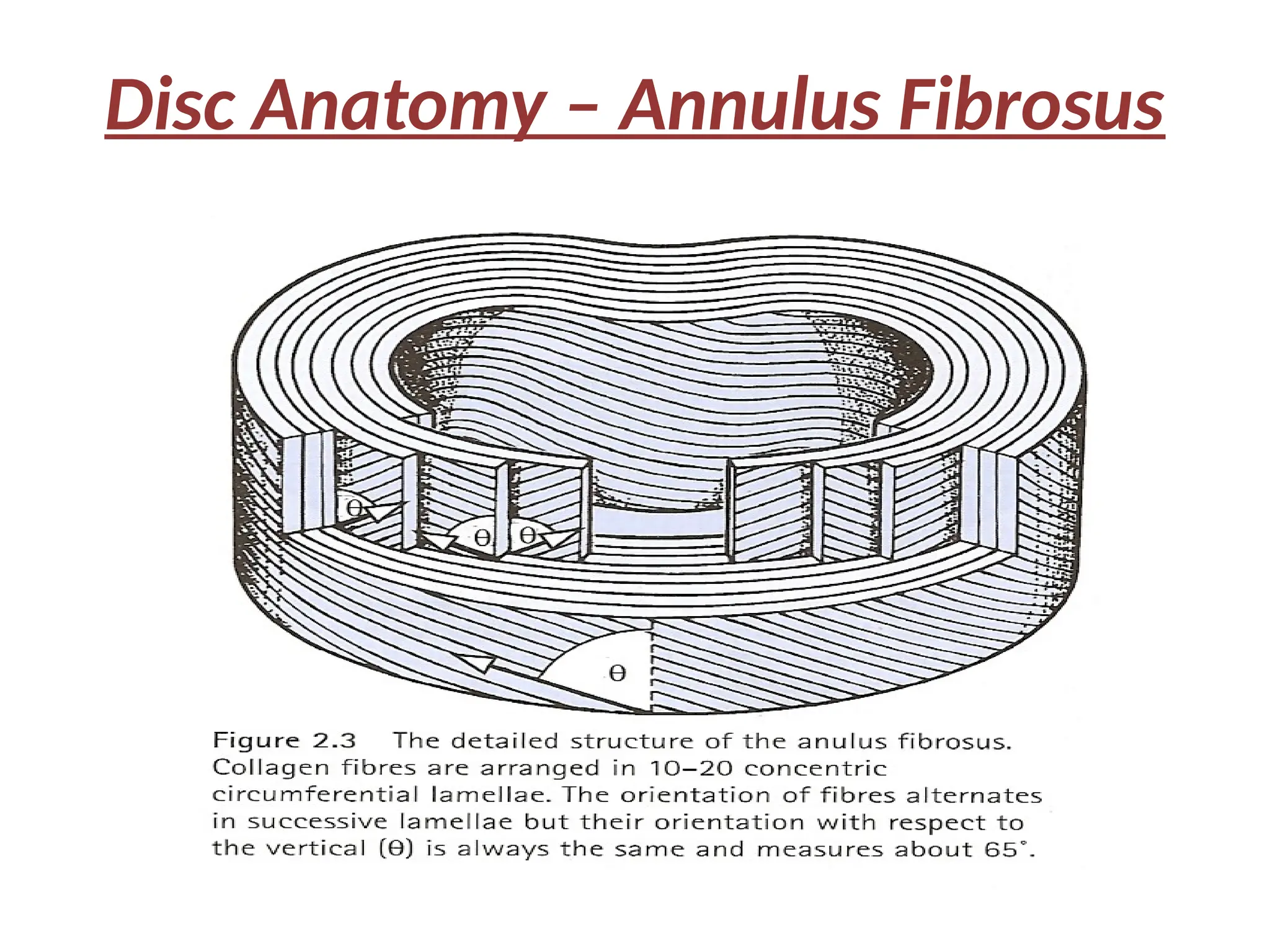
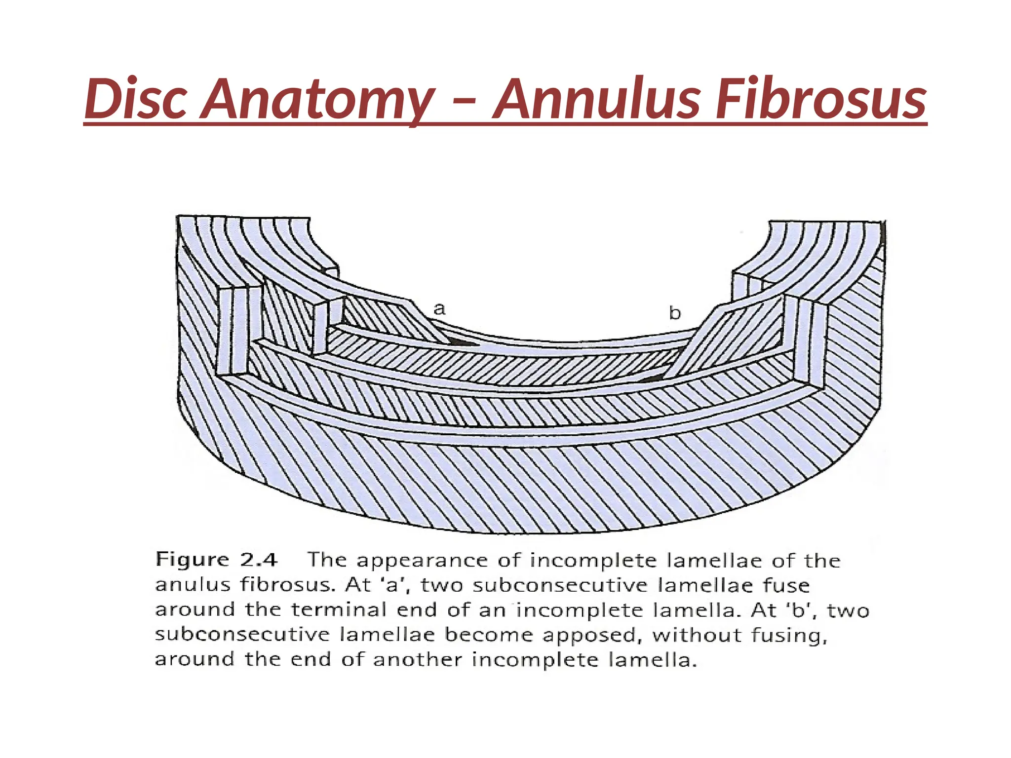
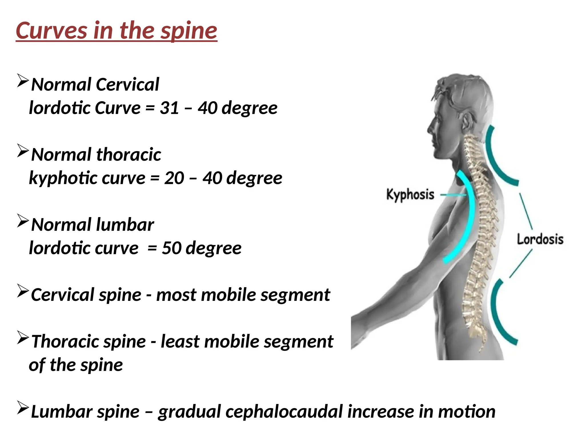
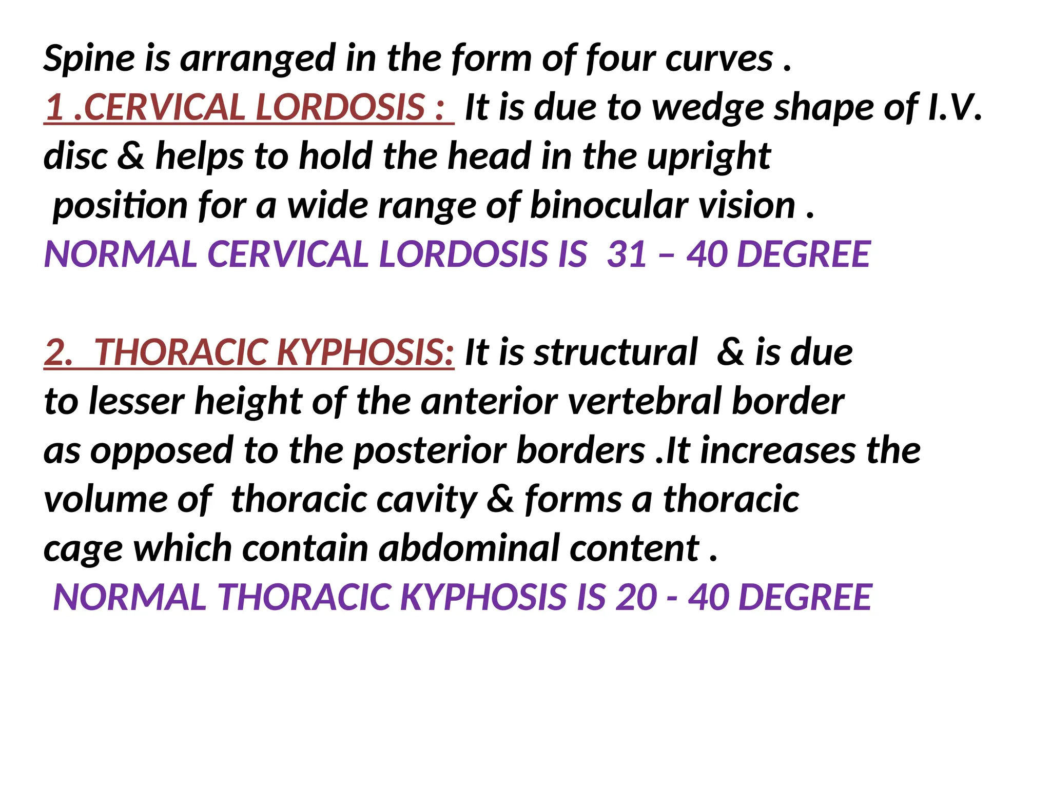
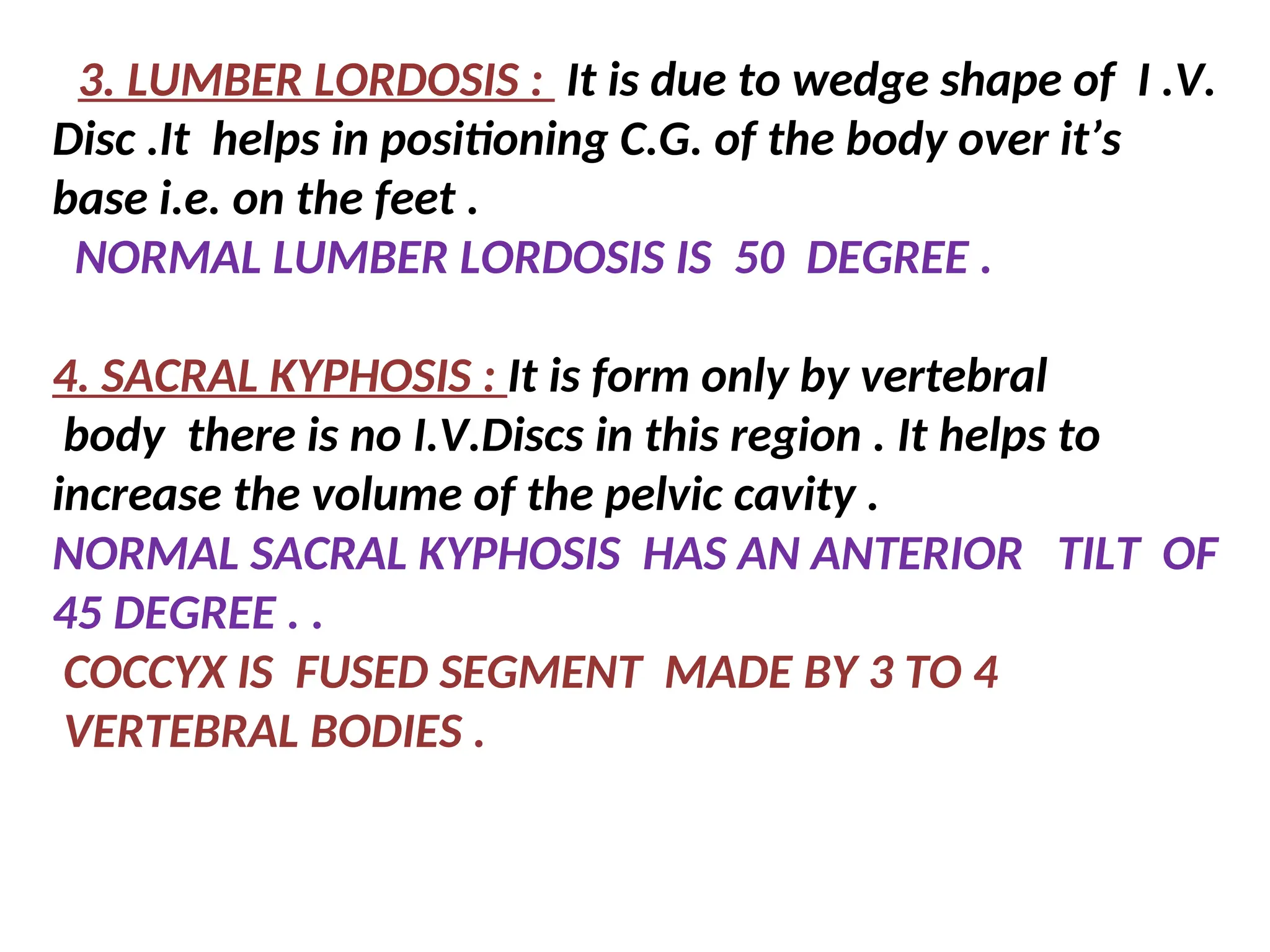
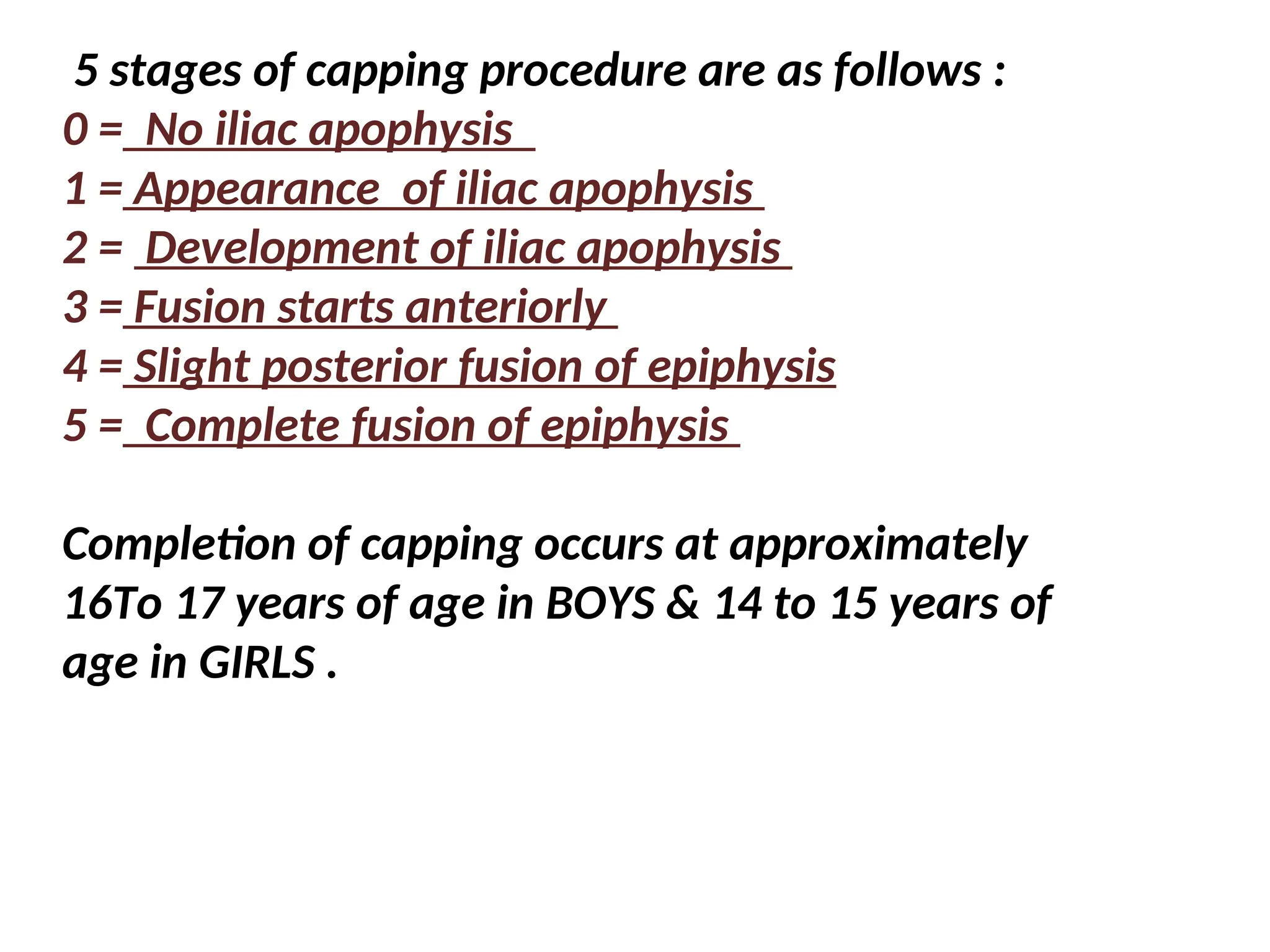
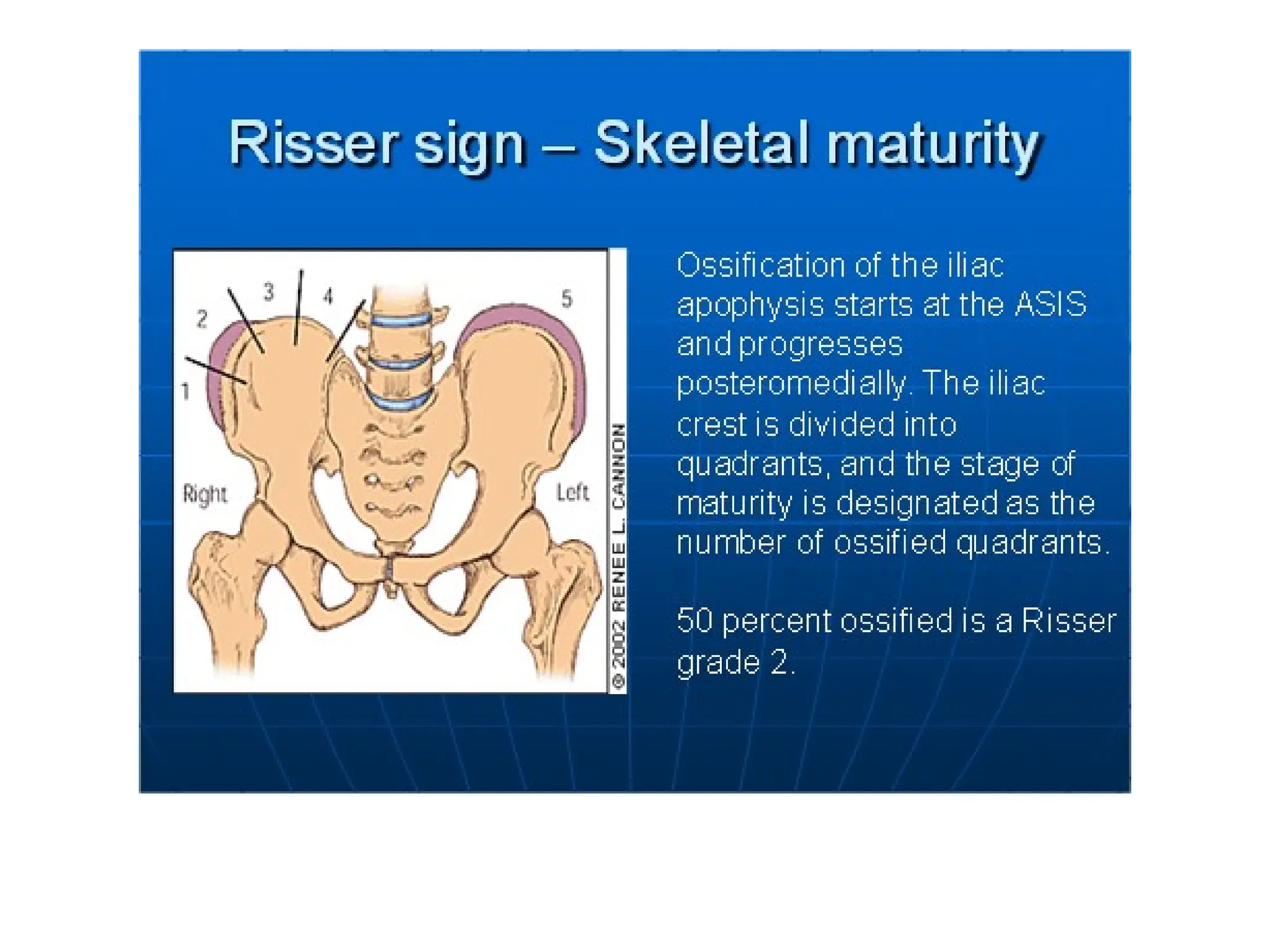
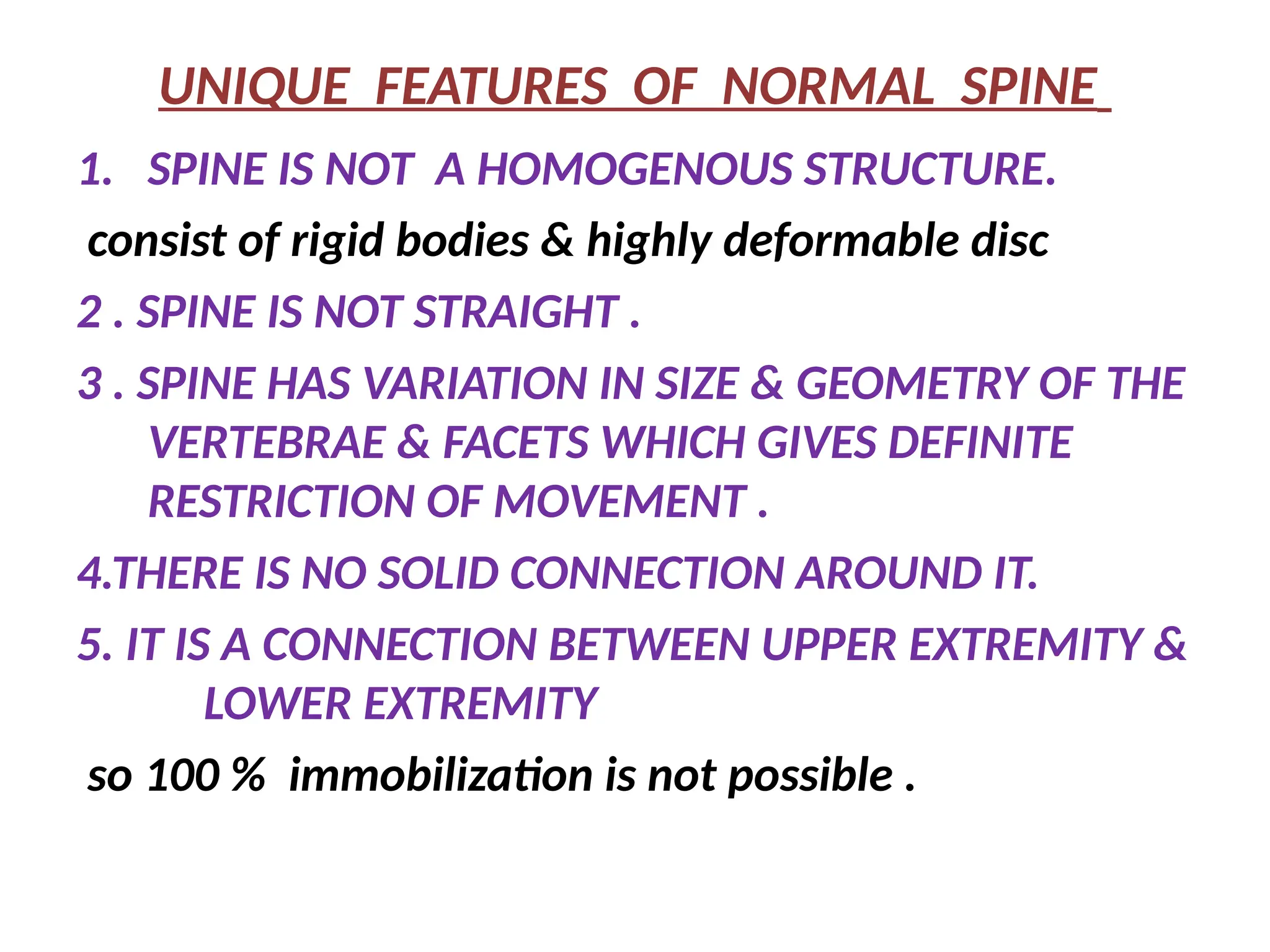
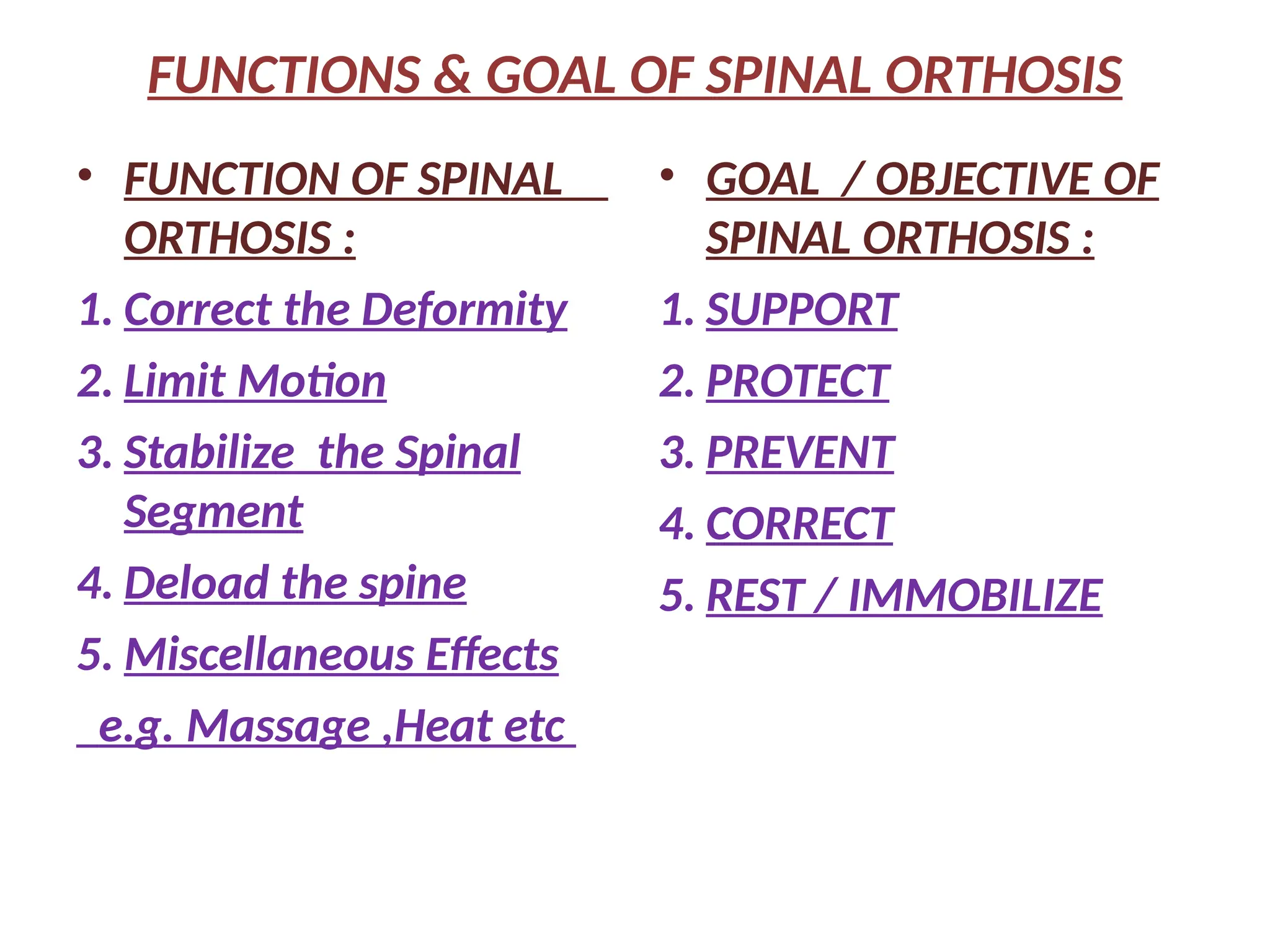
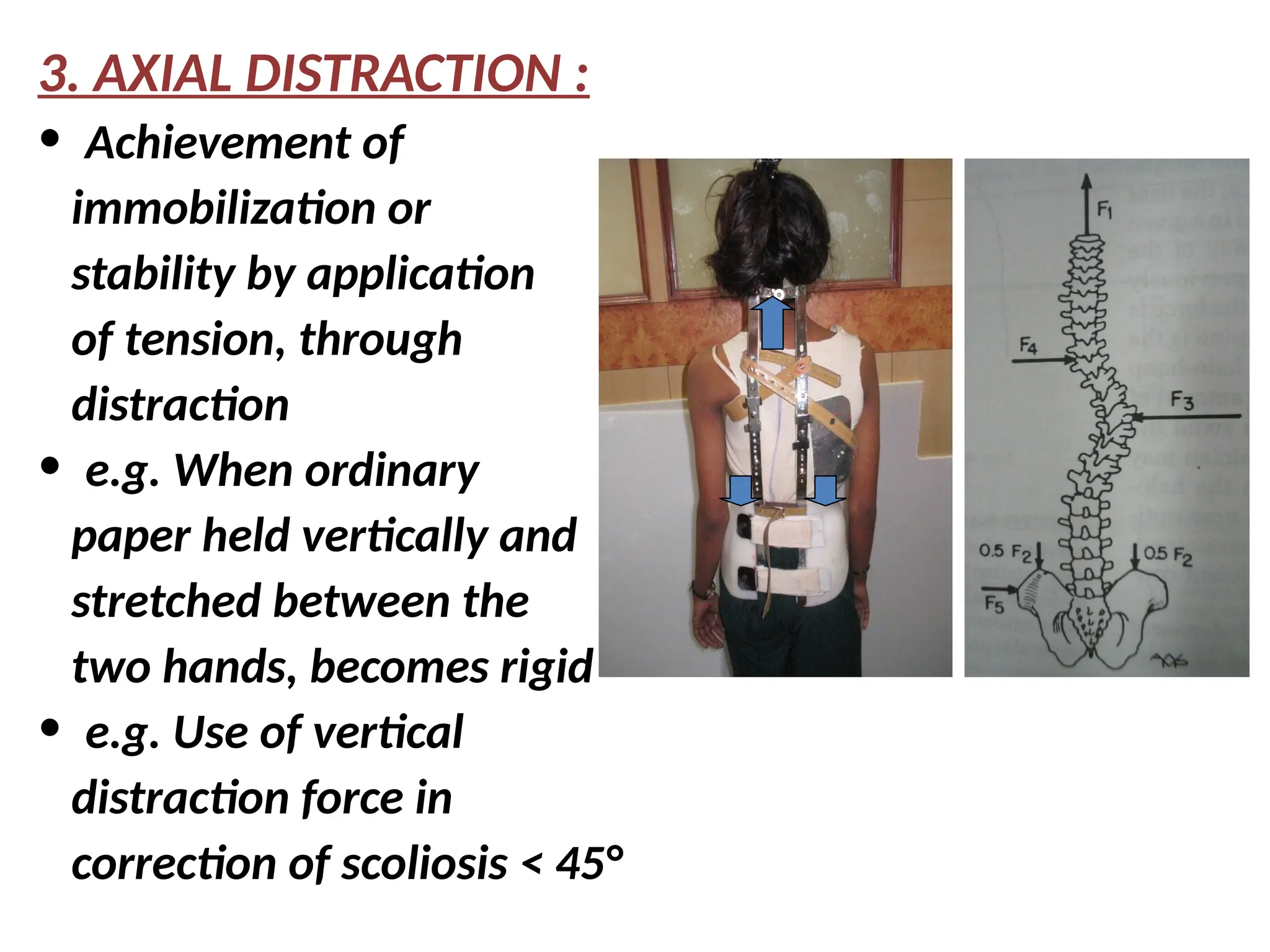
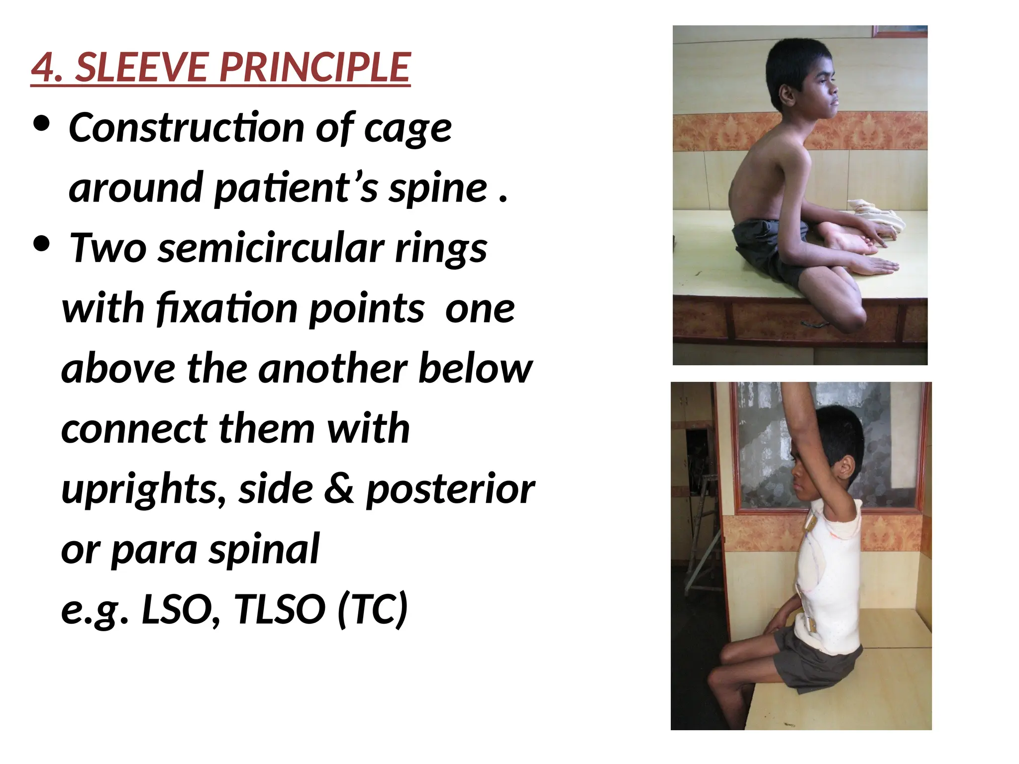
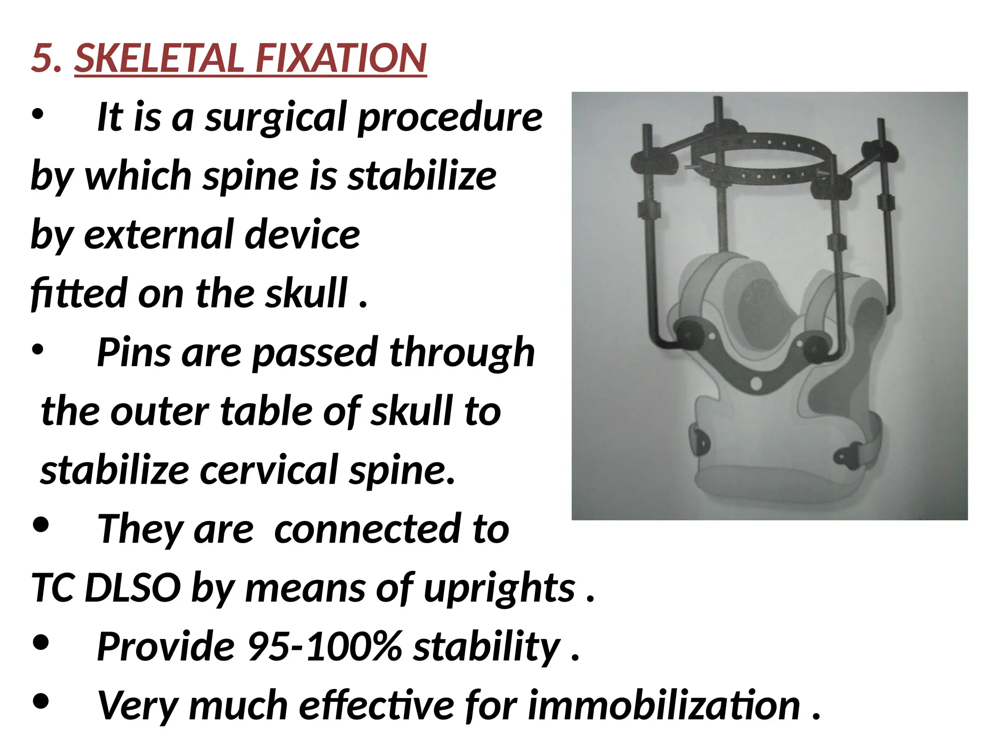
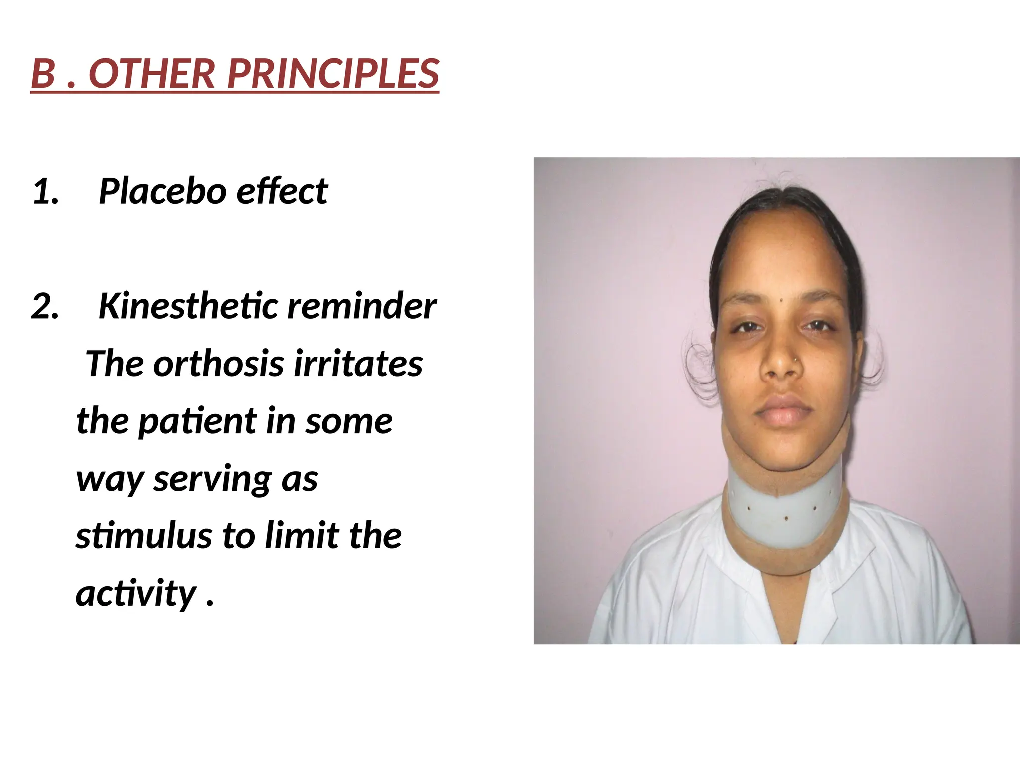
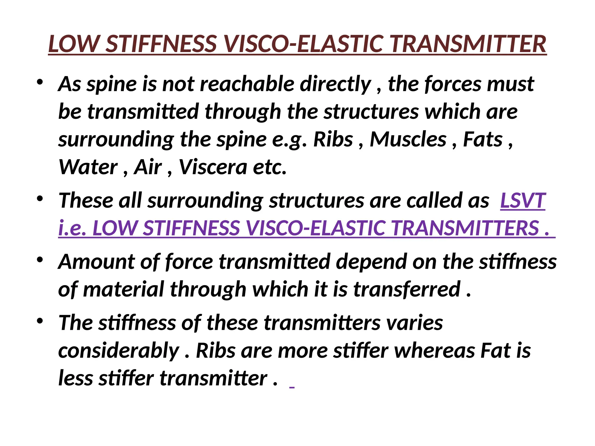
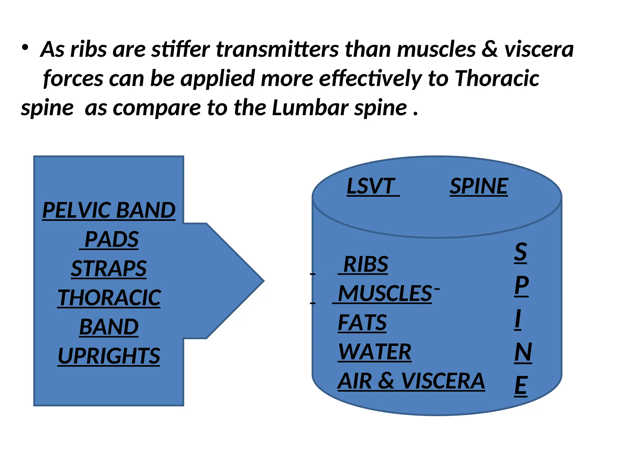
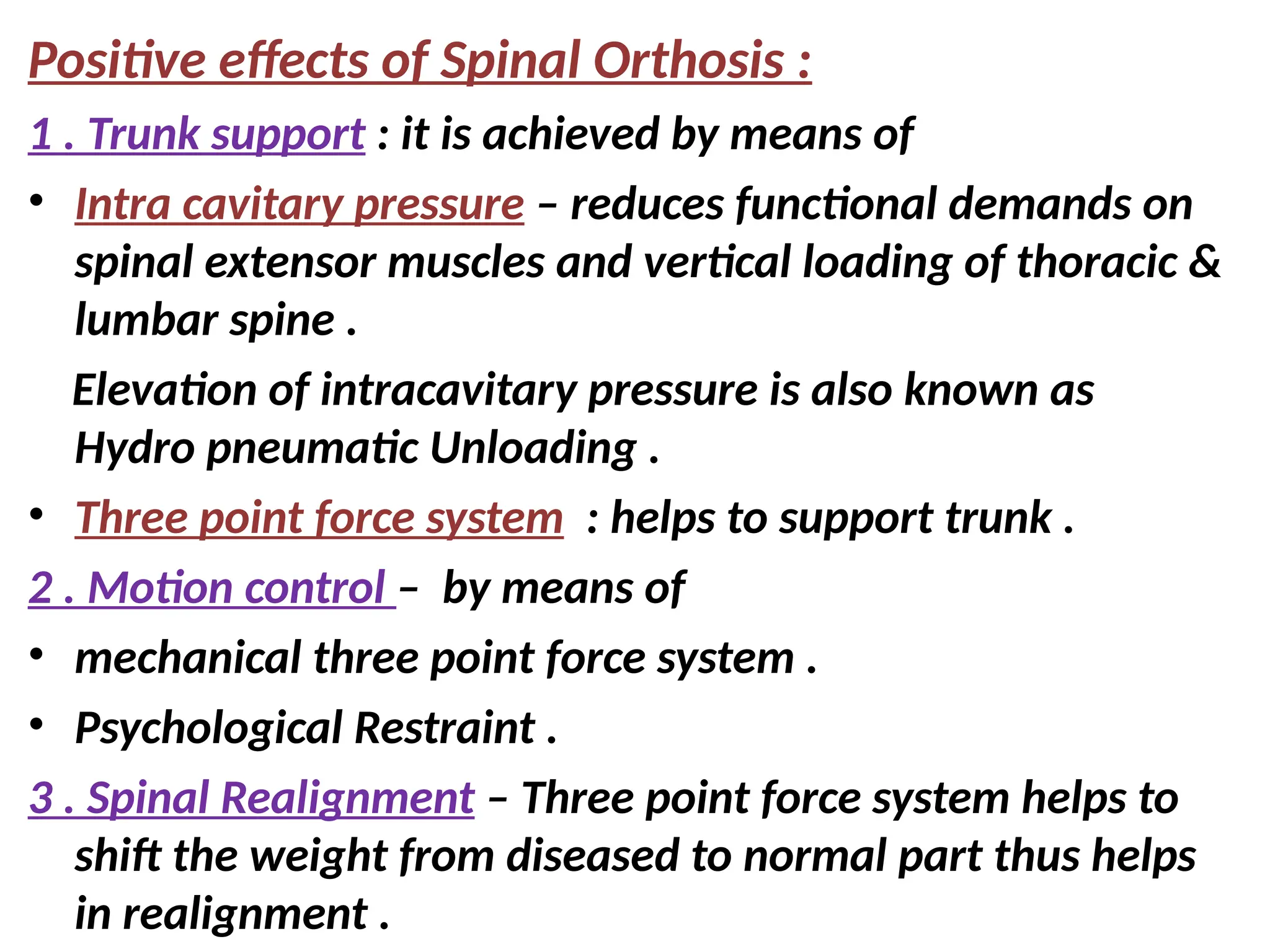
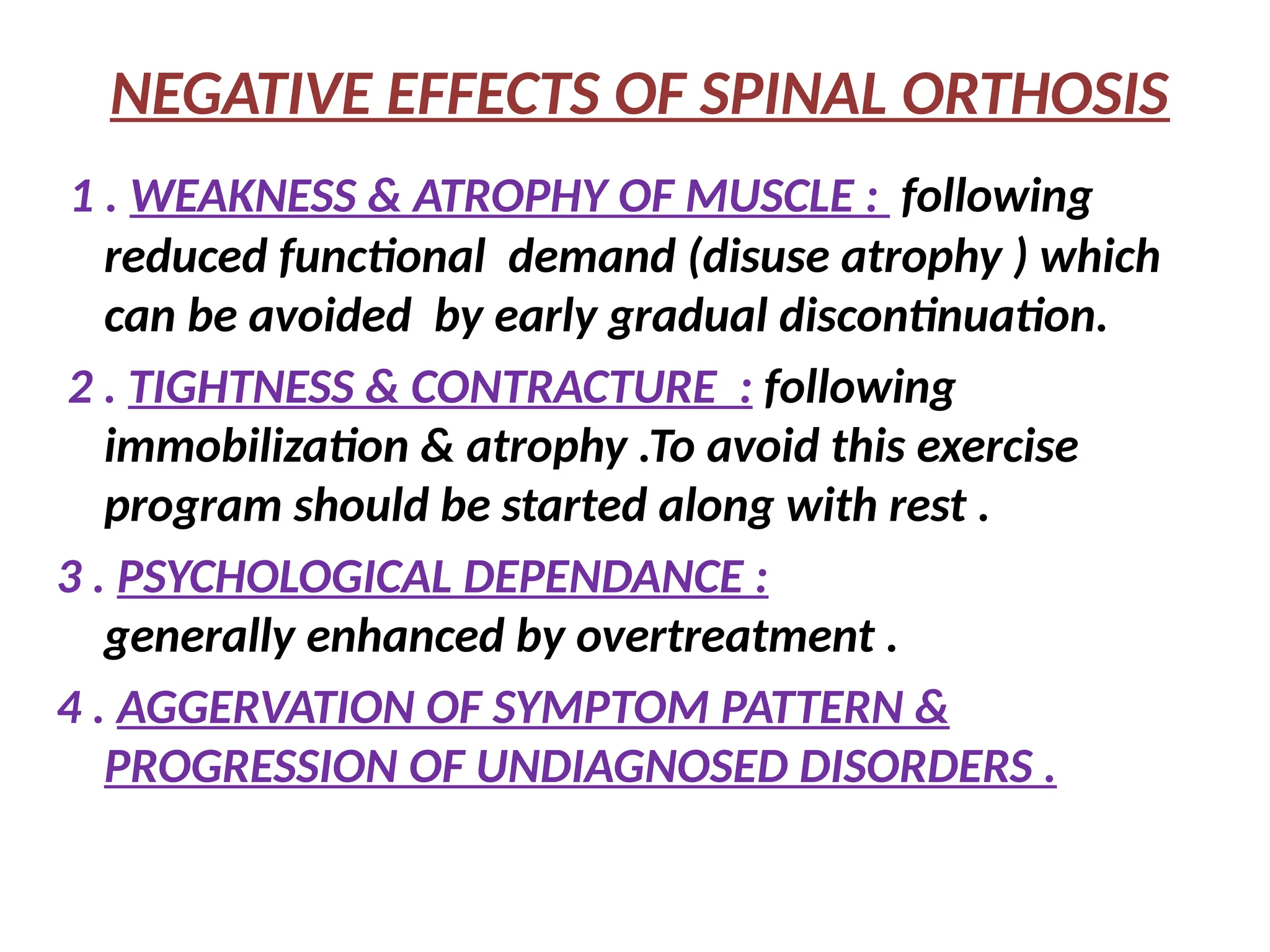
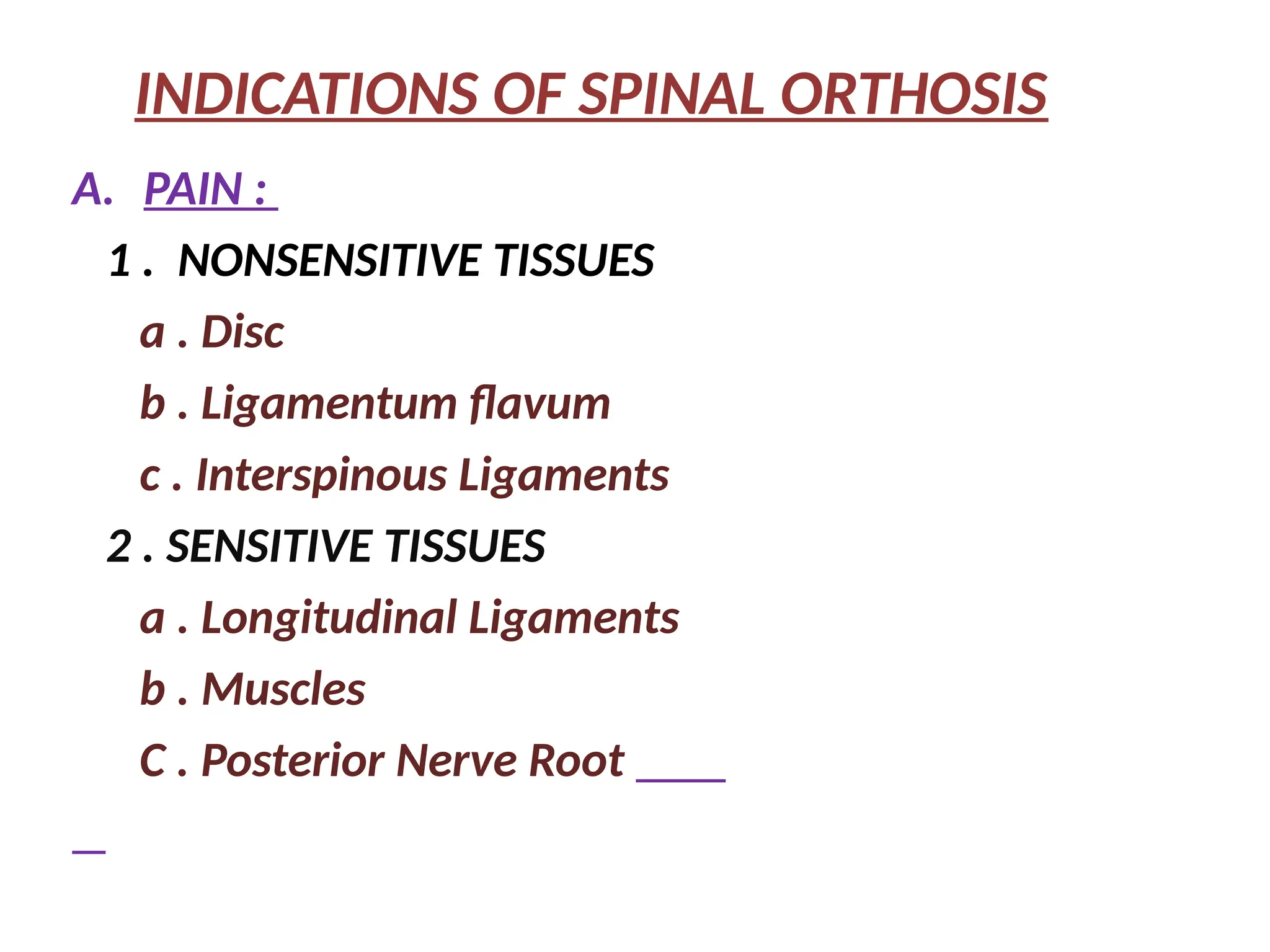
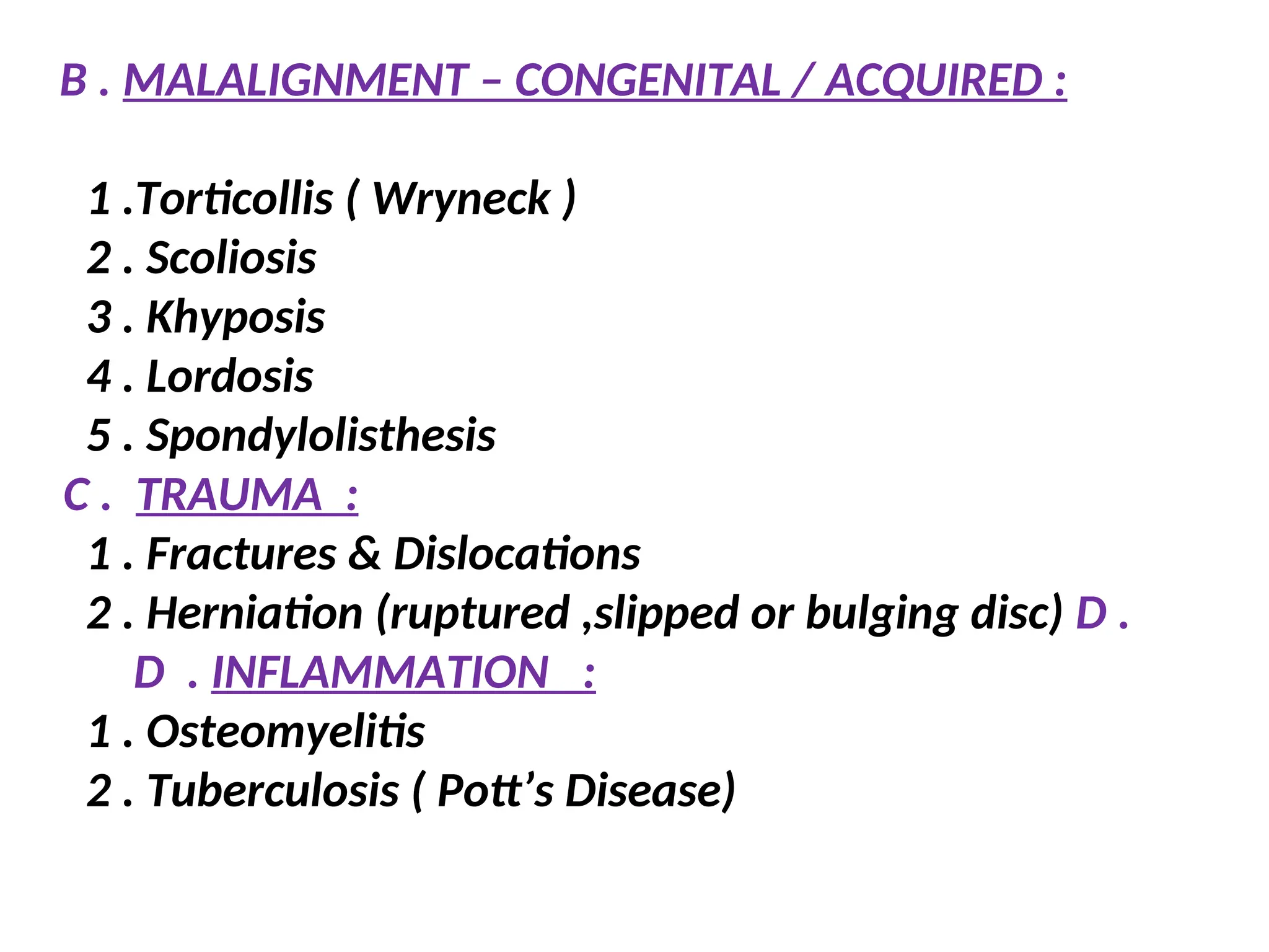
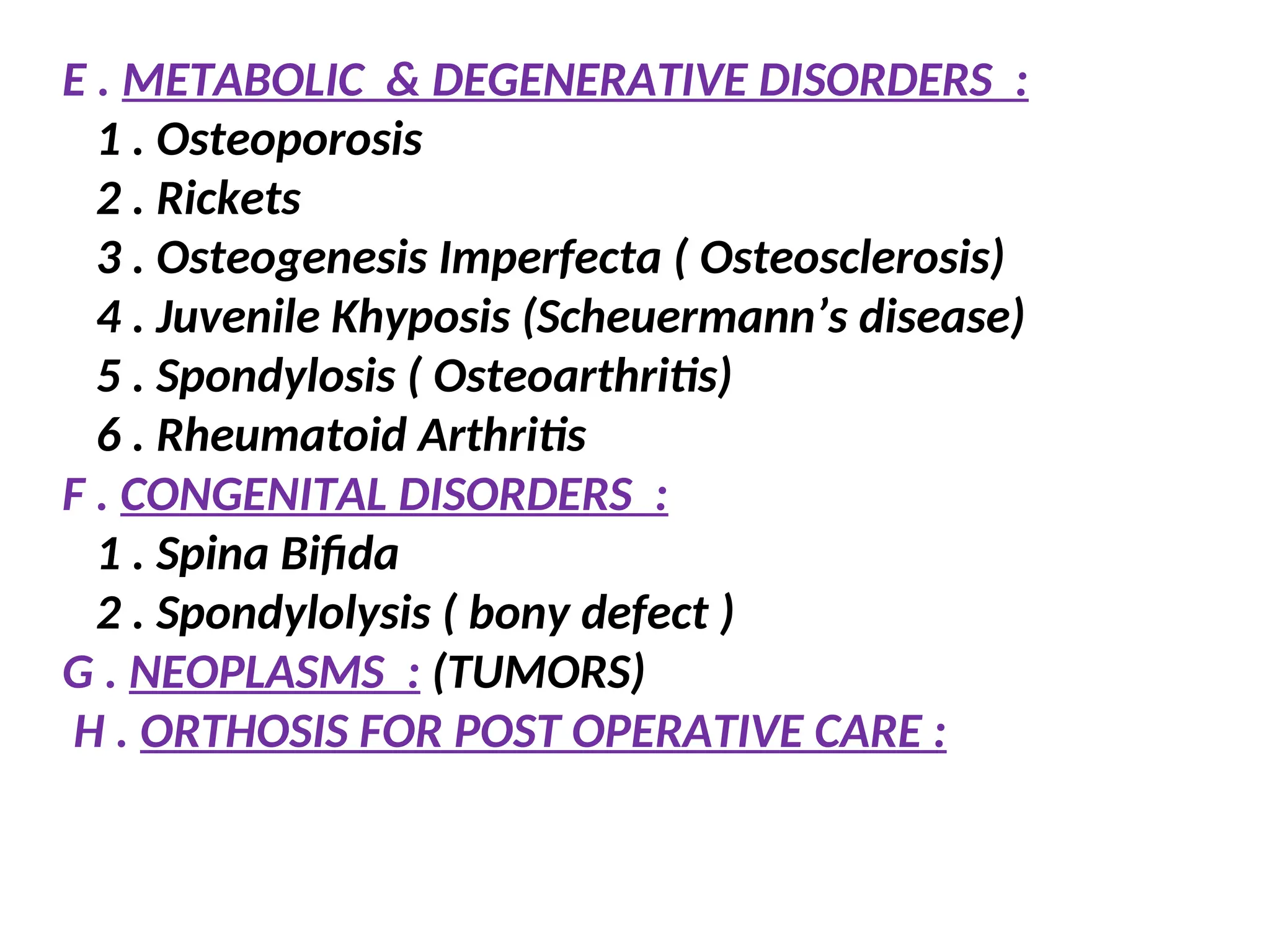
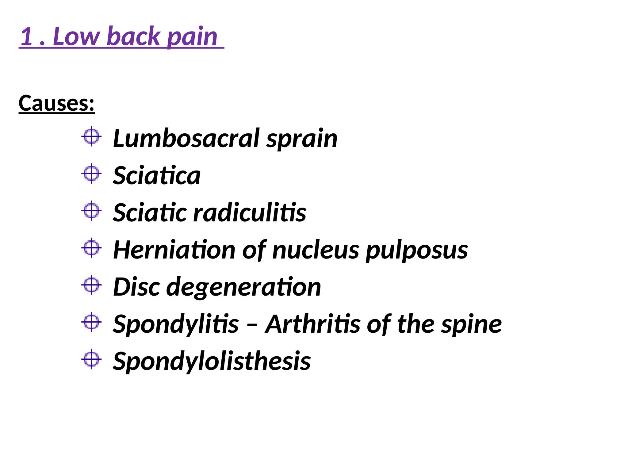
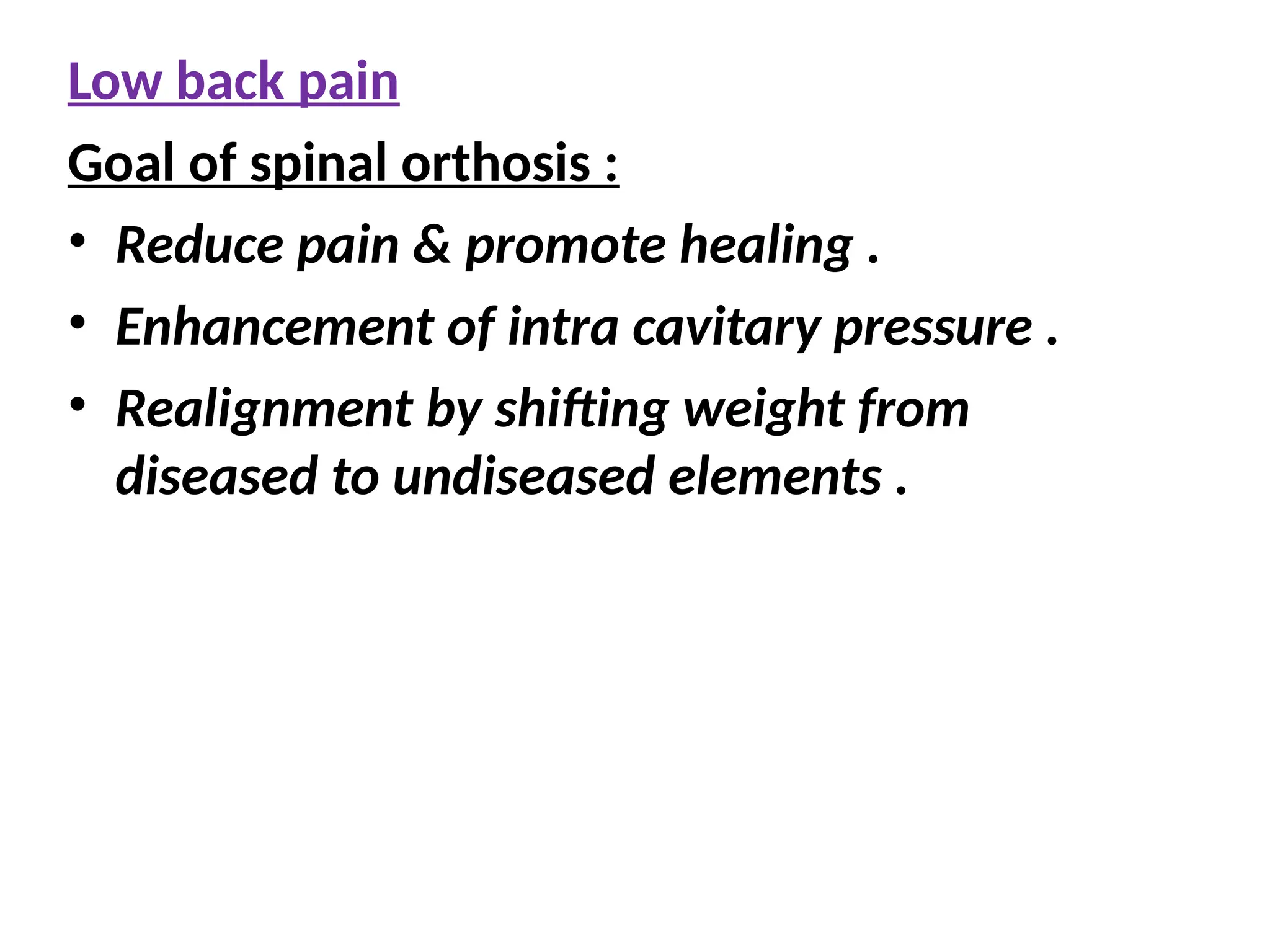
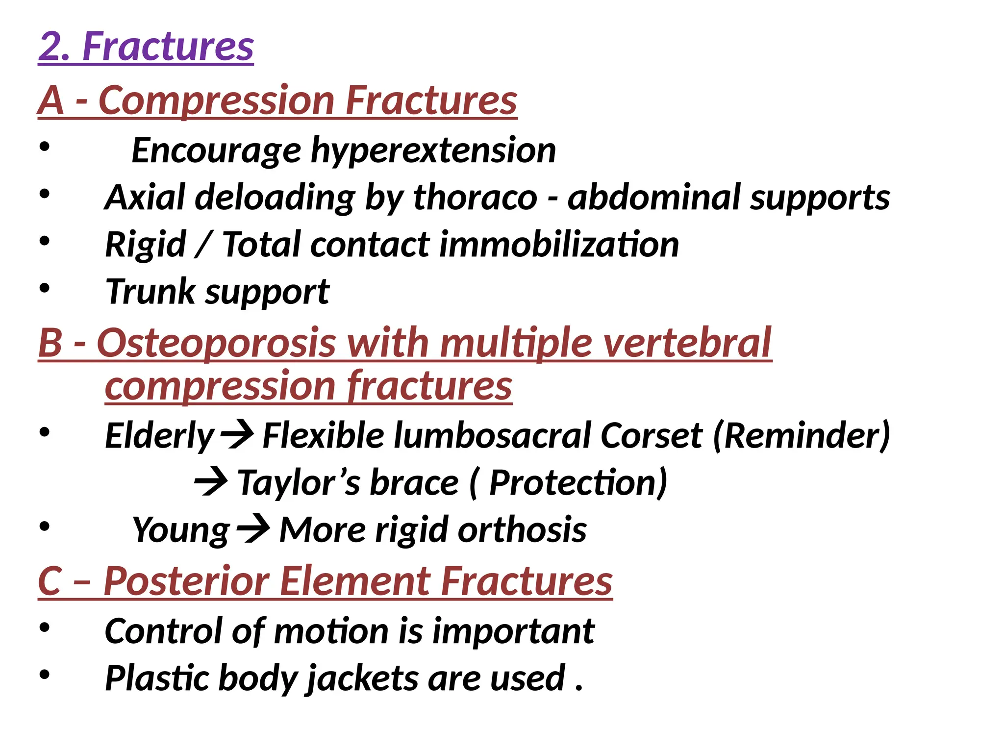
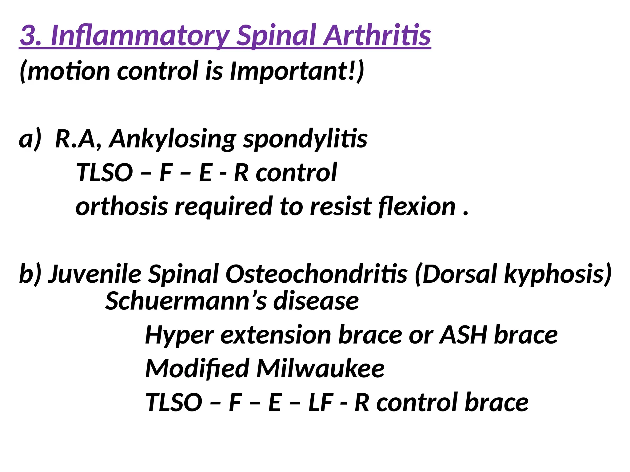
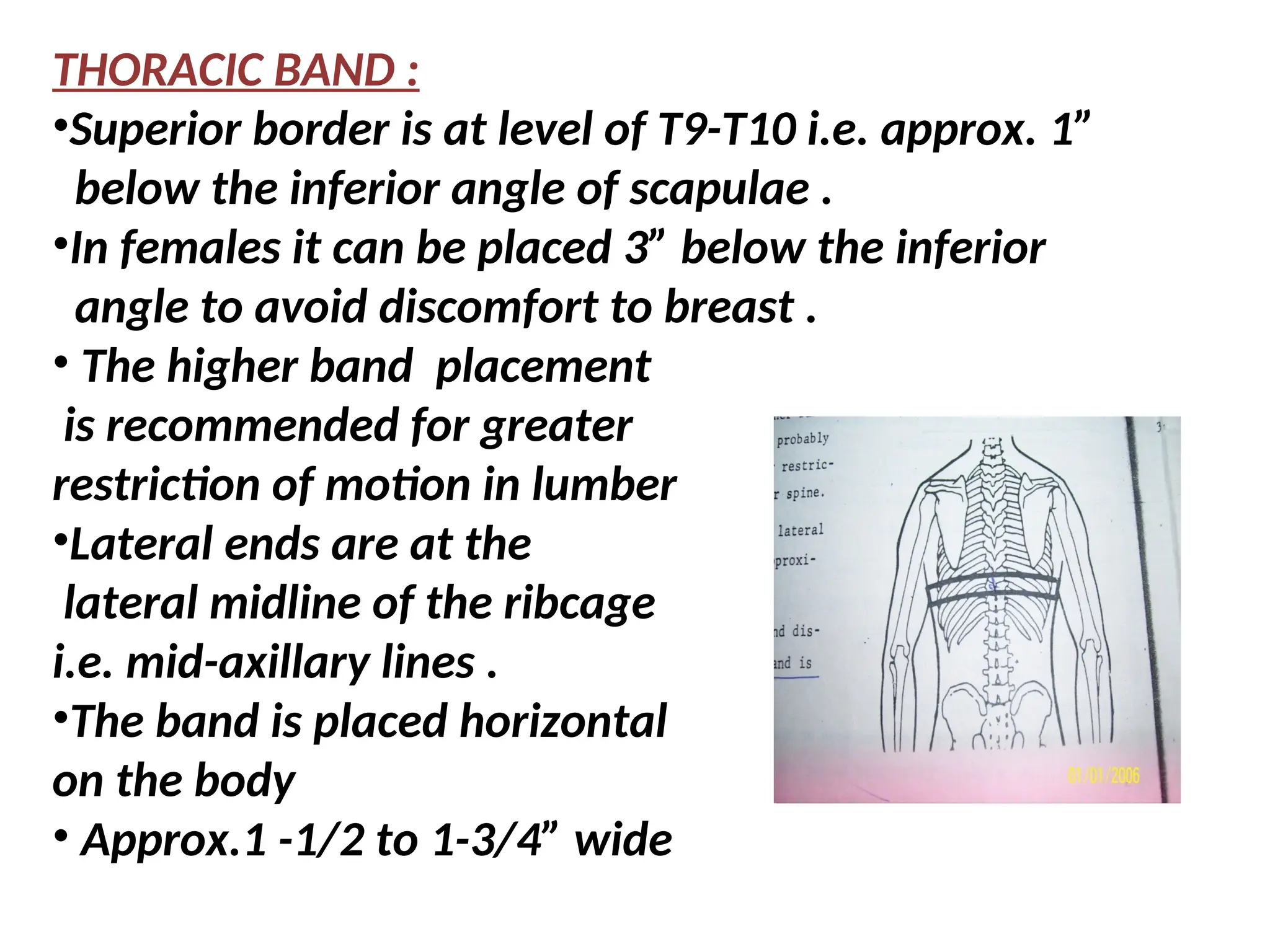
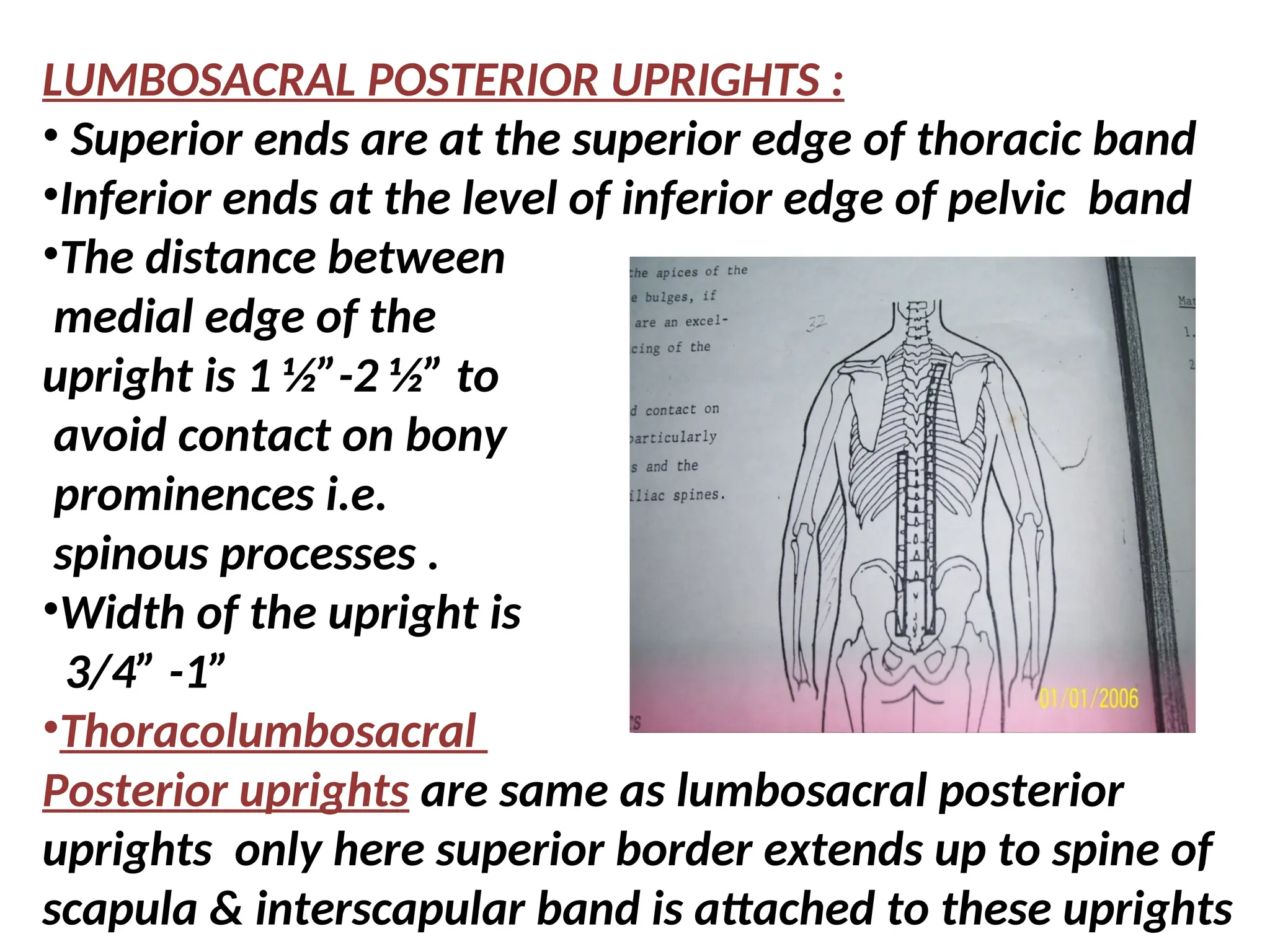
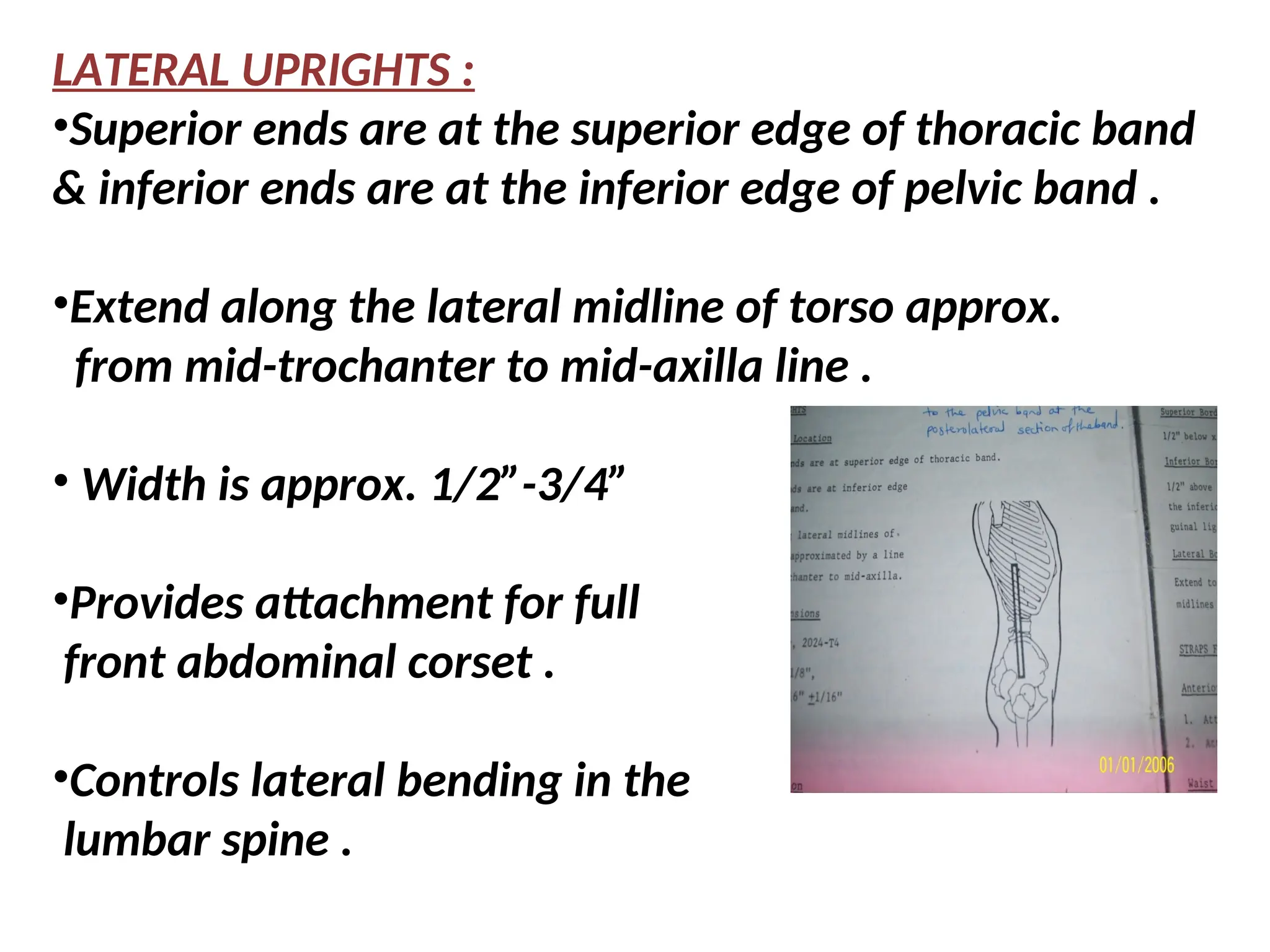
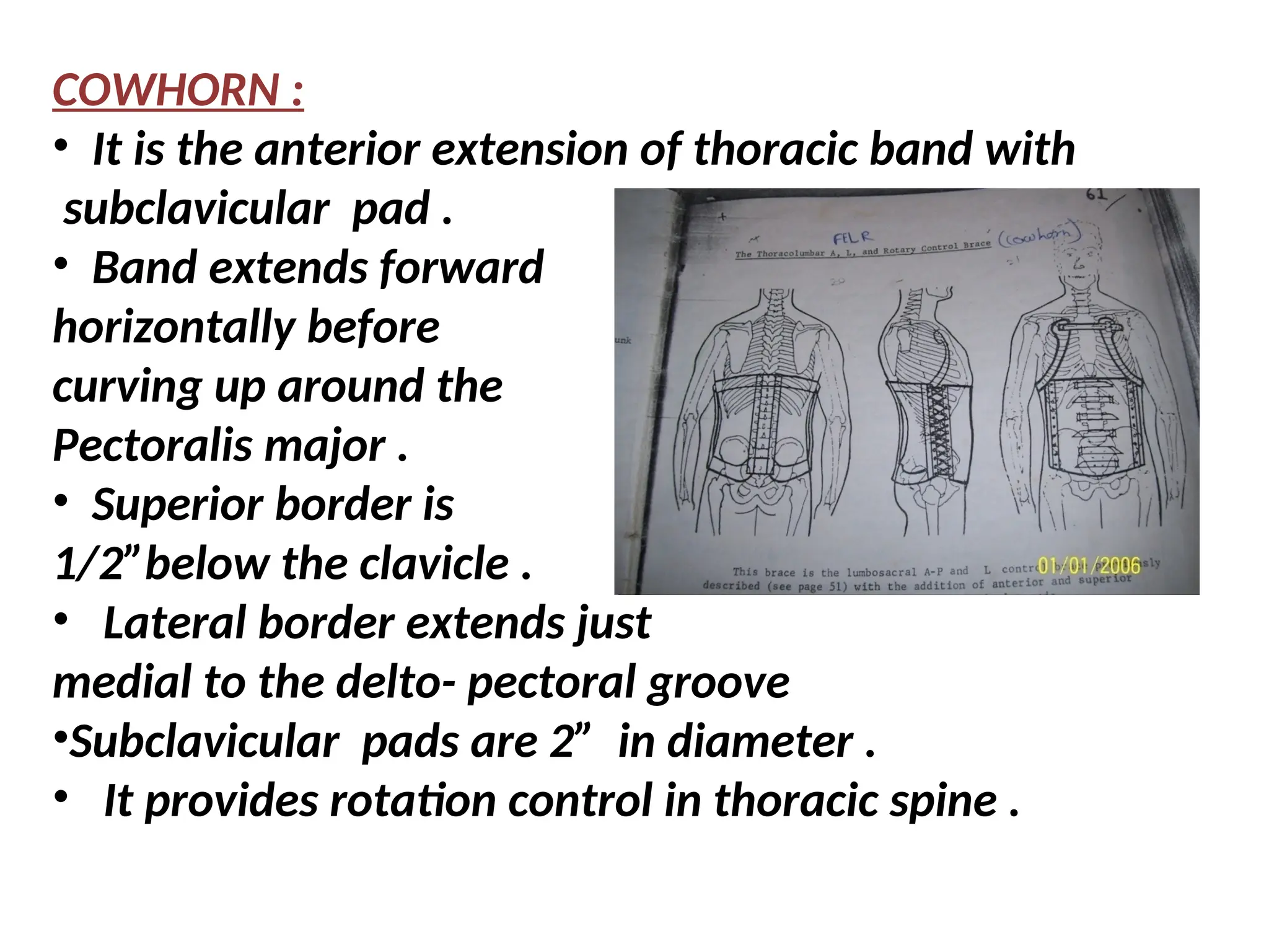
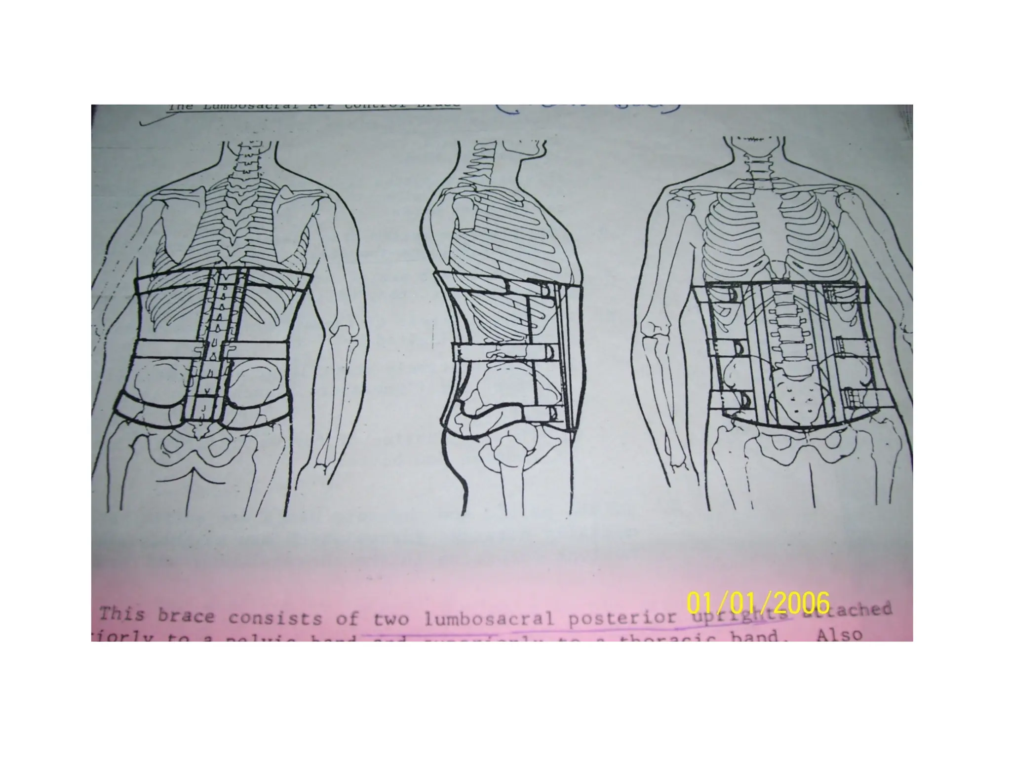
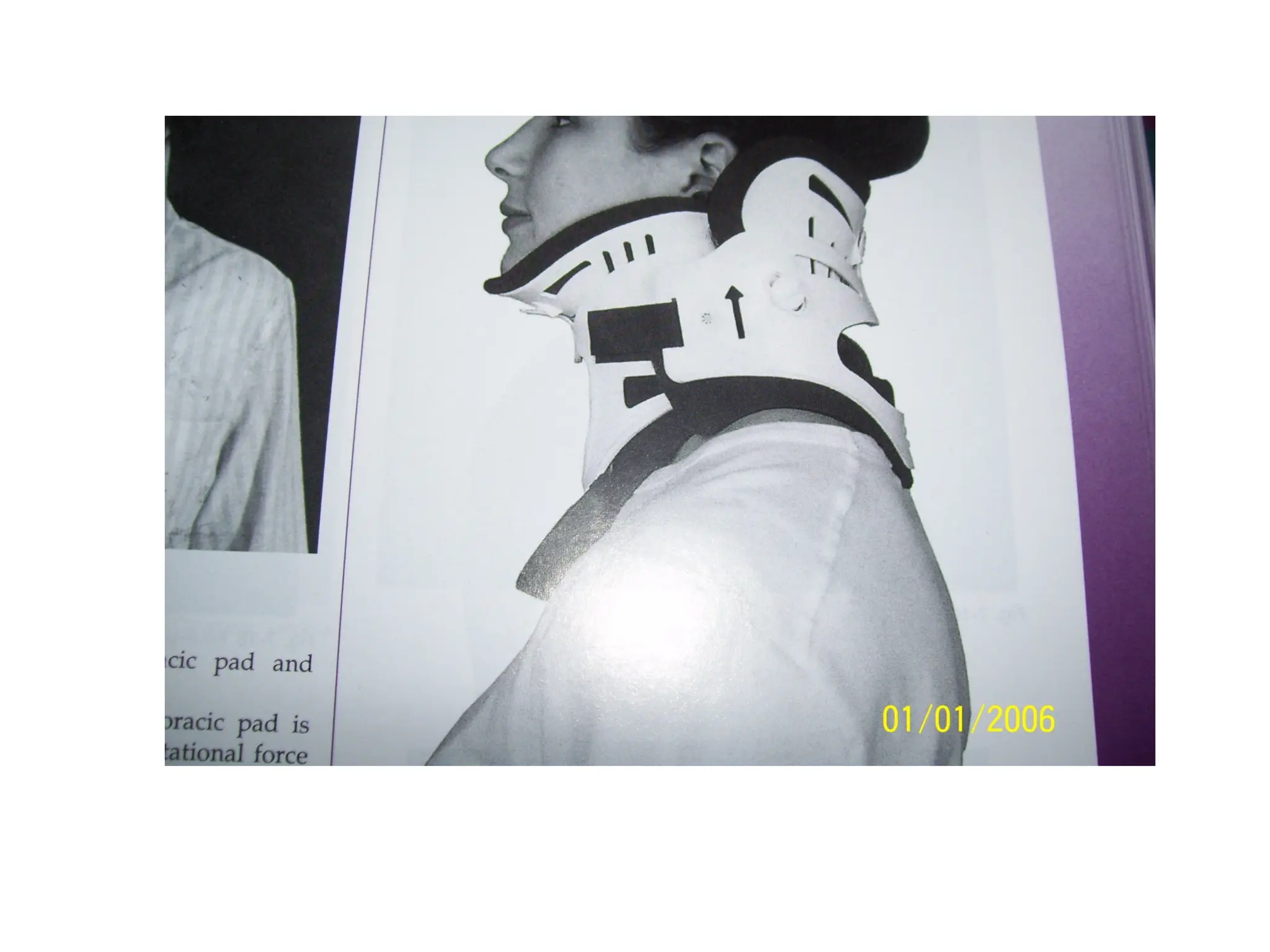
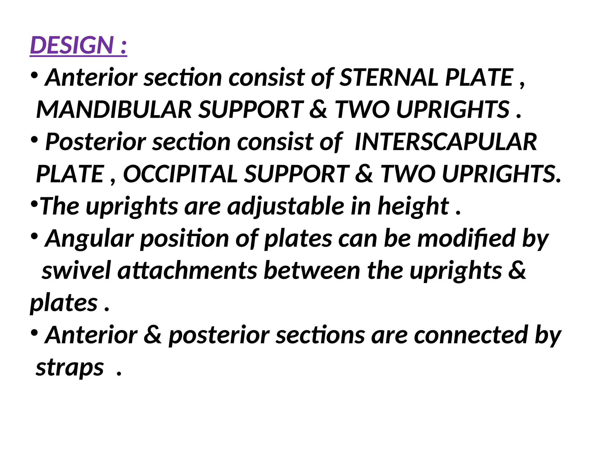
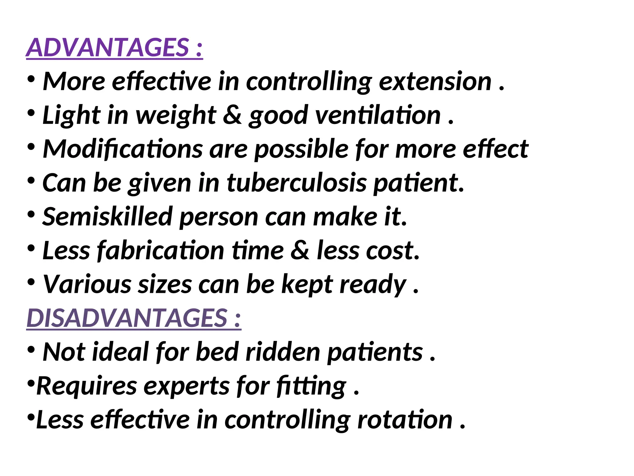
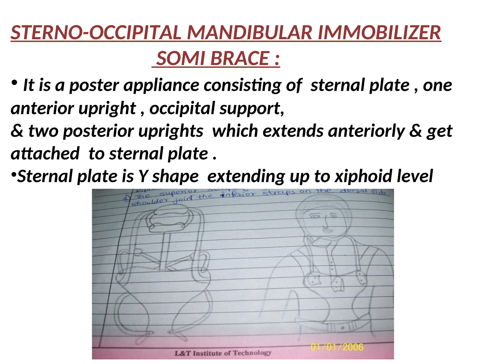
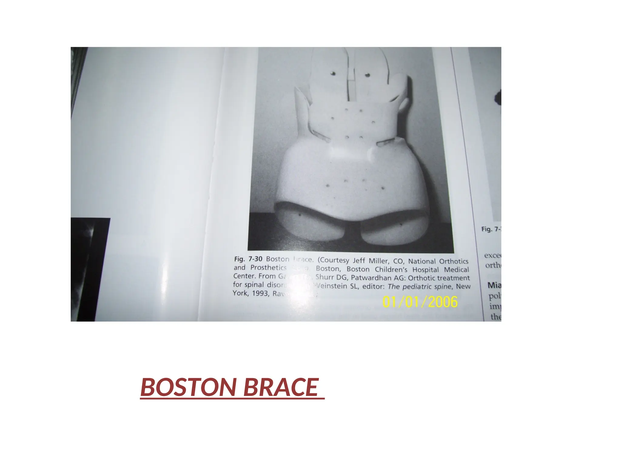
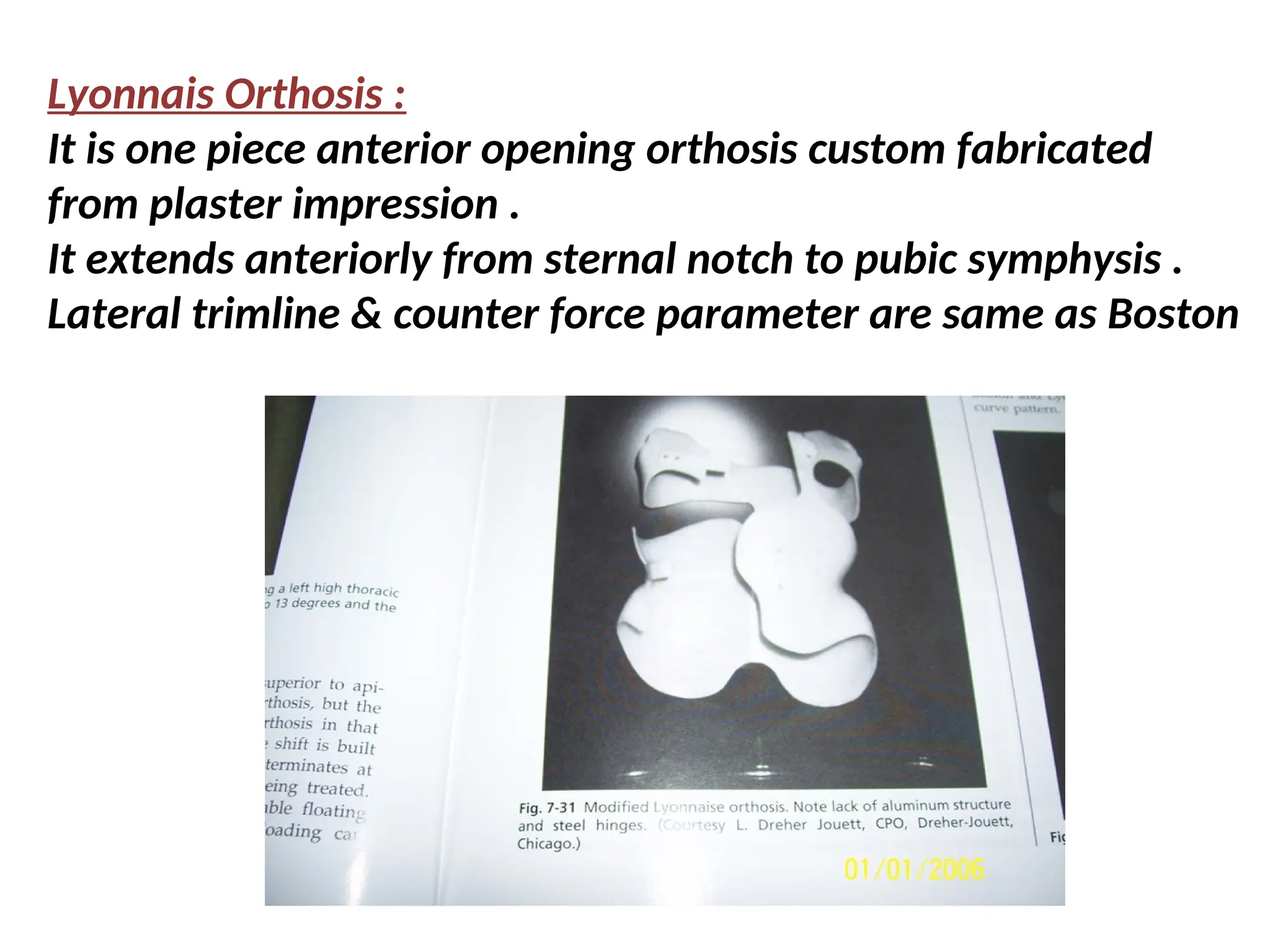
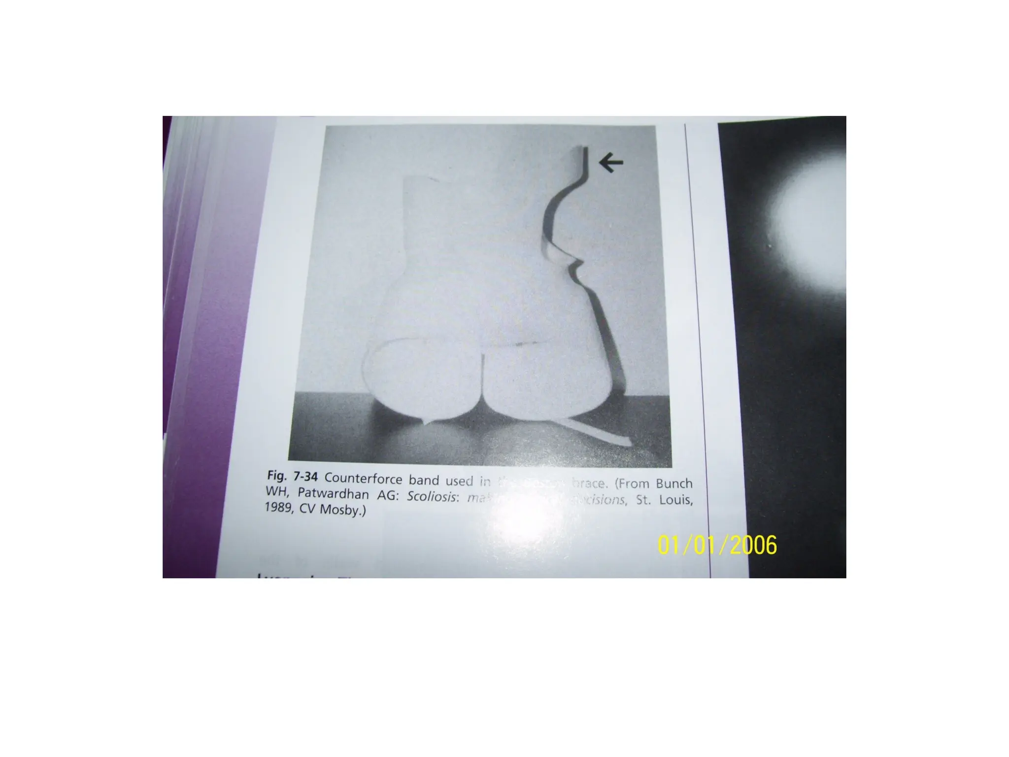
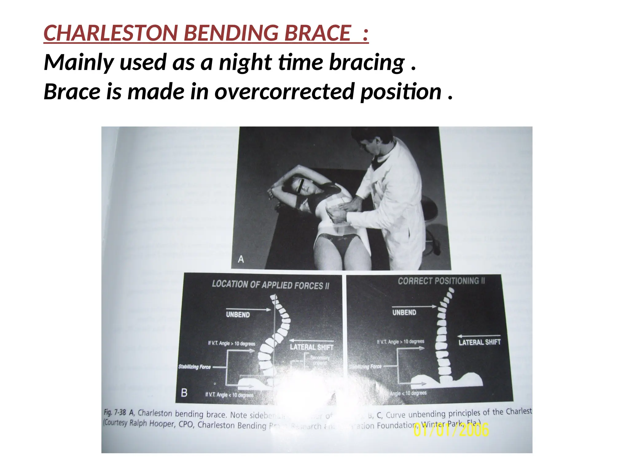
INTRODUCTION
• Spine can be defined as LONG,SLENDER , LIGAMENTOUS
bony structure which is markedly stiffen by rib cage .
• It is a mechanical structure .Vertebrae articulate with
each other in a controlled manner through a complex of
• LEVER (Vertebrae)
• PIVOTS (Facets & Disc)
• PASSIVE RESTRAINTS (Ligaments) &
• ACTIVATION (Muscles)
• It serve as a Sustaining Rod for the maintenance of the
upright position of the body
Spinal column possesses stability in two ways :
•INTRINSIC STABILITY by INTERVERTEBRAL DISC &
LIGAMENTOUS FORCES
• EXTRINSIC STABILITY by MUSCLE SUPPORT especially
of the abdomen & thoracic cage
•It is subjected to many forces such as Compression ,
Shearing ,Tension ,Bending , Twisting etc.
•Combination of rigid structure & elastic structure gives
STABILITY as well as MOBILITY to the spinal column.
•Main function of the spinal column is :
•Protect the spinal cord &
•Transfer the load
BASIC STRUCTURE OF SPINE
CERVICAL
THORASIC
THORACO
LUMBAR
LUMBAR
COCCYX
SACRUM
INTER-
VERTEBRAL
DISC
1ST
LUMBAR
1ST
THORACIC
1ST
CERVICAL
 CERVICAL = 7
 THORACIC = 12
 LUMBAR = 5
 SACRUM = 5
 COCCYX = 4
TOTAL 33 VERTEBRAE &
23 Intervertebral Disc
FUNCTIONAL SPINAL UNIT
 VERTEBRA
 DISCS
A Single FSU
• FSU = functional spinal unit
• Simplest functional unit
of the spine
– Two vertebral bodies
– Ligaments & capsules
– Intervertebral disc
• Often used in testing
http://www.eorthopod.com/images/ContentImages/spine/spine_thoracic/anatomy/thoracic_spine_anatomy12.jpg
BASIC ANATOMY OF SPINAL COLUMN
• Spinal Column is a DYNAMIC structure composed of
STATIC ELEMENTS .
1. VERTEBRAL BODY (RIGID STRUCTURE)
2. INTERVERTEBRAL DISC (ELASTIC SEGMENT)
• Each vertebrae is composed of 4 parts
1. BODY which transmit the forces
2. LAMINA & PEDICLE which encloses the spinal canal
3.SPINOUS & TRANSVERSE PROCESSES which provides
attachment for muscles & ligaments
4. POSTERIOR ARTICULAR PROCESSES OR FACETS
which guides & limit motion between adjacent vertebrae.
Intervertebral Disc Anatomy
• Annulus fibrosus
• Nucleus pulposus
• Vertebral
endplates
Bogduk N, Clinical Anatomy of the Lumbar Spine and Sacrum.
Elsevier/Churchill Livingstone, Edinburgh, Fourth Edition, 2005.
INTERVERTEBRAL DISC
• DISC is composed of two elements
• 1. ANNULUS FIBROSUS
• These are obliquely placed elastic fibers which
permit controlled motion.
• Fibro elastic nature of the interwoven pattern of
annulus fibrosus permits motion between the
individual vertebrae.
• Anterior-posterior movement takes place due to
specific arrangement of fibers.
• Orientation of fibers with respect to vertical axis is
approx. 65 degrees .
Disc Anatomy – Annulus Fibrosus
• 2. NUCLEUS PULPOSUS
• It is a gelatinous substance which forms the center of
the disc .
• It has properties of gel
• I.V.DISC has particular VISCOELASTICITY which shows
sensitivity to the Rate of Loading or Deformation.
• Disc is a shock absorber & force disseminator of spinal
column & allow for direct control motion .
Disc Anatomy - Composition
• Water content in disc is very important for
resistance to compression .
• Throughout a normal day your spine
compresses due to water degress from discs .
• Normal degeneration with age leads to
permanent loss of proteoglycans and water
content .
TRIPOD CONCEPT OF ARTICULATION
• Static elements are arranged through their tripod
articulation & ligamentous support to allow for
intersegmental motion .
• Articular processes & cartilaginous disc form pyramid
like structure having apex directed in upward
direction in which other vertebral body fit .
• This tripod configuration gives very stable structure
of spinal column .
• Each vertebra has three articular surfaces
• TWO FACETS & ONE BODY which articulate with
the vertebrae above it .
VERTEBRAL ARTICULATION
SPINAL CURVES
1.CERVICAL LORDOSIS : 7
2.THORACIC KYPHOSIS: 12
3.LUMBAR LORDOSIS: 5
4.SACRAL KYHPHOSIS: 5
5. COCCYGEAL: 3 TO 4
Total 33 vertebrae
23 intervertebral disc
Total length of spine is
45% of body length
out of this ¼ of length
is formed by I.V. disc
Curves in the spine
Normal Cervical
lordotic Curve = 31 – 40 degree
Normal thoracic
kyphotic curve = 20 – 40 degree
Normal lumbar
lordotic curve = 50 degree
Cervical spine - most mobile segment
Thoracic spine - least mobile segment
of the spine
Lumbar spine – gradual cephalocaudal increase in motion
Spine is arranged in the form of four curves .
1 .CERVICAL LORDOSIS : It is due to wedge shape of I.V.
disc & helps to hold the head in the upright
position for a wide range of binocular vision .
NORMAL CERVICAL LORDOSIS IS 31 – 40 DEGREE
2. THORACIC KYPHOSIS: It is structural & is due
to lesser height of the anterior vertebral border
as opposed to the posterior borders .It increases the
volume of thoracic cavity & forms a thoracic
cage which contain abdominal content .
NORMAL THORACIC KYPHOSIS IS 20 - 40 DEGREE
3. LUMBER LORDOSIS : It is due to wedge shape of I .V.
Disc .It helps in positioning C.G. of the body over it’s
base i.e. on the feet .
NORMAL LUMBER LORDOSIS IS 50 DEGREE .
4. SACRAL KYPHOSIS : It is form only by vertebral
body there is no I.V.Discs in this region . It helps to
increase the volume of the pelvic cavity .
NORMAL SACRAL KYPHOSIS HAS AN ANTERIOR TILT OF
45 DEGREE . .
COCCYX IS FUSED SEGMENT MADE BY 3 TO 4
VERTEBRAL BODIES .
MOVEMENTS OF THE SPINE
• Spine has movements in all three planes because
anterior & posterior intervertebral joint has
universal axes. Motion of one vertebra on the other
is controlled by several factors as follows :
1. DIRECTION OF ARTICULAR PROCESSES MAY
RESTRICT ROTATION AS IN LUMBER AREA .
2. OBLIQUITY OF SPINE CAN HINDER HYPEREXTENSION
AS IN THORACIC AREA .
3.RIBS IMPEDE LATERAL FLEXION IN THORACIC
REGION
4 . THICKNESS , SHAPE & COMPRESSIBILITY OF
THE DISC ALSO AFFECTS THE MOVEMENT .
5 . LIGAMENT S & MUSCLES RESIST EXCESSIVE
MOTION .
• CERVICAL SPINE IS HIGHLY MOBILE .
• THORACIC SPINE IS MORE CONSTRICT & VERY
MINIMAL MOTION DUE TO RIB CAGE
ATTACHMENT
• LUMBER SPINE HAS GRADUALCEPHALOQUADAL
INCREASE IN MOTION .
CERVICAL SPINE : First cervical vertebra forms free
Movable , condyloid joint with the occipital skull bone
permitting the head to flex , extend & lateral flexion on
the neck . Freely movable pivot joint between C 1 & C 2
allows ample rotation .
In THORACIC & LUMBER SPINE facets are at an
approximately 90 degree to each other in their
respective plane i.e. FRONTAL & SAGITTAL PLANE .
So pure lateral bending is impossible in this region .
RANGE OF MOTION
Cervical
Flex = 60 degree
Ext = 80 degree
lateral flex =(45 +45)
Rotation (75 + 75)
Thoracic
Flex = 15 degree
Ext = 15 degree
lateral flex =(15 +15)
Rotation (40 + 40)
Lumbar
Flex = 40 degree
Ext = 25 degree
lateral flex =(20 +20)
Rotation (5 + 5)
cervical
thoracic
lumbar
MOVEMENT CERVICAL THORACIC LUMBER
FLEXION 60 15 40
EXTENSION 80 15 25
LATERAL
FLEXION
45 + 45
= 90
15 + 15
= 30
20 + 20
= 40
ROTATION 75 + 75
= 150
40 + 40
= 80
5 + 5
= 10
VARIOUS MOVEMENTS OCCURING IN THE
CERVICAL , THORACIC & LUMBER SPINES :
VERTEBRAL GROWTH
• Iliac apophysis is an important sign representing the
completion of vertebral growth .Complete
ossification of iliac apophysis coincide with the end
of vertebral growth .
• This is also called as RISSER’S SIGN which helps to
detect the skeletal maturity .
• The ossification process is called as CAPPING .
5 stages of capping procedure are as follows :
0 = No iliac apophysis
1 = Appearance of iliac apophysis
2 = Development of iliac apophysis
3 = Fusion starts anteriorly
4 = Slight posterior fusion of epiphysis
5 = Complete fusion of epiphysis
Completion of capping occurs at approximately
16To 17 years of age in BOYS & 14 to 15 years of
age in GIRLS .
UNIQUE FEATURES OF NORMAL SPINE
1. SPINE IS NOT A HOMOGENOUS STRUCTURE.
consist of rigid bodies & highly deformable disc
2 . SPINE IS NOT STRAIGHT .
3 . SPINE HAS VARIATION IN SIZE & GEOMETRY OF THE
VERTEBRAE & FACETS WHICH GIVES DEFINITE
RESTRICTION OF MOVEMENT .
4.THERE IS NO SOLID CONNECTION AROUND IT.
5. IT IS A CONNECTION BETWEEN UPPER EXTREMITY &
LOWER EXTREMITY
so 100 % immobilization is not possible .
DIFFICULTY IN SPINAL ORTHOTIC MANAGEMENT
1. WE CANNOT REACH THE SPINE .
Only spinous processes can be feel from posterior
aspect , so no direct correcting force can be applied
. All the forces has to be transmitted through
transmitter to the spine
2 . IT IS NOT STRAIGHT so it has to be covered all
around by forming a cage like structure .
3. IT IS A CONNECTION BETWEEN UPPER EXTREMITY
& LOWER EXTREMITY so 100% immobilization is
not possible .
FUNCTIONS & GOAL OF SPINAL ORTHOSIS
• FUNCTION OF SPINAL
ORTHOSIS :
1. Correct the Deformity
2. Limit Motion
3. Stabilize the Spinal
Segment
4. Deload the spine
5. Miscellaneous Effects
e.g. Massage ,Heat etc
• GOAL / OBJECTIVE OF
SPINAL ORTHOSIS :
1. SUPPORT
2. PROTECT
3. PREVENT
4. CORRECT
5. REST / IMMOBILIZE
MECHANICAL PRINCIPLES OF
SPINAL ORTHOSIS
• BALANCED HORIZONTAL FORCES OR THREE POINT
PRESSURE PRINCIPLE .
• FLUID COMPRESSION
• SLEEVE PRINCIPLE
• AXIAL DISTRACTION
• SKELETAL FIXATION
1. BALANCED HORIZONTAL FORCES:
• It is formed by three horizontal forces , two in one
direction & one in opposite direction .
• This force system is suitable for providing efficient
bending moments for correction of lateral curvature
& derotation of vertebrae .
• Depending upon the length of LEVER ARM from the
point of application of the force, the magnitude of
the force will change .
• This is useful for moderate curve correction .
BALANCED HORIZONTAL FORCES( Three point force
systems)
•Example of parallel force system
•∑ FA, FB & FC = 0
•Most suitable for correction of
scoliosis derotation of vertebrae
& immobilization of spine
•Two forces in one direction &
third in opposite direction
• The magnitude of forces
is inversely proportional
to their perpendicular
distances from reaction
point
• Therefore magnitude of
force at A,B &C must be
3:2:1
• Pad size must be proportional
to the magnitude of force
• e.g.. Jewette Brace
A
B
C
2 . FLUID COMPRESSION :
• This can be utilized by compressing lower
abdominal muscles externally with corset or
anterior shell .
• It helps in RESTING & UNLOADING the spine .
• The suspended weight of the abdomen in case of
obese patient is supported by the anterior corset
which reduces the lever arm by shifting muscles
backward & make person erect .
• This helps to unload spine by 30 to 40 %
MECHANISM OF FLUID COMPRESSION :
• Use of soft tissue to support a
compressive load
• Diaphragm & abdominal
muscles help to compress
the contents of trunk
(abdominal cavity).
• The turgor of fluid under
pressure is employed to
support the spine
• e.g. Corset of LSO,
abdominal belt, binder
• Effective in unloading &
resting the lumbar spine
3. AXIAL DISTRACTION :
• Achievement of
immobilization or
stability by application
of tension, through
distraction
• e.g. When ordinary
paper held vertically and
stretched between the
two hands, becomes rigid
• e.g. Use of vertical
distraction force in
correction of scoliosis < 45°
4. SLEEVE PRINCIPLE
• Construction of cage
around patient’s spine .
• Two semicircular rings
with fixation points one
above the another below
connect them with
uprights, side & posterior
or para spinal
e.g. LSO, TLSO (TC)
5. SKELETAL FIXATION
• It is a surgical procedure
by which spine is stabilize
by external device
fitted on the skull .
• Pins are passed through
the outer table of skull to
stabilize cervical spine.
• They are connected to
TC DLSO by means of uprights .
• Provide 95-100% stability .
• Very much effective for immobilization .
B . OTHER PRINCIPLES
1. Placebo effect
2. Kinesthetic reminder
The orthosis irritates
the patient in some
way serving as
stimulus to limit the
activity .
LOW STIFFNESS VISCO-ELASTIC TRANSMITTER
• As spine is not reachable directly , the forces must
be transmitted through the structures which are
surrounding the spine e.g. Ribs , Muscles , Fats ,
Water , Air , Viscera etc.
• These all surrounding structures are called as LSVT
i.e. LOW STIFFNESS VISCO-ELASTIC TRANSMITTERS .
• Amount of force transmitted depend on the stiffness
of material through which it is transferred .
• The stiffness of these transmitters varies
considerably . Ribs are more stiffer whereas Fat is
less stiffer transmitter .
• As ribs are stiffer transmitters than muscles & viscera
forces can be applied more effectively to Thoracic
spine as compare to the Lumbar spine .
PELVIC BAND
PADS
STRAPS
THORACIC
BAND
UPRIGHTS
LSVT SPINE
RIBS
MUSCLES
FATS
WATER
AIR & VISCERA
S
P
I
N
E
Positive effects of Spinal Orthosis :
1 . Trunk support : it is achieved by means of
• Intra cavitary pressure – reduces functional demands on
spinal extensor muscles and vertical loading of thoracic &
lumbar spine .
Elevation of intracavitary pressure is also known as
Hydro pneumatic Unloading .
• Three point force system : helps to support trunk .
2 . Motion control – by means of
• mechanical three point force system .
• Psychological Restraint .
3 . Spinal Realignment – Three point force system helps to
shift the weight from diseased to normal part thus helps
in realignment .
NEGATIVE EFFECTS OF SPINAL ORTHOSIS
1 . WEAKNESS & ATROPHY OF MUSCLE : following
reduced functional demand (disuse atrophy ) which
can be avoided by early gradual discontinuation.
2 . TIGHTNESS & CONTRACTURE : following
immobilization & atrophy .To avoid this exercise
program should be started along with rest .
3 . PSYCHOLOGICAL DEPENDANCE :
generally enhanced by overtreatment .
4 . AGGERVATION OF SYMPTOM PATTERN &
PROGRESSION OF UNDIAGNOSED DISORDERS .
INDICATIONS OF SPINAL ORTHOSIS
A. PAIN :
1 . NONSENSITIVE TISSUES
a . Disc
b . Ligamentum flavum
c . Interspinous Ligaments
2 . SENSITIVE TISSUES
a . Longitudinal Ligaments
b . Muscles
C . Posterior Nerve Root
B . MALALIGNMENT – CONGENITAL / ACQUIRED :
1 .Torticollis ( Wryneck )
2 . Scoliosis
3 . Khyposis
4 . Lordosis
5 . Spondylolisthesis
C . TRAUMA :
1 . Fractures & Dislocations
2 . Herniation (ruptured ,slipped or bulging disc) D .
D . INFLAMMATION :
1 . Osteomyelitis
2 . Tuberculosis ( Pott’s Disease)
E . METABOLIC & DEGENERATIVE DISORDERS :
1 . Osteoporosis
2 . Rickets
3 . Osteogenesis Imperfecta ( Osteosclerosis)
4 . Juvenile Khyposis (Scheuermann’s disease)
5 . Spondylosis ( Osteoarthritis)
6 . Rheumatoid Arthritis
F . CONGENITAL DISORDERS :
1 . Spina Bifida
2 . Spondylolysis ( bony defect )
G . NEOPLASMS : (TUMORS)
H . ORTHOSIS FOR POST OPERATIVE CARE :
INDICATIONS FOR SPINAL ORTHOSES
(THORACO-LUMBAR)
1 . Low back pain
Causes:
Lumbosacral sprain
Sciatica
Sciatic radiculitis
Herniation of nucleus pulposus
Disc degeneration
Spondylitis – Arthritis of the spine
Spondylolisthesis
Low back pain
Goal of spinal orthosis :
• Reduce pain & promote healing .
• Enhancement of intra cavitary pressure .
• Realignment by shifting weight from
diseased to undiseased elements .
2. Fractures
A - Compression Fractures
• Encourage hyperextension
• Axial deloading by thoraco - abdominal supports
• Rigid / Total contact immobilization
• Trunk support
B - Osteoporosis with multiple vertebral
compression fractures
• Elderly Flexible lumbosacral Corset (Reminder)
 Taylor’s brace ( Protection)
• Young More rigid orthosis
C – Posterior Element Fractures
• Control of motion is important
• Plastic body jackets are used .
3. Inflammatory Spinal Arthritis
(motion control is Important!)
a) R.A, Ankylosing spondylitis
TLSO – F – E - R control
orthosis required to resist flexion .
b) Juvenile Spinal Osteochondritis (Dorsal kyphosis)
Schuermann’s disease
Hyper extension brace or ASH brace
Modified Milwaukee
TLSO – F – E – LF - R control brace
C. Infectious diseases
Osteomyelitis
Tuberculosis
Motion control is the prime objective
Body jackets & TLSO – F – E – L - R control
4. Tumors
5. Paralytic Disorders - Poliomyelitis
Paraplegia
Spina Bifida-MMC
6. Deformities e.g. Scoliosis , kyphosis
Kyphoscoliosis
7. Muscular Dystrophy
8. Degenerative disorders
INDICATIONS FOR SPINAL
ORTHOSES
(CERVICAL)
1. Sprains of the cervical spine
Mild – Soft cervical collar
Moderate - Soft - night time
Rigid – day time
Severe - Four Poster
2. Torticollis
Neurologic origin – No bracing
Muscular origin - Orthosis followed by
surgical release of SCM
( Four poster collar or
molded collar)
3.Degenerative disc disease
Motion control is most important
Head is to be positioned in slight flexion
- Soft Cervical collar - Night time
- Rigid Cervical Collar – Day time
- Four poster collar for reducing pain & promote
healing ( in severe cases)
4. Fractures & Dislocations
Relief from gravitational stress
Orthotic treatment is decided by
a) stability of fracture
b) state of the nervous system
c) extent and stability of spinal fusion
( if performed)
Stable fractures
- Initial bed rest & traction
- Poster appliances
- Rigid collar with mandibular
& occipital support
FRACTURE
SITE
Unstable fractures
- Skeletal Traction
- Molded HCTO
- Halo devices attached
to molded DLSO
- Minerva jacket
(Post fusion)
SPINAL ORTHOSIS :
• According to the material used for fabrication of
orthosis they are classified as
1 . FLEXIBLE SPINAL ORTHOSIS :
Use of soft material like canvas , foam etc.
2 . SEMIRIGID SPINAL ORTHOSIS :
Combination of flexible & rigid material like M.S.
Patti covered with leather or plastics covered with
soft material .
3 . RIGID SPINAL ORTHOSIS :
Molded in H.D.P.E. or ORFIT , AP shell type
FLEXIBLE SPINAL ORTHOSIS :
• Provides minimal control of flexion & extension of
the spine .
• Basic flexible spinal orthosis are CORSETS & BELTS
which are generally used for back pain.
• CORSET is an encircling garment with stiffening
reinforcement which attempts to contain soft
tissues under pressure at a desired elevation to
compress bone & other tissue to restrict or prevent
motion or to support the muscle & other soft tissues
.
BELTS are same as corset in the function only it is
Uniform in dimension whereas corset extend down
over the buttocks & upper thighs to provide more
acceptable contour.
VARIOUS TYPE OF BELTS & CORSETS :
1.SACROILIAC BELT :
It is 2-4” wide. Made up of canvas .
Helps to stabilize sacroiliac joints . Mainly used
in post traumatic sacroiliac separation.
2 . SACROILIAC CORSET :
It is 4-6” wide .Useful in elevating intraabdominal
pressure & use for post traumatic stabilization of
pelvic joint .
3 . LUMBOSACRAL BELT :
It is 8-10” wide .Extra paraspinal steel inserts are
provided if additional stiffness is desired .
4 . LUMBOSACRAL CORSET :
It is 8-10” wide & encompasses torso & hips.
5 . HIGH L.S.CORSET :
It is 10-12” wide .
6 . L.S.CORSET WITH BROAD ABDOMINAL APRON:
In this anterior apron is made to encompass full
abdomen for better support .
7 . THORACIC RIB BELT : Given for rib fracture to prevent
maximum rib expansion & dislocation .
IN ALL CASES CORSET SERVE AS A REMINDER TO
RESTRICT MOTION & DELOAD THE SPINE
7. THORACOLUMBOSACRAL CORSET :
Here corset extends up to axilla, shoulder straps
encircle the axilla. Usually reinforced by posterior
rigid or semirigid stays extending the full length of
the orthosis .
FUNCTIONS OF CORSETS:
1 . TRUNK STABILISATION
2 . DELOADING OF SPINE
3 . THREE POINT PRESSURE SYSTEM TEND TO
RESTRICT SPINAL MOTION
SPECIAL CONSIDERATION : Posterior stays should be
shaped so as to flatten (not maintain) lumbar
lordosis .
COMPONENTS OF SEMIRIGID SPINAL ORTHOSIS
• Semi rigid orthosis provides an efficient mean of
obtaining ABDOMINAL COMPRESSION with an
anterior force & at the same time distributing the
counter force over an extensive area which is three
point pressure principle .
• Most of the semi rigid orthosis has similar basic
components which forms stable base of the orthosis
& helps in providing efficient three point pressures .
PELVIC BAND :
• It fall midway between TROCHANTER & ILIAC CREST
•This position avoids bony prominences & provides
attachment for pelvic strap as low as possible consistent
with sitting comfort .
•It also helps to prevent upward
displacement of the brace .
•Lateral ends of the band extend
to the midtrochanteric line to
prevent lateral shifting .
• Posteriorly band crosses
above the inferior edge of
sacrum & below the PSIS .
• Approx . 1-1/2 to 1-3/4 “ wide .
THORACIC BAND :
•Superior border is at level of T9-T10 i.e. approx. 1”
below the inferior angle of scapulae .
•In females it can be placed 3” below the inferior
angle to avoid discomfort to breast .
• The higher band placement
is recommended for greater
restriction of motion in lumber
•Lateral ends are at the
lateral midline of the ribcage
i.e. mid-axillary lines .
•The band is placed horizontal
on the body
• Approx.1 -1/2 to 1-3/4” wide
LUMBOSACRAL POSTERIOR UPRIGHTS :
• Superior ends are at the superior edge of thoracic band
•Inferior ends at the level of inferior edge of pelvic band
•The distance between
medial edge of the
upright is 1 ½”-2 ½” to
avoid contact on bony
prominences i.e.
spinous processes .
•Width of the upright is
3/4” -1”
•Thoracolumbosacral
Posterior uprights are same as lumbosacral posterior
uprights only here superior border extends up to spine of
scapula & interscapular band is attached to these uprights
INTERSCAPULAR BAND :
•Extends from 2” medial to axillary fold horizontally to
the same point on the other side .
• It is placed at the distal 1/3 rd of scapula i.e. approx. 1”
above the inferior angle of scapula .
•This location is designed to place
the axillary strap slightly above
the posterior margin of axillary
fold which helps to prevent
upward displacement of the brace
•It helps in providing 3 point
pressure system .
LATERAL UPRIGHTS :
•Superior ends are at the superior edge of thoracic band
& inferior ends are at the inferior edge of pelvic band .
•Extend along the lateral midline of torso approx.
from mid-trochanter to mid-axilla line .
• Width is approx. 1/2”-3/4”
•Provides attachment for full
front abdominal corset .
•Controls lateral bending in the
lumbar spine .
OBLIQUE LATERAL UPRIGHT :
•Superior ends are pivotably attached to the lateral
upright & approx. 1”below the inferior border of thoracic
band .
•Inferior ends are rigidly attached to the pelvic band at
the postero-lateral section of the band .
•Length of oblique
lateral upright
is 2”more than
lateral upright .
COWHORN :
• It is the anterior extension of thoracic band with
subclavicular pad .
• Band extends forward
horizontally before
curving up around the
Pectoralis major .
• Superior border is
1/2”below the clavicle .
• Lateral border extends just
medial to the delto- pectoral groove
•Subclavicular pads are 2” in diameter .
• It provides rotation control in thoracic spine .
FULL FRONT ABDOMINAL SUPPORT :
•Superior border is 1/2 “ below xiphoid process .
•Inferior border is 1/2 “ above pubic symphysis .
•Extends to lateral uprights or posterior uprights
depending on the design of orthosis.
CORSET FRONT :
It is similar to the full front abdominal support ,
only difference is it is attached to the lateral
upright by laces whereas full front abdominal
support is attached to the posterior uprights
utilizing straps .
Corset front is generally worn over the brace .
LUMBOSACRAL FLEXION EXTENSION CONTROL
ORTHOSIS (CHAIRBACK):
COMPONENTS :
1 . PELVIC BAND
2 . THORACIC BAND
3 . TWO LUMBOSACRAL POSTERIOR UPRIGHTS
4 . FULL FRONT ABDOMINAL SUPPORT
PRESSURE SYSTEMS :
1.EXTENSION CONTROL : Anteriorly directed force by
thoracic band & pelvic band counter-acted by force
from corset .
2.FLEXION CONTROL : Posteriorly directed force from
thoracic strap & pelvic strap counteracted by force from
posterior uprights .
Corset helps in increasing INTRA ABDOMINAL PRESSURE
& hence help in deloading the spine .
Also helps in decreasing the lumber lordosis .
Helps to limit the trunk motion in lumber area .
INDICATIONS :
• LOW BACK PAIN
• DISC HERNIATION
• STABLE # MANAGEMENT
• STABLE MIDLUMBER NONCOMPRESSION #
• TO LIMIT MOTION i.e. KINESTHETIC REMINDER
LSO F-E-L CONTROL (KNIGHT BRACE ):
• Same as CHAIR BACK orthosis only addition is LATERAL
UPRIGHT .
• Function is same as chair back only restriction of
lateral trunk motion is more effective because the end
of the pelvic & thoracic bands are anchored by the
lateral uprights .
INDICATIONS : 1 . TUBERCULOSIS
2 . LOW BACK PAIN
3 . DISC HERNIATION
4 . MID LUMBER STABLE NON
COMPRESSION # TYPE 2
LSO EXTENSION & LATERAL CONTROL
(WILLIAMS ORTHOSIS ) :
• COMPONENTS :
• PELVIC BAND
• THORACIC BAND
• 2 LATERAL UPRIGHTS
• 2 OBLIQUE LATERAL UPRIGHTS
• ABDOMINAL CORSET
• EXTENSION CONTROL : Anteriorly directed force
from pelvic band & thoracic band counter acted by
posteriorly directed force by abdominal corset .
LATERAL UPRIGHTS are pivotably attached to the
Thoracic band but not attached to pelvic band .
OBLIQUE LATERAL UPRIGHTS are pivotably attached
to the lateral uprights & rigidly attached to the
pelvic band so it allows flexion .
Abdominal corset is attached to the lateral upright.
INDICATIONS :
1 . SPONDYLOLYSIS
2 . SPONDYLOLISTHESIS
IN ALL THESE DESIGNS FORCE SYSTEM ARE
DESCRIBED IN SAGITTAL & TRANSVERSE PLANE .
DESIGN
MOVE
MENTS
F E LF R F E LF R
CHAIR-
BACK
S S S - S - S S S - S -
KNIGHT S S S S - S S S S -
WILLIAM F S S S - F S S S -
CONTROL IN
LUMBER AREA
CONTROL IN LUMBO
SACRAL AREA
COMPARATIVE STUDY OF ORTHOSIS MOTION CONTROL :
F : NO RESTRICTION , S : RESTRICTED MOTION
S- : INTERMEDIATE DEGREE OF MOTION CONTROL
THORACO LUMBAR F – E CONTORL ORTHOSIS
(TAYLORS BRACE)
• COMPONENTS :
1. PELVIC BAND
2. TWO THORACOLUMBAR POSTERIOR UPRIGHTS
3. INTERSCAPULAR BAND
4. ABDOMINAL CORSET
5. AXILLARY STRAPS
Thoracolumbar posterior uprights extends up to
the spine of scapula & gives attachment to axillary
straps & interscapular bands .
FUNCTIONS :
1 . FLEXION CONTROL FORCE SYSTEM :
Posteriorly directed force from axillary strap &
pelvic strap counteracted by anteriorly directed
force from posterior uprights .
2 . EXTENSION CONROL FORCE SYSTEM :
Posteriorly directed force from abdominal corset
counter acted by anteriorly directed force pelvic band &
interscapular band .
3 . Increases intraabdominal pressure .
4 . If forces are of sufficient magnitude they help
to maintain hyperextension posture .
INDICATIONS :
1 . POTT’S SPINE OR TUBERCULOSIS
2 . KYPHOSIS SECONDARY TO OSTEOPOROSIS
SPECIAL CONSIDERATION :
• Chest strap can be added .
• Sternal plate can be added to eliminate axillary
strap as it may be uncomfortable to some patient
causing pressure in axilla .
• Sternal plate helps to distribute forces over large area
hence it is more comfortable .
• It has two straps inferior sternal plate strap &
superior sternal plate strap .
TLSO FOR F-E-L CONTROL (KNIGHT TAYLOR )
• It is same as TAYLORS BRACE only addition is
• THORACIC BAND
• 2 LATERAL UPRIGHTS
• It has better control on the lateral flexion in
lumber area .
• INDICATIONS :
• Post surgical / non surgical stable # management .
• Kyphosis secondary to osteoporosis
TLSO FOR F-E-L-ROTARY CONTROL (COWHORN )
• It has same components as KNIGHT TAYLORS
but here thoracic band extend anteriorly &
superiorly and attached to the
subclavicular pads .
• Posterior uprights extends up to thoracic
band
instead of spine of scapula .
• Provides control in all three planes for
thoracic & lumbar spine .
FUNCTIONS :
1 . FLEXION CONTROL FORCE SYSTEM :
Posteriorly directed force from subclavicular pad
& pelvic strap counter acted by anteriorly directed
force from thoracic band .
2 . EXTENSION CONTROL FORCE SYSTEM :
Posteriorly directed force by abdominal support
counter acted by anteriorly directed force from
pelvic band & thoracic band .
3 . TWO FORCE COUPLE CONTROLLING ROTATION :
a .Posteriorly directed force by subclavicular pad
& anteriorly directed force by contra lateral
thoracic band .
b . Posteriorly directed force by one side of pelvic
strap & opposite side of subclavicular pad &
anteriorly directed force by pelvic band .
c . This pressure system tends to limit axial
rotation occurring in the thoracic & lumbar spine .
d . Lateral uprights provides lateral flexion control
in lumbar spine .
INDICATIONS :
# in low thoracic & lumbar spines .
Conventional cow horn Aspen TLSO
TLSO FOR FLEXION CONTROL
(ANTERIOR HYPEREXTENSION BRACE)
• COMPONENTS:
1. Anterior & lateral torso frame
2. Sternal pad
3. Suprapubic pad
4. Two lateral pads
5. Posterior thoracolumbar pad
TWO BASIC DESIGNS ARE :
1. JWETTE BRACE
2. ASH BRACE
JWETTE BRACE
In ASH BRACE anterior lateral torso frame is
replaced by + sign upright to make it more
sophisticated & cosmetically accepted .
ANATOMICAL POSITION OF COMPONENTS :
1 .STERNAL PAD :
Superior border lies 1/2 “ below sternal notch
with sitting comfort .
2 . SUPRAPUBIC PAD :
Inferior border lies 1/2 “ above the pubic symphysis
Lateral border passes medial to the ASIS
3 . LATERAL PADS :
Lies along the lateral midlines of the body
Starts at iliac crest & cover lower rib cage .
4 . THORACOLUMBER PAD :
Posteriorly covers thoracolumbar area .
FUNCTIONS :
Provides three point pressure system to control
Flexion & maintain Hyperextension .
• Posteriorly directed force from Sternal Pad &
Supra-pubic pad counter acted by anteriorly
directed force from posterior thoracolumbar pad .
• Creation of hyperextension posture tends to
increase lumbar lordosis .
SPECIAL CONSIDERATION :
• Only pads should touch the body & not the frame.
•Lateral pad prevent brace from shifting on the body
INDICATION : # of anterior column in thoracic spine .
PLASTIC MOULDED BODY JACKET
• It can be CTLSO , TLSO , LSO
• Effective in maintaining trunk alignment
• Provides total contact expect for bony prominences .
• CTLSO & TLSO gives triplaner control .
• LSO gives sagittal & coronal plane control .
• Anterior & lateral trunk containment elevates intra
cavitary pressure .
• Limits thoracolumbar rotation .
THORACIC LUMBER LS
CONTROL F E LF R F E LF R F E LF R
CHAIR BACK - - - - S S S- S- S S S- S-
KNIGHT LSO - - - - S S S S- S S S S-
WILLIAMS - - - - F S S S- F S S S-
TAYLORS S- S- F F S- S- F F CI CI F F
KNIGHT
TAYLORS
S- S- S- F S S S S- CI CI S S-
COWHORN S F S- S S S S S CI S S S
ASH BRACE S F F/
S-
F/S- S F F/
S-
F/
S-
S- F F/
S-
F/
S-
F = NO RESTRICTION OF MOTION
S = CLINICALLY SIGNIFICANT RESRICTED MOTION
S- = INTERMEDIATED DEGREE OF MOTION RESTRICTION
CI = COMPENSATORY INCREASE IN MOTION
CERVICAL ORTHOSIS :
• Basically cervical orthosis are of 2 types
1 . COLLARS 2 . POST APPLIANCES
FUNCTIONS OF CERVICAL ORTHOSIS :
• Through sensory feedback remind wearer to
limit head & neck motion .
• Provides mechanical restriction of motion .
• Reduce load on cervical spine .
• Retain body heat which aid in healing soft
tissue injury & reduce muscle spasm .
BASIC CERVICAL ORTHOSIS ARE OF THREE TYPE :
1 .COLLARS : These appliances provides control
of flexion & extension . Depending on the
material used for fabrication the amount of
motion restriction will change . Basically they
provide mild to moderate restriction but most
of the time work as KINESTHETIC REMINDER .
2 . POSTER APPLIANCES : These are used for
treatment of # may be preoperative or post
operative . Provides F-E-R control .
3 . MOULDED ORTHOSIS :These are given for
F-E-LF-R control & provides immobilization .
COLLARS : Collars are devices that wrap around
the neck & are adjustable circumferentially .It
may have provision for height adjustment by
single or multiple layers & of variable firmness.
Depending on the material used they can be :
1. SOFT CERVICAL COLLAR : Provides mild amount of
restriction .These are made up of soft foam or sponge
rubber like DUNLOP SPONGE Some time felt or Evazote
can be use .
FUNCTIONS :
1. Provides mechanical restriction for F & E
2 . Work as reminder &
3 . provide warmth .
2 . SEMIRIGID CERVICAL COLLARS :
Materials used : Thin H.D.P.E.sheet lined with
soft cushion foam .
FUNCTIONS :
1 . Provides moderate amount of F-E control
2 . Work as reminder .
3 . Provide warmth to injured area .
INDICATIONS :
1 . Soft tissue injuries .
2 . Muscle strain .
Provides approx. 20 % motion restriction .
3 . PHILADELPHIA COLLAR :
• It is made up of polyethylene foam with
rigid anterior & posterior plastic strips .
• Covers more of the head & neck .
• Superiorly extends over the mandible & at the
occipital level & inferiorly covers proximal thorax
INDICATIONS:
1 .Sprain/strain
2 . Stable mid Cervical injury .
3.Stable bony & ligamentous injury .
ADVANTAGES :
1 . Bihalved & has velcro adjustment ideal
for bed ridden patients .
2 . Easy donning & doffing .
3 . Restrict gross flexion & extension .
4. Now a days available prefabricated .
DISADVANTAGES :
1 . Less effective in controlling rotation &
lateral flexion .
2 . Discomfort able to cardiovascular &
respiratory dysfunction patient .
3 . Requires skill for fabrication .
4. Cost is more .
4 . FOUR POSTER COLLAR :
• It is a poster appliance which provides
F-E-LF-R control .
DESIGN :
• Anterior section consist of STERNAL PLATE ,
MANDIBULAR SUPPORT & TWO UPRIGHTS .
• Posterior section consist of INTERSCAPULAR
PLATE , OCCIPITAL SUPPORT & TWO UPRIGHTS.
•The uprights are adjustable in height .
• Angular position of plates can be modified by
swivel attachments between the uprights &
plates .
• Anterior & posterior sections are connected by
straps .
ADVANTAGES :
• More effective in controlling extension .
• Light in weight & good ventilation .
• Modifications are possible for more effect
• Can be given in tuberculosis patient.
• Semiskilled person can make it.
• Less fabrication time & less cost.
• Various sizes can be kept ready .
DISADVANTAGES :
• Not ideal for bed ridden patients .
•Requires experts for fitting .
•Less effective in controlling rotation .
STERNO-OCCIPITAL MANDIBULAR IMMOBILIZER
SOMI BRACE :
• It is a poster appliance consisting of sternal plate , one
anterior upright , occipital support,
& two posterior uprights which extends anteriorly & get
attached to sternal plate .
•Sternal plate is Y shape extending up to xiphoid level
ADVANTAGES :
• Can be given for # at T1 level .
• Ideal for bed ridden patient .
• Occipital support arises anteriorly from sternal
plate so easy to apply in supine .
•Height can be adjusted by turnbuckles .
• Adequate ventilation .
• Fabrication time is less & less cost .
• can be kept prefabricated of various sizes .
• Semiskilled person can make it .
• Anterior upright can be easily removed for
eating & oral hygiene .
DISADVANTAGES :
• Donning & doffing is difficult .
• Require experts for fitting .
•Control of cervical extension & lateral flexion is
significantly less .
MODIFICATION :
Dacron skull strap can be added which allows
patient to chew yet provide some flexion control .
SOMI brace is frequently used after removal of
HALO .
CUSTOM MOULDED CERVICAL ORTHOSIS:
• It markedly restrict all neck motions .
• Can also restrict thoracic motion if extended
down on thorax .
• Extension beyond upper thorax are classified
as Cervico-Thoracic Orthosis .
• They are of two types :
1 . CUIRASS TYPE 2 . MINERVA TYPE
• It can be made up of high temperature or
low temperature thermoplastics .
1 . CUIRASS TYPE :
In this type orthosis extends superiorly over the
chin & covers mandible & occiput .
Inferior border extend 1” above the inferior angle
of scapula or inferior costal line depending on the
degrees of control required
2 . MINERVA TYPE :
In this entire posterior skull is enclosed in the
orthosis & band is provided around forehead
Lower border extends up to inferior costal margin
& for greater control may be extended as a pelvic
girdle .
HALO DEVICE :
This orthosis provides greatest control of
cervical motion .
COMPONENTS :
1 . Halo Ring : it encircles the skull .
2 . Distraction Rods : for length adjustment
3 . Shoulder Bars : gives attachment for
distraction rod & distal fixation component .
4 . Distal fixation components : it can be vest
body jacket or pelvic girdle or pelvic hoop or
femoral transfixing pins .
COLLAR F E LF R AD
BASIC S-/S F/S- F/S F/S- F
PHILADELPH S-/S S- S- S- F
SOMI S S- S- S- S
4 POSTER S S S- S- S
MOLDED S S S S S
HALO S S S S S
F : No Restriction S: Restricted Movement
S- : Intermediate degree of restriction
MODIFIED ORTHOSIS FOR SCOLIOSIS :
VARIOUS DESIGNS OF LOW PROFILE ORTHOSIS:
MILWUAKEE BRACE :
BOSTON BRACE
CALIBER ORTHOSIS :
Generally given for child below one year of age .
Lyonnais Orthosis :
It is one piece anterior opening orthosis custom fabricated
from plaster impression .
It extends anteriorly from sternal notch to pubic symphysis .
Lateral trimline & counter force parameter are same as Boston
CHARLESTON BENDING BRACE :
Mainly used as a night time bracing .
Brace is made in overcorrected position .
TLSO FOR PATIENT HAVING RESPIRATORY PROBLEM .
ORTHOSIS FOR OSTEOPOROSIS OR
GERIATIC PATIENTS
POSTURE TRIANING SUPPORT ( PTS ) :
Mainly used for osteoporosis also known as Weighted
Kypho Orthosis .
Hyper kyphosis is a common postural deformity in
osteoporosis & secondary to vertebral compression fracture
Kyphotic deformity elongate paraspinal muscles i.e.
extensors & leads to the overall muscle weakness .
PTS consist of sac in which weight is suspended just inferior
to scapula & can be increased maximum to 2.5 lb.
Two mechanism of actions are involved :
1 . Anterior compression force on the spine are reduced by
the counter acting force produced by posterior weights .
2 . Device encourage active back extension & helps in
strengthening the back extensor .
SPINOMED :
Specially designed for osteoporosis .
Functionally same as Knight Taylor Brace .
Consist of metallic back pad which is in total contact & can
be molded to provide the hyperextension of selected
segment .
Posteriorly directed
Force is provided by
Shoulder straps &
Abdominal corset .
Wt. is 450 gms .
Indicated for the
Management of
Chronic pain
resulting from
Vertebral Compression Fracture .

SPINAL ORTHOTICS spinal cord injury .ppt

  • 1.
    INTRODUCTION • Spine canbe defined as LONG,SLENDER , LIGAMENTOUS bony structure which is markedly stiffen by rib cage . • It is a mechanical structure .Vertebrae articulate with each other in a controlled manner through a complex of • LEVER (Vertebrae) • PIVOTS (Facets & Disc) • PASSIVE RESTRAINTS (Ligaments) & • ACTIVATION (Muscles) • It serve as a Sustaining Rod for the maintenance of the upright position of the body
  • 2.
    Spinal column possessesstability in two ways : •INTRINSIC STABILITY by INTERVERTEBRAL DISC & LIGAMENTOUS FORCES • EXTRINSIC STABILITY by MUSCLE SUPPORT especially of the abdomen & thoracic cage •It is subjected to many forces such as Compression , Shearing ,Tension ,Bending , Twisting etc. •Combination of rigid structure & elastic structure gives STABILITY as well as MOBILITY to the spinal column. •Main function of the spinal column is : •Protect the spinal cord & •Transfer the load
  • 3.
    BASIC STRUCTURE OFSPINE CERVICAL THORASIC THORACO LUMBAR LUMBAR COCCYX SACRUM INTER- VERTEBRAL DISC 1ST LUMBAR 1ST THORACIC 1ST CERVICAL  CERVICAL = 7  THORACIC = 12  LUMBAR = 5  SACRUM = 5  COCCYX = 4 TOTAL 33 VERTEBRAE & 23 Intervertebral Disc
  • 4.
    FUNCTIONAL SPINAL UNIT VERTEBRA  DISCS
  • 5.
    A Single FSU •FSU = functional spinal unit • Simplest functional unit of the spine – Two vertebral bodies – Ligaments & capsules – Intervertebral disc • Often used in testing http://www.eorthopod.com/images/ContentImages/spine/spine_thoracic/anatomy/thoracic_spine_anatomy12.jpg
  • 6.
    BASIC ANATOMY OFSPINAL COLUMN • Spinal Column is a DYNAMIC structure composed of STATIC ELEMENTS . 1. VERTEBRAL BODY (RIGID STRUCTURE) 2. INTERVERTEBRAL DISC (ELASTIC SEGMENT) • Each vertebrae is composed of 4 parts 1. BODY which transmit the forces 2. LAMINA & PEDICLE which encloses the spinal canal 3.SPINOUS & TRANSVERSE PROCESSES which provides attachment for muscles & ligaments 4. POSTERIOR ARTICULAR PROCESSES OR FACETS which guides & limit motion between adjacent vertebrae.
  • 8.
    Intervertebral Disc Anatomy •Annulus fibrosus • Nucleus pulposus • Vertebral endplates Bogduk N, Clinical Anatomy of the Lumbar Spine and Sacrum. Elsevier/Churchill Livingstone, Edinburgh, Fourth Edition, 2005.
  • 9.
    INTERVERTEBRAL DISC • DISCis composed of two elements • 1. ANNULUS FIBROSUS • These are obliquely placed elastic fibers which permit controlled motion. • Fibro elastic nature of the interwoven pattern of annulus fibrosus permits motion between the individual vertebrae. • Anterior-posterior movement takes place due to specific arrangement of fibers. • Orientation of fibers with respect to vertical axis is approx. 65 degrees .
  • 10.
    Disc Anatomy –Annulus Fibrosus
  • 11.
    Disc Anatomy –Annulus Fibrosus
  • 12.
    • 2. NUCLEUSPULPOSUS • It is a gelatinous substance which forms the center of the disc . • It has properties of gel • I.V.DISC has particular VISCOELASTICITY which shows sensitivity to the Rate of Loading or Deformation. • Disc is a shock absorber & force disseminator of spinal column & allow for direct control motion .
  • 13.
    Disc Anatomy -Composition • Water content in disc is very important for resistance to compression . • Throughout a normal day your spine compresses due to water degress from discs . • Normal degeneration with age leads to permanent loss of proteoglycans and water content .
  • 14.
    TRIPOD CONCEPT OFARTICULATION • Static elements are arranged through their tripod articulation & ligamentous support to allow for intersegmental motion . • Articular processes & cartilaginous disc form pyramid like structure having apex directed in upward direction in which other vertebral body fit . • This tripod configuration gives very stable structure of spinal column . • Each vertebra has three articular surfaces • TWO FACETS & ONE BODY which articulate with the vertebrae above it .
  • 15.
  • 16.
    SPINAL CURVES 1.CERVICAL LORDOSIS: 7 2.THORACIC KYPHOSIS: 12 3.LUMBAR LORDOSIS: 5 4.SACRAL KYHPHOSIS: 5 5. COCCYGEAL: 3 TO 4 Total 33 vertebrae 23 intervertebral disc Total length of spine is 45% of body length out of this ¼ of length is formed by I.V. disc
  • 17.
    Curves in thespine Normal Cervical lordotic Curve = 31 – 40 degree Normal thoracic kyphotic curve = 20 – 40 degree Normal lumbar lordotic curve = 50 degree Cervical spine - most mobile segment Thoracic spine - least mobile segment of the spine Lumbar spine – gradual cephalocaudal increase in motion
  • 18.
    Spine is arrangedin the form of four curves . 1 .CERVICAL LORDOSIS : It is due to wedge shape of I.V. disc & helps to hold the head in the upright position for a wide range of binocular vision . NORMAL CERVICAL LORDOSIS IS 31 – 40 DEGREE 2. THORACIC KYPHOSIS: It is structural & is due to lesser height of the anterior vertebral border as opposed to the posterior borders .It increases the volume of thoracic cavity & forms a thoracic cage which contain abdominal content . NORMAL THORACIC KYPHOSIS IS 20 - 40 DEGREE
  • 19.
    3. LUMBER LORDOSIS: It is due to wedge shape of I .V. Disc .It helps in positioning C.G. of the body over it’s base i.e. on the feet . NORMAL LUMBER LORDOSIS IS 50 DEGREE . 4. SACRAL KYPHOSIS : It is form only by vertebral body there is no I.V.Discs in this region . It helps to increase the volume of the pelvic cavity . NORMAL SACRAL KYPHOSIS HAS AN ANTERIOR TILT OF 45 DEGREE . . COCCYX IS FUSED SEGMENT MADE BY 3 TO 4 VERTEBRAL BODIES .
  • 20.
    MOVEMENTS OF THESPINE • Spine has movements in all three planes because anterior & posterior intervertebral joint has universal axes. Motion of one vertebra on the other is controlled by several factors as follows : 1. DIRECTION OF ARTICULAR PROCESSES MAY RESTRICT ROTATION AS IN LUMBER AREA . 2. OBLIQUITY OF SPINE CAN HINDER HYPEREXTENSION AS IN THORACIC AREA .
  • 21.
    3.RIBS IMPEDE LATERALFLEXION IN THORACIC REGION 4 . THICKNESS , SHAPE & COMPRESSIBILITY OF THE DISC ALSO AFFECTS THE MOVEMENT . 5 . LIGAMENT S & MUSCLES RESIST EXCESSIVE MOTION . • CERVICAL SPINE IS HIGHLY MOBILE . • THORACIC SPINE IS MORE CONSTRICT & VERY MINIMAL MOTION DUE TO RIB CAGE ATTACHMENT • LUMBER SPINE HAS GRADUALCEPHALOQUADAL INCREASE IN MOTION .
  • 22.
    CERVICAL SPINE :First cervical vertebra forms free Movable , condyloid joint with the occipital skull bone permitting the head to flex , extend & lateral flexion on the neck . Freely movable pivot joint between C 1 & C 2 allows ample rotation . In THORACIC & LUMBER SPINE facets are at an approximately 90 degree to each other in their respective plane i.e. FRONTAL & SAGITTAL PLANE . So pure lateral bending is impossible in this region .
  • 23.
    RANGE OF MOTION Cervical Flex= 60 degree Ext = 80 degree lateral flex =(45 +45) Rotation (75 + 75) Thoracic Flex = 15 degree Ext = 15 degree lateral flex =(15 +15) Rotation (40 + 40) Lumbar Flex = 40 degree Ext = 25 degree lateral flex =(20 +20) Rotation (5 + 5) cervical thoracic lumbar
  • 24.
    MOVEMENT CERVICAL THORACICLUMBER FLEXION 60 15 40 EXTENSION 80 15 25 LATERAL FLEXION 45 + 45 = 90 15 + 15 = 30 20 + 20 = 40 ROTATION 75 + 75 = 150 40 + 40 = 80 5 + 5 = 10 VARIOUS MOVEMENTS OCCURING IN THE CERVICAL , THORACIC & LUMBER SPINES :
  • 25.
    VERTEBRAL GROWTH • Iliacapophysis is an important sign representing the completion of vertebral growth .Complete ossification of iliac apophysis coincide with the end of vertebral growth . • This is also called as RISSER’S SIGN which helps to detect the skeletal maturity . • The ossification process is called as CAPPING .
  • 26.
    5 stages ofcapping procedure are as follows : 0 = No iliac apophysis 1 = Appearance of iliac apophysis 2 = Development of iliac apophysis 3 = Fusion starts anteriorly 4 = Slight posterior fusion of epiphysis 5 = Complete fusion of epiphysis Completion of capping occurs at approximately 16To 17 years of age in BOYS & 14 to 15 years of age in GIRLS .
  • 28.
    UNIQUE FEATURES OFNORMAL SPINE 1. SPINE IS NOT A HOMOGENOUS STRUCTURE. consist of rigid bodies & highly deformable disc 2 . SPINE IS NOT STRAIGHT . 3 . SPINE HAS VARIATION IN SIZE & GEOMETRY OF THE VERTEBRAE & FACETS WHICH GIVES DEFINITE RESTRICTION OF MOVEMENT . 4.THERE IS NO SOLID CONNECTION AROUND IT. 5. IT IS A CONNECTION BETWEEN UPPER EXTREMITY & LOWER EXTREMITY so 100 % immobilization is not possible .
  • 29.
    DIFFICULTY IN SPINALORTHOTIC MANAGEMENT 1. WE CANNOT REACH THE SPINE . Only spinous processes can be feel from posterior aspect , so no direct correcting force can be applied . All the forces has to be transmitted through transmitter to the spine 2 . IT IS NOT STRAIGHT so it has to be covered all around by forming a cage like structure . 3. IT IS A CONNECTION BETWEEN UPPER EXTREMITY & LOWER EXTREMITY so 100% immobilization is not possible .
  • 30.
    FUNCTIONS & GOALOF SPINAL ORTHOSIS • FUNCTION OF SPINAL ORTHOSIS : 1. Correct the Deformity 2. Limit Motion 3. Stabilize the Spinal Segment 4. Deload the spine 5. Miscellaneous Effects e.g. Massage ,Heat etc • GOAL / OBJECTIVE OF SPINAL ORTHOSIS : 1. SUPPORT 2. PROTECT 3. PREVENT 4. CORRECT 5. REST / IMMOBILIZE
  • 31.
    MECHANICAL PRINCIPLES OF SPINALORTHOSIS • BALANCED HORIZONTAL FORCES OR THREE POINT PRESSURE PRINCIPLE . • FLUID COMPRESSION • SLEEVE PRINCIPLE • AXIAL DISTRACTION • SKELETAL FIXATION
  • 32.
    1. BALANCED HORIZONTALFORCES: • It is formed by three horizontal forces , two in one direction & one in opposite direction . • This force system is suitable for providing efficient bending moments for correction of lateral curvature & derotation of vertebrae . • Depending upon the length of LEVER ARM from the point of application of the force, the magnitude of the force will change . • This is useful for moderate curve correction .
  • 33.
    BALANCED HORIZONTAL FORCES(Three point force systems) •Example of parallel force system •∑ FA, FB & FC = 0 •Most suitable for correction of scoliosis derotation of vertebrae & immobilization of spine •Two forces in one direction & third in opposite direction
  • 35.
    • The magnitudeof forces is inversely proportional to their perpendicular distances from reaction point • Therefore magnitude of force at A,B &C must be 3:2:1 • Pad size must be proportional to the magnitude of force • e.g.. Jewette Brace A B C
  • 36.
    2 . FLUIDCOMPRESSION : • This can be utilized by compressing lower abdominal muscles externally with corset or anterior shell . • It helps in RESTING & UNLOADING the spine . • The suspended weight of the abdomen in case of obese patient is supported by the anterior corset which reduces the lever arm by shifting muscles backward & make person erect . • This helps to unload spine by 30 to 40 %
  • 37.
    MECHANISM OF FLUIDCOMPRESSION : • Use of soft tissue to support a compressive load • Diaphragm & abdominal muscles help to compress the contents of trunk (abdominal cavity). • The turgor of fluid under pressure is employed to support the spine • e.g. Corset of LSO, abdominal belt, binder • Effective in unloading & resting the lumbar spine
  • 38.
    3. AXIAL DISTRACTION: • Achievement of immobilization or stability by application of tension, through distraction • e.g. When ordinary paper held vertically and stretched between the two hands, becomes rigid • e.g. Use of vertical distraction force in correction of scoliosis < 45°
  • 39.
    4. SLEEVE PRINCIPLE •Construction of cage around patient’s spine . • Two semicircular rings with fixation points one above the another below connect them with uprights, side & posterior or para spinal e.g. LSO, TLSO (TC)
  • 40.
    5. SKELETAL FIXATION •It is a surgical procedure by which spine is stabilize by external device fitted on the skull . • Pins are passed through the outer table of skull to stabilize cervical spine. • They are connected to TC DLSO by means of uprights . • Provide 95-100% stability . • Very much effective for immobilization .
  • 41.
    B . OTHERPRINCIPLES 1. Placebo effect 2. Kinesthetic reminder The orthosis irritates the patient in some way serving as stimulus to limit the activity .
  • 42.
    LOW STIFFNESS VISCO-ELASTICTRANSMITTER • As spine is not reachable directly , the forces must be transmitted through the structures which are surrounding the spine e.g. Ribs , Muscles , Fats , Water , Air , Viscera etc. • These all surrounding structures are called as LSVT i.e. LOW STIFFNESS VISCO-ELASTIC TRANSMITTERS . • Amount of force transmitted depend on the stiffness of material through which it is transferred . • The stiffness of these transmitters varies considerably . Ribs are more stiffer whereas Fat is less stiffer transmitter .
  • 43.
    • As ribsare stiffer transmitters than muscles & viscera forces can be applied more effectively to Thoracic spine as compare to the Lumbar spine . PELVIC BAND PADS STRAPS THORACIC BAND UPRIGHTS LSVT SPINE RIBS MUSCLES FATS WATER AIR & VISCERA S P I N E
  • 45.
    Positive effects ofSpinal Orthosis : 1 . Trunk support : it is achieved by means of • Intra cavitary pressure – reduces functional demands on spinal extensor muscles and vertical loading of thoracic & lumbar spine . Elevation of intracavitary pressure is also known as Hydro pneumatic Unloading . • Three point force system : helps to support trunk . 2 . Motion control – by means of • mechanical three point force system . • Psychological Restraint . 3 . Spinal Realignment – Three point force system helps to shift the weight from diseased to normal part thus helps in realignment .
  • 46.
    NEGATIVE EFFECTS OFSPINAL ORTHOSIS 1 . WEAKNESS & ATROPHY OF MUSCLE : following reduced functional demand (disuse atrophy ) which can be avoided by early gradual discontinuation. 2 . TIGHTNESS & CONTRACTURE : following immobilization & atrophy .To avoid this exercise program should be started along with rest . 3 . PSYCHOLOGICAL DEPENDANCE : generally enhanced by overtreatment . 4 . AGGERVATION OF SYMPTOM PATTERN & PROGRESSION OF UNDIAGNOSED DISORDERS .
  • 47.
    INDICATIONS OF SPINALORTHOSIS A. PAIN : 1 . NONSENSITIVE TISSUES a . Disc b . Ligamentum flavum c . Interspinous Ligaments 2 . SENSITIVE TISSUES a . Longitudinal Ligaments b . Muscles C . Posterior Nerve Root
  • 48.
    B . MALALIGNMENT– CONGENITAL / ACQUIRED : 1 .Torticollis ( Wryneck ) 2 . Scoliosis 3 . Khyposis 4 . Lordosis 5 . Spondylolisthesis C . TRAUMA : 1 . Fractures & Dislocations 2 . Herniation (ruptured ,slipped or bulging disc) D . D . INFLAMMATION : 1 . Osteomyelitis 2 . Tuberculosis ( Pott’s Disease)
  • 49.
    E . METABOLIC& DEGENERATIVE DISORDERS : 1 . Osteoporosis 2 . Rickets 3 . Osteogenesis Imperfecta ( Osteosclerosis) 4 . Juvenile Khyposis (Scheuermann’s disease) 5 . Spondylosis ( Osteoarthritis) 6 . Rheumatoid Arthritis F . CONGENITAL DISORDERS : 1 . Spina Bifida 2 . Spondylolysis ( bony defect ) G . NEOPLASMS : (TUMORS) H . ORTHOSIS FOR POST OPERATIVE CARE :
  • 50.
    INDICATIONS FOR SPINALORTHOSES (THORACO-LUMBAR)
  • 51.
    1 . Lowback pain Causes: Lumbosacral sprain Sciatica Sciatic radiculitis Herniation of nucleus pulposus Disc degeneration Spondylitis – Arthritis of the spine Spondylolisthesis
  • 52.
    Low back pain Goalof spinal orthosis : • Reduce pain & promote healing . • Enhancement of intra cavitary pressure . • Realignment by shifting weight from diseased to undiseased elements .
  • 53.
    2. Fractures A -Compression Fractures • Encourage hyperextension • Axial deloading by thoraco - abdominal supports • Rigid / Total contact immobilization • Trunk support B - Osteoporosis with multiple vertebral compression fractures • Elderly Flexible lumbosacral Corset (Reminder)  Taylor’s brace ( Protection) • Young More rigid orthosis C – Posterior Element Fractures • Control of motion is important • Plastic body jackets are used .
  • 54.
    3. Inflammatory SpinalArthritis (motion control is Important!) a) R.A, Ankylosing spondylitis TLSO – F – E - R control orthosis required to resist flexion . b) Juvenile Spinal Osteochondritis (Dorsal kyphosis) Schuermann’s disease Hyper extension brace or ASH brace Modified Milwaukee TLSO – F – E – LF - R control brace
  • 55.
    C. Infectious diseases Osteomyelitis Tuberculosis Motioncontrol is the prime objective Body jackets & TLSO – F – E – L - R control
  • 56.
    4. Tumors 5. ParalyticDisorders - Poliomyelitis Paraplegia Spina Bifida-MMC 6. Deformities e.g. Scoliosis , kyphosis Kyphoscoliosis 7. Muscular Dystrophy 8. Degenerative disorders
  • 57.
  • 58.
    1. Sprains ofthe cervical spine Mild – Soft cervical collar Moderate - Soft - night time Rigid – day time Severe - Four Poster
  • 59.
    2. Torticollis Neurologic origin– No bracing Muscular origin - Orthosis followed by surgical release of SCM ( Four poster collar or molded collar)
  • 60.
    3.Degenerative disc disease Motioncontrol is most important Head is to be positioned in slight flexion - Soft Cervical collar - Night time - Rigid Cervical Collar – Day time - Four poster collar for reducing pain & promote healing ( in severe cases)
  • 61.
    4. Fractures &Dislocations Relief from gravitational stress Orthotic treatment is decided by a) stability of fracture b) state of the nervous system c) extent and stability of spinal fusion ( if performed)
  • 62.
    Stable fractures - Initialbed rest & traction - Poster appliances - Rigid collar with mandibular & occipital support FRACTURE SITE
  • 63.
    Unstable fractures - SkeletalTraction - Molded HCTO - Halo devices attached to molded DLSO - Minerva jacket (Post fusion)
  • 64.
    SPINAL ORTHOSIS : •According to the material used for fabrication of orthosis they are classified as 1 . FLEXIBLE SPINAL ORTHOSIS : Use of soft material like canvas , foam etc. 2 . SEMIRIGID SPINAL ORTHOSIS : Combination of flexible & rigid material like M.S. Patti covered with leather or plastics covered with soft material . 3 . RIGID SPINAL ORTHOSIS : Molded in H.D.P.E. or ORFIT , AP shell type
  • 65.
    FLEXIBLE SPINAL ORTHOSIS: • Provides minimal control of flexion & extension of the spine . • Basic flexible spinal orthosis are CORSETS & BELTS which are generally used for back pain. • CORSET is an encircling garment with stiffening reinforcement which attempts to contain soft tissues under pressure at a desired elevation to compress bone & other tissue to restrict or prevent motion or to support the muscle & other soft tissues .
  • 66.
    BELTS are sameas corset in the function only it is Uniform in dimension whereas corset extend down over the buttocks & upper thighs to provide more acceptable contour. VARIOUS TYPE OF BELTS & CORSETS : 1.SACROILIAC BELT : It is 2-4” wide. Made up of canvas . Helps to stabilize sacroiliac joints . Mainly used in post traumatic sacroiliac separation. 2 . SACROILIAC CORSET : It is 4-6” wide .Useful in elevating intraabdominal pressure & use for post traumatic stabilization of pelvic joint .
  • 67.
    3 . LUMBOSACRALBELT : It is 8-10” wide .Extra paraspinal steel inserts are provided if additional stiffness is desired . 4 . LUMBOSACRAL CORSET : It is 8-10” wide & encompasses torso & hips. 5 . HIGH L.S.CORSET : It is 10-12” wide . 6 . L.S.CORSET WITH BROAD ABDOMINAL APRON: In this anterior apron is made to encompass full abdomen for better support . 7 . THORACIC RIB BELT : Given for rib fracture to prevent maximum rib expansion & dislocation . IN ALL CASES CORSET SERVE AS A REMINDER TO RESTRICT MOTION & DELOAD THE SPINE
  • 68.
    7. THORACOLUMBOSACRAL CORSET: Here corset extends up to axilla, shoulder straps encircle the axilla. Usually reinforced by posterior rigid or semirigid stays extending the full length of the orthosis . FUNCTIONS OF CORSETS: 1 . TRUNK STABILISATION 2 . DELOADING OF SPINE 3 . THREE POINT PRESSURE SYSTEM TEND TO RESTRICT SPINAL MOTION SPECIAL CONSIDERATION : Posterior stays should be shaped so as to flatten (not maintain) lumbar lordosis .
  • 70.
    COMPONENTS OF SEMIRIGIDSPINAL ORTHOSIS • Semi rigid orthosis provides an efficient mean of obtaining ABDOMINAL COMPRESSION with an anterior force & at the same time distributing the counter force over an extensive area which is three point pressure principle . • Most of the semi rigid orthosis has similar basic components which forms stable base of the orthosis & helps in providing efficient three point pressures .
  • 71.
    PELVIC BAND : •It fall midway between TROCHANTER & ILIAC CREST •This position avoids bony prominences & provides attachment for pelvic strap as low as possible consistent with sitting comfort . •It also helps to prevent upward displacement of the brace . •Lateral ends of the band extend to the midtrochanteric line to prevent lateral shifting . • Posteriorly band crosses above the inferior edge of sacrum & below the PSIS . • Approx . 1-1/2 to 1-3/4 “ wide .
  • 72.
    THORACIC BAND : •Superiorborder is at level of T9-T10 i.e. approx. 1” below the inferior angle of scapulae . •In females it can be placed 3” below the inferior angle to avoid discomfort to breast . • The higher band placement is recommended for greater restriction of motion in lumber •Lateral ends are at the lateral midline of the ribcage i.e. mid-axillary lines . •The band is placed horizontal on the body • Approx.1 -1/2 to 1-3/4” wide
  • 73.
    LUMBOSACRAL POSTERIOR UPRIGHTS: • Superior ends are at the superior edge of thoracic band •Inferior ends at the level of inferior edge of pelvic band •The distance between medial edge of the upright is 1 ½”-2 ½” to avoid contact on bony prominences i.e. spinous processes . •Width of the upright is 3/4” -1” •Thoracolumbosacral Posterior uprights are same as lumbosacral posterior uprights only here superior border extends up to spine of scapula & interscapular band is attached to these uprights
  • 75.
    INTERSCAPULAR BAND : •Extendsfrom 2” medial to axillary fold horizontally to the same point on the other side . • It is placed at the distal 1/3 rd of scapula i.e. approx. 1” above the inferior angle of scapula . •This location is designed to place the axillary strap slightly above the posterior margin of axillary fold which helps to prevent upward displacement of the brace •It helps in providing 3 point pressure system .
  • 76.
    LATERAL UPRIGHTS : •Superiorends are at the superior edge of thoracic band & inferior ends are at the inferior edge of pelvic band . •Extend along the lateral midline of torso approx. from mid-trochanter to mid-axilla line . • Width is approx. 1/2”-3/4” •Provides attachment for full front abdominal corset . •Controls lateral bending in the lumbar spine .
  • 77.
    OBLIQUE LATERAL UPRIGHT: •Superior ends are pivotably attached to the lateral upright & approx. 1”below the inferior border of thoracic band . •Inferior ends are rigidly attached to the pelvic band at the postero-lateral section of the band . •Length of oblique lateral upright is 2”more than lateral upright .
  • 78.
    COWHORN : • Itis the anterior extension of thoracic band with subclavicular pad . • Band extends forward horizontally before curving up around the Pectoralis major . • Superior border is 1/2”below the clavicle . • Lateral border extends just medial to the delto- pectoral groove •Subclavicular pads are 2” in diameter . • It provides rotation control in thoracic spine .
  • 79.
    FULL FRONT ABDOMINALSUPPORT : •Superior border is 1/2 “ below xiphoid process . •Inferior border is 1/2 “ above pubic symphysis . •Extends to lateral uprights or posterior uprights depending on the design of orthosis. CORSET FRONT : It is similar to the full front abdominal support , only difference is it is attached to the lateral upright by laces whereas full front abdominal support is attached to the posterior uprights utilizing straps . Corset front is generally worn over the brace .
  • 80.
    LUMBOSACRAL FLEXION EXTENSIONCONTROL ORTHOSIS (CHAIRBACK): COMPONENTS : 1 . PELVIC BAND 2 . THORACIC BAND 3 . TWO LUMBOSACRAL POSTERIOR UPRIGHTS 4 . FULL FRONT ABDOMINAL SUPPORT PRESSURE SYSTEMS : 1.EXTENSION CONTROL : Anteriorly directed force by thoracic band & pelvic band counter-acted by force from corset .
  • 82.
    2.FLEXION CONTROL :Posteriorly directed force from thoracic strap & pelvic strap counteracted by force from posterior uprights . Corset helps in increasing INTRA ABDOMINAL PRESSURE & hence help in deloading the spine . Also helps in decreasing the lumber lordosis . Helps to limit the trunk motion in lumber area . INDICATIONS : • LOW BACK PAIN • DISC HERNIATION • STABLE # MANAGEMENT • STABLE MIDLUMBER NONCOMPRESSION # • TO LIMIT MOTION i.e. KINESTHETIC REMINDER
  • 83.
    LSO F-E-L CONTROL(KNIGHT BRACE ): • Same as CHAIR BACK orthosis only addition is LATERAL UPRIGHT . • Function is same as chair back only restriction of lateral trunk motion is more effective because the end of the pelvic & thoracic bands are anchored by the lateral uprights . INDICATIONS : 1 . TUBERCULOSIS 2 . LOW BACK PAIN 3 . DISC HERNIATION 4 . MID LUMBER STABLE NON COMPRESSION # TYPE 2
  • 85.
    LSO EXTENSION &LATERAL CONTROL (WILLIAMS ORTHOSIS ) : • COMPONENTS : • PELVIC BAND • THORACIC BAND • 2 LATERAL UPRIGHTS • 2 OBLIQUE LATERAL UPRIGHTS • ABDOMINAL CORSET • EXTENSION CONTROL : Anteriorly directed force from pelvic band & thoracic band counter acted by posteriorly directed force by abdominal corset .
  • 87.
    LATERAL UPRIGHTS arepivotably attached to the Thoracic band but not attached to pelvic band . OBLIQUE LATERAL UPRIGHTS are pivotably attached to the lateral uprights & rigidly attached to the pelvic band so it allows flexion . Abdominal corset is attached to the lateral upright. INDICATIONS : 1 . SPONDYLOLYSIS 2 . SPONDYLOLISTHESIS IN ALL THESE DESIGNS FORCE SYSTEM ARE DESCRIBED IN SAGITTAL & TRANSVERSE PLANE .
  • 89.
    DESIGN MOVE MENTS F E LFR F E LF R CHAIR- BACK S S S - S - S S S - S - KNIGHT S S S S - S S S S - WILLIAM F S S S - F S S S - CONTROL IN LUMBER AREA CONTROL IN LUMBO SACRAL AREA COMPARATIVE STUDY OF ORTHOSIS MOTION CONTROL : F : NO RESTRICTION , S : RESTRICTED MOTION S- : INTERMEDIATE DEGREE OF MOTION CONTROL
  • 90.
    THORACO LUMBAR F– E CONTORL ORTHOSIS (TAYLORS BRACE) • COMPONENTS : 1. PELVIC BAND 2. TWO THORACOLUMBAR POSTERIOR UPRIGHTS 3. INTERSCAPULAR BAND 4. ABDOMINAL CORSET 5. AXILLARY STRAPS Thoracolumbar posterior uprights extends up to the spine of scapula & gives attachment to axillary straps & interscapular bands .
  • 91.
    FUNCTIONS : 1 .FLEXION CONTROL FORCE SYSTEM : Posteriorly directed force from axillary strap & pelvic strap counteracted by anteriorly directed force from posterior uprights . 2 . EXTENSION CONROL FORCE SYSTEM : Posteriorly directed force from abdominal corset counter acted by anteriorly directed force pelvic band & interscapular band . 3 . Increases intraabdominal pressure . 4 . If forces are of sufficient magnitude they help to maintain hyperextension posture .
  • 93.
    INDICATIONS : 1 .POTT’S SPINE OR TUBERCULOSIS 2 . KYPHOSIS SECONDARY TO OSTEOPOROSIS SPECIAL CONSIDERATION : • Chest strap can be added . • Sternal plate can be added to eliminate axillary strap as it may be uncomfortable to some patient causing pressure in axilla . • Sternal plate helps to distribute forces over large area hence it is more comfortable . • It has two straps inferior sternal plate strap & superior sternal plate strap .
  • 94.
    TLSO FOR F-E-LCONTROL (KNIGHT TAYLOR ) • It is same as TAYLORS BRACE only addition is • THORACIC BAND • 2 LATERAL UPRIGHTS • It has better control on the lateral flexion in lumber area . • INDICATIONS : • Post surgical / non surgical stable # management . • Kyphosis secondary to osteoporosis
  • 95.
    TLSO FOR F-E-L-ROTARYCONTROL (COWHORN ) • It has same components as KNIGHT TAYLORS but here thoracic band extend anteriorly & superiorly and attached to the subclavicular pads . • Posterior uprights extends up to thoracic band instead of spine of scapula . • Provides control in all three planes for thoracic & lumbar spine .
  • 97.
    FUNCTIONS : 1 .FLEXION CONTROL FORCE SYSTEM : Posteriorly directed force from subclavicular pad & pelvic strap counter acted by anteriorly directed force from thoracic band . 2 . EXTENSION CONTROL FORCE SYSTEM : Posteriorly directed force by abdominal support counter acted by anteriorly directed force from pelvic band & thoracic band . 3 . TWO FORCE COUPLE CONTROLLING ROTATION : a .Posteriorly directed force by subclavicular pad & anteriorly directed force by contra lateral thoracic band .
  • 98.
    b . Posteriorlydirected force by one side of pelvic strap & opposite side of subclavicular pad & anteriorly directed force by pelvic band . c . This pressure system tends to limit axial rotation occurring in the thoracic & lumbar spine . d . Lateral uprights provides lateral flexion control in lumbar spine . INDICATIONS : # in low thoracic & lumbar spines .
  • 99.
  • 100.
    TLSO FOR FLEXIONCONTROL (ANTERIOR HYPEREXTENSION BRACE) • COMPONENTS: 1. Anterior & lateral torso frame 2. Sternal pad 3. Suprapubic pad 4. Two lateral pads 5. Posterior thoracolumbar pad TWO BASIC DESIGNS ARE : 1. JWETTE BRACE 2. ASH BRACE
  • 101.
    JWETTE BRACE In ASHBRACE anterior lateral torso frame is replaced by + sign upright to make it more sophisticated & cosmetically accepted .
  • 103.
    ANATOMICAL POSITION OFCOMPONENTS : 1 .STERNAL PAD : Superior border lies 1/2 “ below sternal notch with sitting comfort . 2 . SUPRAPUBIC PAD : Inferior border lies 1/2 “ above the pubic symphysis Lateral border passes medial to the ASIS 3 . LATERAL PADS : Lies along the lateral midlines of the body Starts at iliac crest & cover lower rib cage . 4 . THORACOLUMBER PAD : Posteriorly covers thoracolumbar area .
  • 104.
    FUNCTIONS : Provides threepoint pressure system to control Flexion & maintain Hyperextension . • Posteriorly directed force from Sternal Pad & Supra-pubic pad counter acted by anteriorly directed force from posterior thoracolumbar pad . • Creation of hyperextension posture tends to increase lumbar lordosis . SPECIAL CONSIDERATION : • Only pads should touch the body & not the frame. •Lateral pad prevent brace from shifting on the body INDICATION : # of anterior column in thoracic spine .
  • 105.
    PLASTIC MOULDED BODYJACKET • It can be CTLSO , TLSO , LSO • Effective in maintaining trunk alignment • Provides total contact expect for bony prominences . • CTLSO & TLSO gives triplaner control . • LSO gives sagittal & coronal plane control . • Anterior & lateral trunk containment elevates intra cavitary pressure . • Limits thoracolumbar rotation .
  • 107.
    THORACIC LUMBER LS CONTROLF E LF R F E LF R F E LF R CHAIR BACK - - - - S S S- S- S S S- S- KNIGHT LSO - - - - S S S S- S S S S- WILLIAMS - - - - F S S S- F S S S- TAYLORS S- S- F F S- S- F F CI CI F F KNIGHT TAYLORS S- S- S- F S S S S- CI CI S S- COWHORN S F S- S S S S S CI S S S ASH BRACE S F F/ S- F/S- S F F/ S- F/ S- S- F F/ S- F/ S- F = NO RESTRICTION OF MOTION S = CLINICALLY SIGNIFICANT RESRICTED MOTION S- = INTERMEDIATED DEGREE OF MOTION RESTRICTION CI = COMPENSATORY INCREASE IN MOTION
  • 108.
    CERVICAL ORTHOSIS : •Basically cervical orthosis are of 2 types 1 . COLLARS 2 . POST APPLIANCES FUNCTIONS OF CERVICAL ORTHOSIS : • Through sensory feedback remind wearer to limit head & neck motion . • Provides mechanical restriction of motion . • Reduce load on cervical spine . • Retain body heat which aid in healing soft tissue injury & reduce muscle spasm .
  • 109.
    BASIC CERVICAL ORTHOSISARE OF THREE TYPE : 1 .COLLARS : These appliances provides control of flexion & extension . Depending on the material used for fabrication the amount of motion restriction will change . Basically they provide mild to moderate restriction but most of the time work as KINESTHETIC REMINDER . 2 . POSTER APPLIANCES : These are used for treatment of # may be preoperative or post operative . Provides F-E-R control . 3 . MOULDED ORTHOSIS :These are given for F-E-LF-R control & provides immobilization .
  • 110.
    COLLARS : Collarsare devices that wrap around the neck & are adjustable circumferentially .It may have provision for height adjustment by single or multiple layers & of variable firmness. Depending on the material used they can be : 1. SOFT CERVICAL COLLAR : Provides mild amount of restriction .These are made up of soft foam or sponge rubber like DUNLOP SPONGE Some time felt or Evazote can be use . FUNCTIONS : 1. Provides mechanical restriction for F & E 2 . Work as reminder & 3 . provide warmth .
  • 112.
    2 . SEMIRIGIDCERVICAL COLLARS : Materials used : Thin H.D.P.E.sheet lined with soft cushion foam . FUNCTIONS : 1 . Provides moderate amount of F-E control 2 . Work as reminder . 3 . Provide warmth to injured area . INDICATIONS : 1 . Soft tissue injuries . 2 . Muscle strain . Provides approx. 20 % motion restriction .
  • 114.
    3 . PHILADELPHIACOLLAR : • It is made up of polyethylene foam with rigid anterior & posterior plastic strips . • Covers more of the head & neck . • Superiorly extends over the mandible & at the occipital level & inferiorly covers proximal thorax INDICATIONS: 1 .Sprain/strain 2 . Stable mid Cervical injury . 3.Stable bony & ligamentous injury .
  • 116.
    ADVANTAGES : 1 .Bihalved & has velcro adjustment ideal for bed ridden patients . 2 . Easy donning & doffing . 3 . Restrict gross flexion & extension . 4. Now a days available prefabricated . DISADVANTAGES : 1 . Less effective in controlling rotation & lateral flexion . 2 . Discomfort able to cardiovascular & respiratory dysfunction patient . 3 . Requires skill for fabrication . 4. Cost is more .
  • 117.
    4 . FOURPOSTER COLLAR : • It is a poster appliance which provides F-E-LF-R control .
  • 118.
    DESIGN : • Anteriorsection consist of STERNAL PLATE , MANDIBULAR SUPPORT & TWO UPRIGHTS . • Posterior section consist of INTERSCAPULAR PLATE , OCCIPITAL SUPPORT & TWO UPRIGHTS. •The uprights are adjustable in height . • Angular position of plates can be modified by swivel attachments between the uprights & plates . • Anterior & posterior sections are connected by straps .
  • 120.
    ADVANTAGES : • Moreeffective in controlling extension . • Light in weight & good ventilation . • Modifications are possible for more effect • Can be given in tuberculosis patient. • Semiskilled person can make it. • Less fabrication time & less cost. • Various sizes can be kept ready . DISADVANTAGES : • Not ideal for bed ridden patients . •Requires experts for fitting . •Less effective in controlling rotation .
  • 121.
    STERNO-OCCIPITAL MANDIBULAR IMMOBILIZER SOMIBRACE : • It is a poster appliance consisting of sternal plate , one anterior upright , occipital support, & two posterior uprights which extends anteriorly & get attached to sternal plate . •Sternal plate is Y shape extending up to xiphoid level
  • 122.
    ADVANTAGES : • Canbe given for # at T1 level . • Ideal for bed ridden patient . • Occipital support arises anteriorly from sternal plate so easy to apply in supine . •Height can be adjusted by turnbuckles . • Adequate ventilation . • Fabrication time is less & less cost . • can be kept prefabricated of various sizes . • Semiskilled person can make it . • Anterior upright can be easily removed for eating & oral hygiene .
  • 123.
    DISADVANTAGES : • Donning& doffing is difficult . • Require experts for fitting . •Control of cervical extension & lateral flexion is significantly less . MODIFICATION : Dacron skull strap can be added which allows patient to chew yet provide some flexion control . SOMI brace is frequently used after removal of HALO .
  • 125.
    CUSTOM MOULDED CERVICALORTHOSIS: • It markedly restrict all neck motions . • Can also restrict thoracic motion if extended down on thorax . • Extension beyond upper thorax are classified as Cervico-Thoracic Orthosis . • They are of two types : 1 . CUIRASS TYPE 2 . MINERVA TYPE • It can be made up of high temperature or low temperature thermoplastics .
  • 126.
    1 . CUIRASSTYPE : In this type orthosis extends superiorly over the chin & covers mandible & occiput . Inferior border extend 1” above the inferior angle of scapula or inferior costal line depending on the degrees of control required 2 . MINERVA TYPE : In this entire posterior skull is enclosed in the orthosis & band is provided around forehead Lower border extends up to inferior costal margin & for greater control may be extended as a pelvic girdle .
  • 128.
    HALO DEVICE : Thisorthosis provides greatest control of cervical motion . COMPONENTS : 1 . Halo Ring : it encircles the skull . 2 . Distraction Rods : for length adjustment 3 . Shoulder Bars : gives attachment for distraction rod & distal fixation component . 4 . Distal fixation components : it can be vest body jacket or pelvic girdle or pelvic hoop or femoral transfixing pins .
  • 130.
    COLLAR F ELF R AD BASIC S-/S F/S- F/S F/S- F PHILADELPH S-/S S- S- S- F SOMI S S- S- S- S 4 POSTER S S S- S- S MOLDED S S S S S HALO S S S S S F : No Restriction S: Restricted Movement S- : Intermediate degree of restriction
  • 131.
    MODIFIED ORTHOSIS FORSCOLIOSIS : VARIOUS DESIGNS OF LOW PROFILE ORTHOSIS:
  • 132.
  • 133.
  • 134.
    CALIBER ORTHOSIS : Generallygiven for child below one year of age .
  • 135.
    Lyonnais Orthosis : Itis one piece anterior opening orthosis custom fabricated from plaster impression . It extends anteriorly from sternal notch to pubic symphysis . Lateral trimline & counter force parameter are same as Boston
  • 137.
    CHARLESTON BENDING BRACE: Mainly used as a night time bracing . Brace is made in overcorrected position .
  • 138.
    TLSO FOR PATIENTHAVING RESPIRATORY PROBLEM .
  • 139.
    ORTHOSIS FOR OSTEOPOROSISOR GERIATIC PATIENTS
  • 140.
    POSTURE TRIANING SUPPORT( PTS ) : Mainly used for osteoporosis also known as Weighted Kypho Orthosis . Hyper kyphosis is a common postural deformity in osteoporosis & secondary to vertebral compression fracture Kyphotic deformity elongate paraspinal muscles i.e. extensors & leads to the overall muscle weakness . PTS consist of sac in which weight is suspended just inferior to scapula & can be increased maximum to 2.5 lb. Two mechanism of actions are involved : 1 . Anterior compression force on the spine are reduced by the counter acting force produced by posterior weights . 2 . Device encourage active back extension & helps in strengthening the back extensor .
  • 142.
    SPINOMED : Specially designedfor osteoporosis . Functionally same as Knight Taylor Brace . Consist of metallic back pad which is in total contact & can be molded to provide the hyperextension of selected segment . Posteriorly directed Force is provided by Shoulder straps & Abdominal corset . Wt. is 450 gms . Indicated for the Management of Chronic pain resulting from Vertebral Compression Fracture .