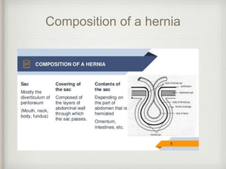
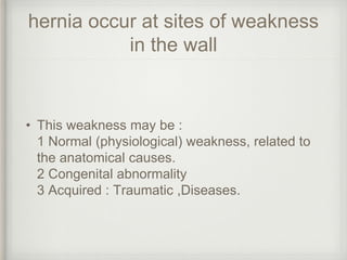
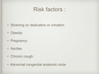
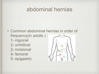
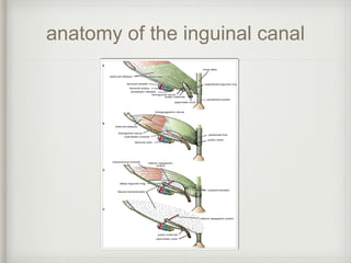
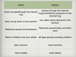
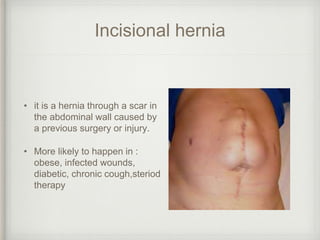
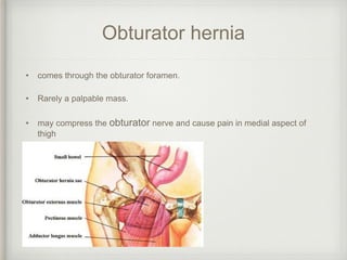
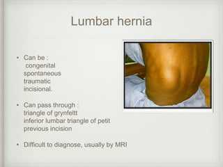
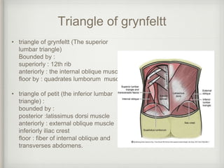
HERNIA
Lina abbas
Definition
• a protrusion of a viscus through an abnormal opening in the
wall of a cavity in which it is contained.
Composition of a hernia
hernia occur at sites of weakness
in the wall
• This weakness may be :
1 Normal (physiological) weakness, related to
the anatomical causes.
2 Congenital abnormality
3 Acquired : Traumatic ,Diseases.
Risk factors :
• Straining on dedication or urination
• Obesity
• Pregnancy
• Ascites
• Chronic cough
• Abnormal congenital anatomic route
abdominal hernias
• Common abdominal hernias in order of
frequency(in adults ) :
1- inguinal
2- umbilical
3- incisional
4- femoral
5- epigastric
Abdominal hernias
• rare hernias include :
1- spigelian
2- obturator
3- lumbar
4- gluteal
Inguinal hernia
• Most common site of hernias
• two types :
1. Direct : does not pass the internal inguinal
canal ring. Inter the canal directly through its
weekend posterior wall.
2. indirect : go through the internal ring traveling
to the external ring. may enter the scroyum
Anatomy of the inguinal canal
• it is an oblique passage through the lower part
of the anterior abdominal wall.
• it is 1.5 inch passes from the deep inguinal ring
downward medially to the superficial ring.
• lies immediately above
the the inguinal ligament
anatomy of the inguinal canal
• the deep ring :oval opening in fascia transversalis
0.5 inch above the inguinal ligament midway between ASIS and
the symphsis pubis ,its margins gives attachment to the internal
spermatic fascia/internal covering of round ligament.
• The superficial ring triangular defect in the aponeurosis of external
oblique muscle immediately above and medial to the pubic
tubercle, its margins gives attachment to the external spermatic
fascia
• walls :
anterior :external oblique aponeurosis
posterior :conjoint tendon medially, fascia transversals laterally
superior: lower fibers of internal oblique and transverses
abodminos muscle
floor : inguinal and lacunar ligaments
anatomy of the inguinal canal
Contents of the inguinal canal
• in males :spermatic cord[& its coverings + the ilioinguinal nerve.
the spermatic cord contains :
1 vas deferens and the artery to vas deferens
2 pampiniform plexus
3 testicular artery
4 cremasteric artery
5 fascial layers: external spermatic, cremasteric, and internal spermatic fascia
6 genital branch of the genitofemoral nerve
• in females : round ligament of the uterus + the ilioinguinal nerve.
Hesselbachs triangel
• triangular area in the lower abdominal wall , it is the site of direct
inguinal hernia its boundaries are
inferior : inguinal ligament
medial : rectus abdominos
lateral :inferior epigastric vessels
direct Indirect
Does not passthrough the internal
ring
passes through the internal
ring(controlled by pressure on the
internal ring
does not go down in the scortum
can (often does) descend in the
scortum
Reduced upward and backward
Reduced upward lately and back
ward
Rare in children and your adults all ages groups including children
less common most common
more medial more lateral
Pantaloon hernia
Femoral hernia
• it is a herniation through the
femoral canal
• they are more common in females
• The hernia appear below the
inguinal ligament and lateral to the
pubic tubercle . Directly behind the
skin crease of the groin
Anatomy of the femoral canal
• It is the small medial compartment in the femoral sheat that
contain the lymph vessels.
• it is 0.5inch long starts at : the femoral ring.to the saphenous
opening
Umbilical hernia
• May me congenital : the
umbilical scar fails to form .
or acquired: the umbilical
scar is stretched by an
increased intra-abdominal
pressure.
• may be from the umbilical
defect itself or come
through a defect adjacent
to the umbilical scar(may
be called paraumblical).
Epigastric hernia
• hernia in a defect in the lines alba
between the xiphisternum and the
umbilicus.
• Patient does not often notice the
underlying lump.
• it is common in children and
associated with divarication of the
rectus abdominis muscle.
Incisional hernia
• it is a hernia through a scar in
the abdominal wall caused by
a previous surgery or injury.
• More likely to happen in :
obese, infected wounds,
diabetic, chronic cough,steriod
therapy
Spigelian hernia
• is a hernia through the
Spigelian fascia, which is the
aponeurotic layer between the
rectus abdominis muscle
medially, and the semilunar line
laterally.
• below the umblicus and above
the inguinal area .
• rare. seen in obese patients,
difficult to diagnose.
Obturator hernia
• comes through the obturator foramen.
• Rarely a palpable mass.
• may compress the obturator nerve and cause pain in medial aspect of
thigh
Lumbar hernia
• Can be :
congenital
spontaneous
traumatic
incisional.
• Can pass through :
triangle of grynfeltt
inferior lumbar triangle of petit
previous incision
• Difficult to diagnose, usually by MRI
Triangle of grynfeltt
• triangle of grynfeltt (The superior
lumbar triangle)
Bounded by :
superiorly : 12th rib
anteriorly : the internal oblique muscle
floor by : quadrates lumborum muscle
• triangle of petit (the inferior lumbar
triangle) :
bounded by :
posterior :latissimus dorsi muscle
anteriorly : external oblique muscle
inferiorly iliac crest
floor : fiber of internal oblique and
transverses abdomens.
Esophageal hiatal hernia
• Herniation through the esophageal hiatus of
the diaphragm.
• 3 types are :
type 1: sliding hernia : award dislocation of
cardia in the posterior mediastinum
type 2 : rolling paraesophgeal hernia :upward
dislocation of gastric fundus with normal
cardia.
type 3: combined sliding rolling. Upward
dislocation of both cardia and funds
Esophageal hiatal hernia
hernias may be :
• Irreducible :contents cannot be replaced into the
abdomen.
• Incarcerated : contents are imprisoned in the
hernia. Usually by adhesion. they are alive and
functioning normally.
• Obstructed : the lumen in obstructed but the blood
supply is not.
• Strangulation: obstruction of blood supply, leads to
ischemia and infarction
Other types of hernia
Sliding herniaRechters hernia
Other types of hernia
maydle’s hernia
Surgical repair of hernia
• Open surgery :
1. Tissue repair
2. mesh repair
• Laparoscopic

Hernia

  • 1.
  • 2.
    Definition • a protrusionof a viscus through an abnormal opening in the wall of a cavity in which it is contained.
  • 3.
  • 4.
    hernia occur atsites of weakness in the wall • This weakness may be : 1 Normal (physiological) weakness, related to the anatomical causes. 2 Congenital abnormality 3 Acquired : Traumatic ,Diseases.
  • 5.
    Risk factors : •Straining on dedication or urination • Obesity • Pregnancy • Ascites • Chronic cough • Abnormal congenital anatomic route
  • 6.
    abdominal hernias • Commonabdominal hernias in order of frequency(in adults ) : 1- inguinal 2- umbilical 3- incisional 4- femoral 5- epigastric
  • 7.
    Abdominal hernias • rarehernias include : 1- spigelian 2- obturator 3- lumbar 4- gluteal
  • 8.
    Inguinal hernia • Mostcommon site of hernias • two types : 1. Direct : does not pass the internal inguinal canal ring. Inter the canal directly through its weekend posterior wall. 2. indirect : go through the internal ring traveling to the external ring. may enter the scroyum
  • 9.
    Anatomy of theinguinal canal • it is an oblique passage through the lower part of the anterior abdominal wall. • it is 1.5 inch passes from the deep inguinal ring downward medially to the superficial ring. • lies immediately above the the inguinal ligament
  • 10.
    anatomy of theinguinal canal • the deep ring :oval opening in fascia transversalis 0.5 inch above the inguinal ligament midway between ASIS and the symphsis pubis ,its margins gives attachment to the internal spermatic fascia/internal covering of round ligament. • The superficial ring triangular defect in the aponeurosis of external oblique muscle immediately above and medial to the pubic tubercle, its margins gives attachment to the external spermatic fascia • walls : anterior :external oblique aponeurosis posterior :conjoint tendon medially, fascia transversals laterally superior: lower fibers of internal oblique and transverses abodminos muscle floor : inguinal and lacunar ligaments
  • 11.
    anatomy of theinguinal canal
  • 12.
    Contents of theinguinal canal • in males :spermatic cord[& its coverings + the ilioinguinal nerve. the spermatic cord contains : 1 vas deferens and the artery to vas deferens 2 pampiniform plexus 3 testicular artery 4 cremasteric artery 5 fascial layers: external spermatic, cremasteric, and internal spermatic fascia 6 genital branch of the genitofemoral nerve • in females : round ligament of the uterus + the ilioinguinal nerve.
  • 13.
    Hesselbachs triangel • triangulararea in the lower abdominal wall , it is the site of direct inguinal hernia its boundaries are inferior : inguinal ligament medial : rectus abdominos lateral :inferior epigastric vessels
  • 14.
    direct Indirect Does notpassthrough the internal ring passes through the internal ring(controlled by pressure on the internal ring does not go down in the scortum can (often does) descend in the scortum Reduced upward and backward Reduced upward lately and back ward Rare in children and your adults all ages groups including children less common most common more medial more lateral
  • 15.
  • 16.
    Femoral hernia • itis a herniation through the femoral canal • they are more common in females • The hernia appear below the inguinal ligament and lateral to the pubic tubercle . Directly behind the skin crease of the groin
  • 17.
    Anatomy of thefemoral canal • It is the small medial compartment in the femoral sheat that contain the lymph vessels. • it is 0.5inch long starts at : the femoral ring.to the saphenous opening
  • 18.
    Umbilical hernia • Mayme congenital : the umbilical scar fails to form . or acquired: the umbilical scar is stretched by an increased intra-abdominal pressure. • may be from the umbilical defect itself or come through a defect adjacent to the umbilical scar(may be called paraumblical).
  • 19.
    Epigastric hernia • herniain a defect in the lines alba between the xiphisternum and the umbilicus. • Patient does not often notice the underlying lump. • it is common in children and associated with divarication of the rectus abdominis muscle.
  • 20.
    Incisional hernia • itis a hernia through a scar in the abdominal wall caused by a previous surgery or injury. • More likely to happen in : obese, infected wounds, diabetic, chronic cough,steriod therapy
  • 21.
    Spigelian hernia • isa hernia through the Spigelian fascia, which is the aponeurotic layer between the rectus abdominis muscle medially, and the semilunar line laterally. • below the umblicus and above the inguinal area . • rare. seen in obese patients, difficult to diagnose.
  • 22.
    Obturator hernia • comesthrough the obturator foramen. • Rarely a palpable mass. • may compress the obturator nerve and cause pain in medial aspect of thigh
  • 23.
    Lumbar hernia • Canbe : congenital spontaneous traumatic incisional. • Can pass through : triangle of grynfeltt inferior lumbar triangle of petit previous incision • Difficult to diagnose, usually by MRI
  • 24.
    Triangle of grynfeltt •triangle of grynfeltt (The superior lumbar triangle) Bounded by : superiorly : 12th rib anteriorly : the internal oblique muscle floor by : quadrates lumborum muscle • triangle of petit (the inferior lumbar triangle) : bounded by : posterior :latissimus dorsi muscle anteriorly : external oblique muscle inferiorly iliac crest floor : fiber of internal oblique and transverses abdomens.
  • 25.
    Esophageal hiatal hernia •Herniation through the esophageal hiatus of the diaphragm. • 3 types are : type 1: sliding hernia : award dislocation of cardia in the posterior mediastinum type 2 : rolling paraesophgeal hernia :upward dislocation of gastric fundus with normal cardia. type 3: combined sliding rolling. Upward dislocation of both cardia and funds
  • 26.
  • 27.
    hernias may be: • Irreducible :contents cannot be replaced into the abdomen. • Incarcerated : contents are imprisoned in the hernia. Usually by adhesion. they are alive and functioning normally. • Obstructed : the lumen in obstructed but the blood supply is not. • Strangulation: obstruction of blood supply, leads to ischemia and infarction
  • 28.
    Other types ofhernia Sliding herniaRechters hernia
  • 29.
    Other types ofhernia maydle’s hernia
  • 30.
    Surgical repair ofhernia • Open surgery : 1. Tissue repair 2. mesh repair • Laparoscopic