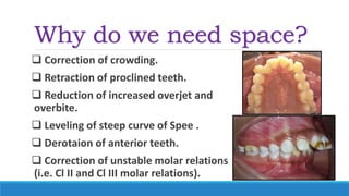
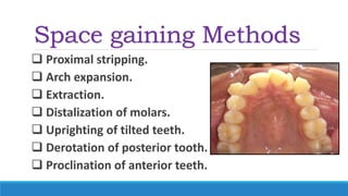
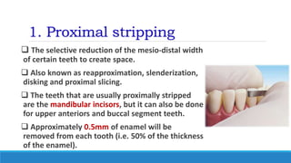
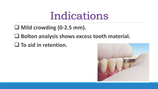
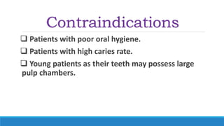
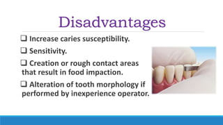
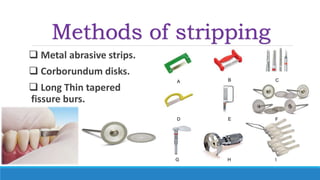
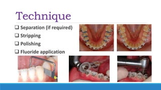
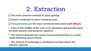
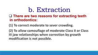
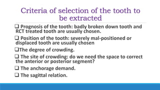
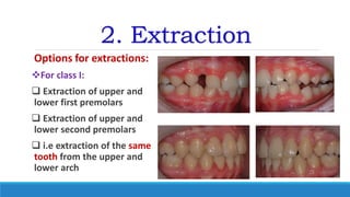
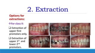
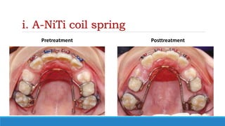
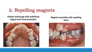
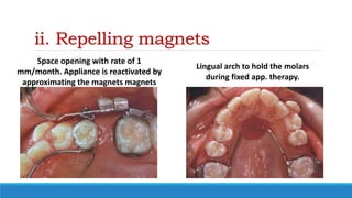
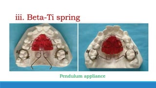
Space Gaining Methods
‫الرحيم‬‫الرحمن‬‫هللا‬ ‫بسم‬
Ibn Sina University
Faculty of Dentistry
Department of Orthodontics
Mohanad Elsherif
BDS (U of K), MFD RCSI, MFDS RCPS(Glasg), MSc (Orthodontics), M.Orth. RCSEd
Importance of space in
Ortho
Why do we need space?
 Correction of crowding.
 Retraction of proclined teeth.
 Reduction of increased overjet and
overbite.
 Leveling of steep curve of Spee .
 Derotaion of anterior teeth.
 Correction of unstable molar relations
(i.e. Cl II and Cl III molar relations).
Space gaining Methods
 Proximal stripping.
 Arch expansion.
 Extraction.
 Distalization of molars.
 Uprighting of tilted teeth.
 Derotation of posterior tooth.
 Proclination of anterior teeth.
1. Proximal stripping
 The selective reduction of the mesio-distal width
of certain teeth to create space.
 Also known as reapproximation, slenderization,
disking and proximal slicing.
 The teeth that are usually proximally stripped
are the mandibular incisors, but it can also be done
for upper anteriors and buccal segment teeth.
 Approximately 0.5mm of enamel will be
removed from each tooth (i.e. 50% of the thickness
of the enamel).
Indications
 Mild crowding (0-2.5 mm).
 Bolton analysis shows excess tooth material.
 To aid in retention.
Contraindications
 Patients with poor oral hygiene.
 Patients with high caries rate.
 Young patients as their teeth may possess large
pulp chambers.
Advantages
 Avoid extraction.
 More stable results.
Disadvantages
 Increase caries susceptibility.
 Sensitivity.
 Creation or rough contact areas
that result in food impaction.
 Alteration of tooth morphology if
performed by inexperience operator.
Methods of stripping
 Metal abrasive strips.
 Corborundum disks.
 Long Thin tapered
fissure burs.
Technique
 Separation (if required)
 Stripping
 Polishing
 Fluoride application
2. Extraction
 The most common method of space gaining.
 Used in moderate to sever crowding cases.
 First premolars are the most commonly extracted tooth (Why?).
 It lies in the middle of the arch so its extraction will provide space
for both anterior and posterior segment.
 The contact between the canine and second premolar is usually
stable following space closure.
 The amount of anchorage is satisfactory to help retract the
anterior segment.
b. Extraction
 There are two reasons for extracting teeth
in orthodontics:
(1) To correct moderate to sever crowding.
(2) To allow camouflage of moderate Class II or Class
III jaw relationships when correction by growth
modification is not possible.
Criteria of selection of the tooth to
be extracted
 Prognosis of the tooth: badly broken down tooth and
RCT treated tooth are usually chosen.
 Position of the tooth: severely mal-positioned or
displaced tooth are usually chosen
The degree of crowding.
 The site of crowding: do we need the space to correct
the anterior or posterior segment?
 The anchorage demand.
 The sagittal relation.
2. Extraction
Options for extractions:
For class I:
 Extraction of upper and
lower first premolars
 Extraction of upper and
lower second premolars
 i.e extraction of the same
tooth from the upper and
lower arch
2. Extraction
Options for
extractions:
For class II:
 Extraction of
upper first
premolars only.
 Extraction of
upper 1st and
lower 2nd
premolars.
2. Extraction
Options for extractions:
 For class III
 Extraction of lower first
premolars only.
 Extraction of upper 2nd
and lower 1st premolars.
 Extraction of one lower
incisor.
3. Arch expansion
 The term jaw expansion refer to widening of
posterior teeth.
 First introduced by Emerson C. Angell in 1860.
 Walter Coffin in 1877 introduced the coffin spring.
 Karhkaus and Andrew Hass in 1950s introduced
the Hass appliance.
3. Arch
expansion
 Types of arch expansions
1. According to the type:
A. Skeletal expansion:
 Achieved by opening the mid-palatal suture.
 The expansion screw is activated at a rate of
(0.25-0.5 mm) per day.
 The treatment usually takes around two weeks
(midline diastema occur when suture open).
 Ineffective after age of 15-16 years due to
ossification of the mid-platal suture
 Surgically assisted rapid palatal expansion is
indicated after age of 16 years.
3. Arch
expansion
 Types of arch expansions
2. According to the type:
b. Dental expansion:
 Achieved by buccal tipping of
teeth.
 The expansion screw is activated
at rated of (1-2 mm) per months.
 The treatment usually takes
around 5 months.
3. Arch expansion
 Types of arch expansions
3. According to the speed
 Rapid expansion: 0.25- 0.5 mm/ per day ( i.e. 1-2 turn
per day)
 Slow expansion: ( 1-2 mm/per month) ( 1-2 turn per
week)
 Semi-rapid: Start with rapid activation rate for the first
week then continue at slow expansion rate
3. Arch expansion
Comparison between slow and rapid expansion
Feature Slow expansion Rapid expansion
Type of expansion Mostly dental Skeletal
Rate of expansion Slow Rapid
Type of tissue reaction More physiological More traumatic
Force used Milder force Greater forces
Frequency of activation Less frequent More frequent
Duration of treatment Long Short
Type of appliance Either fixed or removable Mostly fixed appliance
Age Any age Before fusion of mid-palatal
suture
Retention Lesser chance of relapse More chance of relapse
4. Distalizaion of molars
 It is the distal movement of molars to gain space.
 Usually done in mixed dentition before the eruption
of 2nd molars.
 Not more than 2-3 mm can be achieved without
extraction of second molars or third molars.
Distal movement of molar teeth can be
accomplished by:
 Extraoral appliance: Headgear.
 Intraoral distalizing appliances.
 Skeletal anchorage.
a. Extraoral appliance: headgear
 The appliance is worn for
10- 12 hours per day.
 The force is usually about
200-400 mg per side.
 Unilateral distalization can
be done by using asymmetric
headgear.
b. Intraoral distalizing appliance
 Gain its anchorage from the palate
 The relative stability of the anterior palate, both the soft
tissue rugae and the cortical bone beneath them
 Once palatal anchorage has been established, there are
several possibilities for generating the molar distalizing force.
 Austenite nickel–titanium (A-NiTi) coil springs compressed against
the molars  advantages: continues force less bulky.
 Repelling magnets  disadvantiges: Bulky, force decay as the teeth
moves.
 Beta Ti springs  Example: Pendulum appliance
i. A-NiTi coil spring
Pretreatment Posttreatment
ii. Repelling magnets
Palatal anchorage with stabilizing
lingual arch from premolars
Magnet assembly with repelling
force
ii. Repelling magnets
Space opening with rate of 1
mm/month. Appliance is reactivated by
approximating the magnets magnets
Lingual arch to hold the molars
during fixed app. therapy.
iii. Beta-Ti spring
Pendulum appliance
The pendulum appliance?
Canine is blocked out with ½ molar
relationship
Pendulum appliance
with both a jackscrew for transverse expansion and
molar distalizing springs (this modification is called
the T-Rex appliance).
The pendulum appliance?
Appliance removed, noticed the
significance palatal irritation
Lingual holding arch to stabilize the
molars as fixed appliance treatment
proceeds
Treatment
results
c. Skeletal Anchorage
 At this point, the advantage of
skeletal anchorage for distalization
of molars is so great that it is rapidly
replacing the previous methods.
 Skeletal anchorage or not, two
objects cannot occupy the same
space at the same time, therefore
extraction of 7s or 8s may be
indicated.
5. Derotation of posterior teeth
 Rotated posterior tooth
occupy more space
 Derotation of these teeth
gain space
 This is better achieved by
fixed appliance
 Headgear and trans-platal
arch can also be used to
derotate molar teeth.
6. Uprighting of molars
 Premature loss of primary
second molar leads to mesial
tipping of first molar and
space loss.
 Uprighting of this tooth
gain space.
 Either removable or fixed
appliances can be used for
this purposes.
7. Proclination of Incisors
This can be done only if the incisors are already
retorclined (e.g. in class II div 2 patients)
Declaration
 The author wish to declare that; these presentations are his original work, all
materials and pictures collection, typing and slide design has been done by the
author.
 Most of these materials has been done for undergraduate students, although
postgraduate students may find some useful basic and advanced information.
 The universities title at the front page indicate where the lecture was first
presented. The author was working as a lecturer of orthodontics at Ibn Sina
University, Sudan International University, and as a Master student in Orthodontics at
University of Khartoum.
 The author declare that all materials and photos in these presentations has been
collected from different textbooks, papers and online websites. These pictures are
presented here for education and demonstration purposes only. The author are not
attempting to plagiarize or reproduced unauthorized material, and the intellectual
properties of these photos belong to their original authors.
Declaration
 As the authors reviews several textbooks, papers and other references during
preparation of these materials, it was impossible to cite every textbook and journal
article, the main textbooks that has been reviewed during preparation of these
presentations were:
Contemporary Orthodontics 5th edition; Proffit, William R, Henry W. Fields, and
David M. Sarver.
Handbook of Orthodontics. 1st edition; Cobourne, Martyn T, and Andrew T. DiBiase.
Essentials of orthodontics: Diagnosis and Treatment; Robert N. Staley, Neil T. Reske
Orthodontics: Current Principles & Techniques 5th edition; Graber, Lee W, Robert L.
Vanarsdall, and Katherine W. L. Vig
Orthodontics: The Art and Science. 3rd Edition. Bhalajhi, S.I.
Declaration
 For the purposes of dissemination and sharing of knowledge, these
lectures were given to several colleagues and students. It were also
uploaded to SlideShare website by the author. Colleagues and students
may download, use, and modify these materials as they see fit for non-
profit purposes. The author retain the copyright of the original work.
 The author wish to thank his family, teachers, colleagues and students
for their love and support throughout his career. I also wish to express
my sincere gratitude to all orthodontic pillars for their tremendous
contribution to our specialty.
 Finally, the author welcome any advices and enquires through his
email address: Mohanad-07@hotmail.com
Thank you

Space gaining methods

  • 1.
    Space Gaining Methods ‫الرحيم‬‫الرحمن‬‫هللا‬‫بسم‬ Ibn Sina University Faculty of Dentistry Department of Orthodontics Mohanad Elsherif BDS (U of K), MFD RCSI, MFDS RCPS(Glasg), MSc (Orthodontics), M.Orth. RCSEd
  • 2.
  • 3.
    Why do weneed space?  Correction of crowding.  Retraction of proclined teeth.  Reduction of increased overjet and overbite.  Leveling of steep curve of Spee .  Derotaion of anterior teeth.  Correction of unstable molar relations (i.e. Cl II and Cl III molar relations).
  • 4.
    Space gaining Methods Proximal stripping.  Arch expansion.  Extraction.  Distalization of molars.  Uprighting of tilted teeth.  Derotation of posterior tooth.  Proclination of anterior teeth.
  • 5.
    1. Proximal stripping The selective reduction of the mesio-distal width of certain teeth to create space.  Also known as reapproximation, slenderization, disking and proximal slicing.  The teeth that are usually proximally stripped are the mandibular incisors, but it can also be done for upper anteriors and buccal segment teeth.  Approximately 0.5mm of enamel will be removed from each tooth (i.e. 50% of the thickness of the enamel).
  • 6.
    Indications  Mild crowding(0-2.5 mm).  Bolton analysis shows excess tooth material.  To aid in retention.
  • 7.
    Contraindications  Patients withpoor oral hygiene.  Patients with high caries rate.  Young patients as their teeth may possess large pulp chambers.
  • 8.
  • 9.
    Disadvantages  Increase cariessusceptibility.  Sensitivity.  Creation or rough contact areas that result in food impaction.  Alteration of tooth morphology if performed by inexperience operator.
  • 10.
    Methods of stripping Metal abrasive strips.  Corborundum disks.  Long Thin tapered fissure burs.
  • 11.
    Technique  Separation (ifrequired)  Stripping  Polishing  Fluoride application
  • 12.
    2. Extraction  Themost common method of space gaining.  Used in moderate to sever crowding cases.  First premolars are the most commonly extracted tooth (Why?).  It lies in the middle of the arch so its extraction will provide space for both anterior and posterior segment.  The contact between the canine and second premolar is usually stable following space closure.  The amount of anchorage is satisfactory to help retract the anterior segment.
  • 13.
    b. Extraction  Thereare two reasons for extracting teeth in orthodontics: (1) To correct moderate to sever crowding. (2) To allow camouflage of moderate Class II or Class III jaw relationships when correction by growth modification is not possible.
  • 14.
    Criteria of selectionof the tooth to be extracted  Prognosis of the tooth: badly broken down tooth and RCT treated tooth are usually chosen.  Position of the tooth: severely mal-positioned or displaced tooth are usually chosen The degree of crowding.  The site of crowding: do we need the space to correct the anterior or posterior segment?  The anchorage demand.  The sagittal relation.
  • 15.
    2. Extraction Options forextractions: For class I:  Extraction of upper and lower first premolars  Extraction of upper and lower second premolars  i.e extraction of the same tooth from the upper and lower arch
  • 16.
    2. Extraction Options for extractions: Forclass II:  Extraction of upper first premolars only.  Extraction of upper 1st and lower 2nd premolars.
  • 17.
    2. Extraction Options forextractions:  For class III  Extraction of lower first premolars only.  Extraction of upper 2nd and lower 1st premolars.  Extraction of one lower incisor.
  • 18.
    3. Arch expansion The term jaw expansion refer to widening of posterior teeth.  First introduced by Emerson C. Angell in 1860.  Walter Coffin in 1877 introduced the coffin spring.  Karhkaus and Andrew Hass in 1950s introduced the Hass appliance.
  • 19.
    3. Arch expansion  Typesof arch expansions 1. According to the type: A. Skeletal expansion:  Achieved by opening the mid-palatal suture.  The expansion screw is activated at a rate of (0.25-0.5 mm) per day.  The treatment usually takes around two weeks (midline diastema occur when suture open).  Ineffective after age of 15-16 years due to ossification of the mid-platal suture  Surgically assisted rapid palatal expansion is indicated after age of 16 years.
  • 20.
    3. Arch expansion  Typesof arch expansions 2. According to the type: b. Dental expansion:  Achieved by buccal tipping of teeth.  The expansion screw is activated at rated of (1-2 mm) per months.  The treatment usually takes around 5 months.
  • 21.
    3. Arch expansion Types of arch expansions 3. According to the speed  Rapid expansion: 0.25- 0.5 mm/ per day ( i.e. 1-2 turn per day)  Slow expansion: ( 1-2 mm/per month) ( 1-2 turn per week)  Semi-rapid: Start with rapid activation rate for the first week then continue at slow expansion rate
  • 22.
    3. Arch expansion Comparisonbetween slow and rapid expansion Feature Slow expansion Rapid expansion Type of expansion Mostly dental Skeletal Rate of expansion Slow Rapid Type of tissue reaction More physiological More traumatic Force used Milder force Greater forces Frequency of activation Less frequent More frequent Duration of treatment Long Short Type of appliance Either fixed or removable Mostly fixed appliance Age Any age Before fusion of mid-palatal suture Retention Lesser chance of relapse More chance of relapse
  • 23.
    4. Distalizaion ofmolars  It is the distal movement of molars to gain space.  Usually done in mixed dentition before the eruption of 2nd molars.  Not more than 2-3 mm can be achieved without extraction of second molars or third molars. Distal movement of molar teeth can be accomplished by:  Extraoral appliance: Headgear.  Intraoral distalizing appliances.  Skeletal anchorage.
  • 24.
    a. Extraoral appliance:headgear  The appliance is worn for 10- 12 hours per day.  The force is usually about 200-400 mg per side.  Unilateral distalization can be done by using asymmetric headgear.
  • 25.
    b. Intraoral distalizingappliance  Gain its anchorage from the palate  The relative stability of the anterior palate, both the soft tissue rugae and the cortical bone beneath them  Once palatal anchorage has been established, there are several possibilities for generating the molar distalizing force.  Austenite nickel–titanium (A-NiTi) coil springs compressed against the molars  advantages: continues force less bulky.  Repelling magnets  disadvantiges: Bulky, force decay as the teeth moves.  Beta Ti springs  Example: Pendulum appliance
  • 26.
    i. A-NiTi coilspring Pretreatment Posttreatment
  • 27.
    ii. Repelling magnets Palatalanchorage with stabilizing lingual arch from premolars Magnet assembly with repelling force
  • 28.
    ii. Repelling magnets Spaceopening with rate of 1 mm/month. Appliance is reactivated by approximating the magnets magnets Lingual arch to hold the molars during fixed app. therapy.
  • 29.
  • 30.
    The pendulum appliance? Canineis blocked out with ½ molar relationship Pendulum appliance with both a jackscrew for transverse expansion and molar distalizing springs (this modification is called the T-Rex appliance).
  • 31.
    The pendulum appliance? Applianceremoved, noticed the significance palatal irritation Lingual holding arch to stabilize the molars as fixed appliance treatment proceeds
  • 32.
  • 33.
    c. Skeletal Anchorage At this point, the advantage of skeletal anchorage for distalization of molars is so great that it is rapidly replacing the previous methods.  Skeletal anchorage or not, two objects cannot occupy the same space at the same time, therefore extraction of 7s or 8s may be indicated.
  • 34.
    5. Derotation ofposterior teeth  Rotated posterior tooth occupy more space  Derotation of these teeth gain space  This is better achieved by fixed appliance  Headgear and trans-platal arch can also be used to derotate molar teeth.
  • 35.
    6. Uprighting ofmolars  Premature loss of primary second molar leads to mesial tipping of first molar and space loss.  Uprighting of this tooth gain space.  Either removable or fixed appliances can be used for this purposes.
  • 36.
    7. Proclination ofIncisors This can be done only if the incisors are already retorclined (e.g. in class II div 2 patients)
  • 37.
    Declaration  The authorwish to declare that; these presentations are his original work, all materials and pictures collection, typing and slide design has been done by the author.  Most of these materials has been done for undergraduate students, although postgraduate students may find some useful basic and advanced information.  The universities title at the front page indicate where the lecture was first presented. The author was working as a lecturer of orthodontics at Ibn Sina University, Sudan International University, and as a Master student in Orthodontics at University of Khartoum.  The author declare that all materials and photos in these presentations has been collected from different textbooks, papers and online websites. These pictures are presented here for education and demonstration purposes only. The author are not attempting to plagiarize or reproduced unauthorized material, and the intellectual properties of these photos belong to their original authors.
  • 38.
    Declaration  As theauthors reviews several textbooks, papers and other references during preparation of these materials, it was impossible to cite every textbook and journal article, the main textbooks that has been reviewed during preparation of these presentations were: Contemporary Orthodontics 5th edition; Proffit, William R, Henry W. Fields, and David M. Sarver. Handbook of Orthodontics. 1st edition; Cobourne, Martyn T, and Andrew T. DiBiase. Essentials of orthodontics: Diagnosis and Treatment; Robert N. Staley, Neil T. Reske Orthodontics: Current Principles & Techniques 5th edition; Graber, Lee W, Robert L. Vanarsdall, and Katherine W. L. Vig Orthodontics: The Art and Science. 3rd Edition. Bhalajhi, S.I.
  • 39.
    Declaration  For thepurposes of dissemination and sharing of knowledge, these lectures were given to several colleagues and students. It were also uploaded to SlideShare website by the author. Colleagues and students may download, use, and modify these materials as they see fit for non- profit purposes. The author retain the copyright of the original work.  The author wish to thank his family, teachers, colleagues and students for their love and support throughout his career. I also wish to express my sincere gratitude to all orthodontic pillars for their tremendous contribution to our specialty.  Finally, the author welcome any advices and enquires through his email address: Mohanad-07@hotmail.com
  • 40.