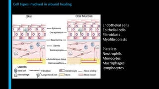
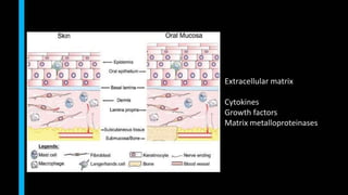
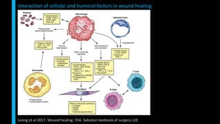
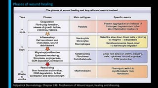
SlideShare a Scribd company logo
SOFT TISSUE HEALING
RAKESH CHANDRAN
Technically wound healing is a term that should be used only in the context of true
regeneration, when the original architecture and structure of an organ or anatomic part
is completely restored to the way it was before injury.
Regeneration is often used in periodontal literature to describe instances where the
structure and functional relationships of damaged periodontal tissues appear to be
renewed. The most common use of term regeneration describes new attachment or
formation of new cementum, alveolar bone and intervening periodontal ligament.
Repair is the process where in damaged tissues are replaced by tissues that do not
duplicate the function of original tissues.
Definitions
• Cellular and humoral factors involved in soft tissue wound healing
• Phases of wound healing
• Soft tissue healing with regard to specific periodontal procedures
Presentation outline
Endothelial cells
Epithelial cells
Fibroblasts
Myofibroblasts
Platelets
Neutrophils
Monocytes
Macrophages
Lymphocytes
Cell types involved in wound healing
Endothelial cells
• endothelial cells of the original blood vessels
• circulating endothelial progenitors
Fibroblasts
• connective tissue in the wound edges
• monocyte-derived fibrocytes
• from vessel-derived pericytes
• possibly also by a process termed epithelialmesenchymal transition
Epithelial cells
• keratinocytes at the wound edges
A proportion of the fibroblasts achieve a phenotype that resembles smooth muscle cells
(myofibroblasts) which can draw the wound edges together and are thus critical
components of wound healing.
Cellular origins
Sculean et al 2014. Soft tissue wound healing around teeth and dental implants. J Clin Periodontol
Extracellular matrix
Cytokines
Growth factors
Matrix metalloproteinases
• In wound healing, the extracellular matrix (ECM) comprises four main components:
1. structural proteins (i.e., collagen, elastin)
2. multidomain adhesive glycoproteins (i.e., fibronectin, vitronectin, laminin)
3. matricellular proteins, including secreted protein acidic and rich in cysteine
(SPARC), thrombospondin 1 and 2, tenascins, osteopontin
4. glycosaminoglycans (GAG), including hyaluronic acid and proteoglycans, that is,
syndecans, perlecan (such as chondroitin sulfate and heparan sulfate)
Extracellular matrix
Fiztpatrick Dermatology. Chapter 248. Mechanism of Wound repair, healing and dressing.
• Three main classes of collagens are present normally in connective tissue:
(1) fibrillar collagens (types I, III and V)
(2) basement membrane collagen (type IV)
(3) other interstitial collagens (types VI, VII, and VIII)
Collagen
Fiztpatrick Dermatology. Chapter 248. Mechanism of Wound repair, healing and dressing.
• particularly abundant component of the provisional matrix
• thought to be at least in part responsible for the “scarless” healing of embryonic
wounds.
• Fibroblasts from early granulation tissue produce large amounts of hyaluronic acid,
and proliferating cells express CD44, which is the receptor for this GAG molecule.
• offers less resistance to cell movement and stimulates cell motility by altering cell-
matrix adhesion.
• creates a highly hydrated structure leading to tissue swelling and interstitial spaces,
and thus an environment more conducive to cell movement.
• as remodeling occurs, hyaluronic acid is degraded by hyaluronidase and replaced by
sulfated proteoglycans, which contribute a stronger structural role in late granulation
tissue formation and in scars, while being less able to stimulate cellular movement.
• Two major proteoglycans, chondroitin-4-sulfate and dermatan sulfate, are produced
by mature scar fibroblasts.
Hyaluronic acid
Fiztpatrick Dermatology. Chapter 248. Mechanism of Wound repair, healing and dressing.
Cytokine Activity in Wound Healing
Leong et al 2017. Wound healing. Ch6. Sabiston textbook of surgery 12E
Actions of growth factors and cytokines are often context specific and not the same in every biologic situation
Growth Factors That Affect Wound Healing
Leong et al 2017. Wound healing. Ch6. Sabiston textbook of surgery 12E
Growth Factors That Affect Wound Healing
Leong et al 2017. Wound healing. Ch6. Sabiston textbook of surgery 12E
Matrix metalloproteinases
Fiztpatrick Dermatology. Chapter 248. Mechanism of Wound repair, healing and dressing.
Phases of wound healing
Fiztpatrick Dermatology. Chapter 248. Mechanism of Wound repair, healing and dressing.
Interaction of cellular and humoral factors in wound healing.
Leong et al 2017. Wound healing. Ch6. Sabiston textbook of surgery 12E
SPM-mediated switching from inflammation to resolution
Freire 2017 Resolution of Inflammation and Periodontal Diseases Carranza Elsevier
Specialized Pro-Resolving Lipid Mediators
• Resolution is initiated when innate immune cells (neutrophils and monocytes) are
“told” to clear the injury and restore to homeostasis.
• Resolution mediators are oxidative fatty acids, constituted by a family of lipid
mediators and have agonist cellular functions.
• There are four main families of specialized lipid mediators: lipoxins, resolvins,
protectins, and maresins.
• In addition to lipid mediators, regulatory cytokines and cells also work in a temporally
and spatially coordinated micro-environment to activate resolution signals.
• Among all cellular functions activated by resolution signals, induction of phagocytosis,
clearance of infection, and exit to the lymphatics are the main cellular actions.
• This allows the return to homeostasis and activation of tissue healing.
SPM-mediated switching from inflammation to resolution
Freire 2017 Resolution of Inflammation and Periodontal Diseases Carranza Elsevier
Phases of wound healing
Fiztpatrick Dermatology. Chapter 248. Mechanism of Wound repair, healing and dressing.
• normal fibroblasts express vimentin and fibroblast-specific protein 1 (FSP1)
• activation of fibroblasts takes place when tissue injury occurs
• injured epithelial cells and infiltrating mononuclear cells such as monocytes and
macrophages release various factors like TGF-ß, EGF, PDGF and FGF2
• fibroblasts are activated by direct cell–cell communication and contacts with
leukocytes through adhesion molecules such as intercellular-adhesion molecule 1
(ICAM1) or vascular-cell adhesion molecule 1 (VCAM1).
• The activated fibroblasts secrete various mediators which play a key role in
connective tissue remodelling. These include proteases such as MMP2, MMP-3 and
MMP-9. In addition to these, activated fibroblasts secrete various growth factors
such as TGF-ß, IGF, nerve growth factor (NGF) and keratinocyte growth factor (KGF)
which can induce proliferative signals within adjacent epithelial cells.
Fibroblast activation
Fiztpatrick Dermatology. Chapter 248. Mechanism of Wound repair, healing and dressing.
Phases of wound healing
Fiztpatrick Dermatology. Chapter 248. Mechanism of Wound repair, healing and dressing.
• MMP-1 (collagenase-1 or interstitial collagenase) is important in keratinocyte
migration and epithelialization.
• MMP-9 plays a fundamental role in “cutting” Type IV and type VII collagen, which are
essential components of the basement membrane and anchoring fibrils, and it also
promotes inflammation and neutrophil migration.
• MMP-10 (stromelysin) breaks down other noncollagenous ECM components and
facilitates migration
Proliferation/migration and remodeling phases
Fiztpatrick Dermatology. Chapter 248. Mechanism of Wound repair, healing and dressing.
The integrin family of adhesion receptors.
Akhil 2000. Biology of wound healing. Perio 2000.
Integrins Ligands
, ; 21 Fibrillar collagen, laminin.
31 Fibronectin, entactin, epilgrin, laminin, denatured collagen.
41 Fibronectin, VCAM-1
51 Fibronectin (RGD)
61; 71; 64 Laminin
81 Fibronectin, vitronectin
91 Tenascin
v1; v5 Fibronectin, vitronectin
v3; 11b3 Vitronectin (RGD), fibronectin, fibrinogen, von Willebrand factor,
thrombospondin, denatured collagen
v6 Fibronectin, tenascin
47 Fibronectin (IIIcs)
v8 Vitronectin
M2 Factor X, fibrinogen
X2 Fibrinogen
• In the normal tissues, keratinocytes use the integrins α6β4 to bind to laminin in the
basal lamina, and these integrins have intracellular links with the keratin cytoskeletal
network.
• In preparation for migration, the keratinocytes at the edge of the surgical wound have
to dissolve the hemidesmosome attachment and begin to express other integrins that
are more suitable for the wound environment.
• The migrating keratinocytes will start expressing the integrins α5β1 for binding to
fibronectin and αVβ6 for binding to tenascin, the integrin αVβ5 for binding to
vitronectin and, finally, reorganize the distribution of the integrins α2β1 (collagen
receptor).
• This mobilization and expression of new integrins facilitates adhesion of keratinocytes
to matrix molecules in the provisional matrix as well as the adjacent wound debris.
This can explain the lag time between wounding and the initiation of epithelial
migration. It is generally believed that the basal cells provide the majority of
migrating keratinocytes, although there is some evidence that some of the suprabasal
cells may also migrate.
Integrins
Akhil 2000. Biology of wound healing. Perio 2000.
Phases of wound healing
Fiztpatrick Dermatology. Chapter 248. Mechanism of Wound repair, healing and dressing.
The linear progression seen in acute wounds is not seen in chronic wounds.
Fiztpatrick Dermatology 8th Ed. Chapter 249. Wound repair: Mechanisms and Practical considerations
Acute vs Chronic wounds
Mechanisms involved in the development and persistence of chronic wounds
Leong et al 2017. Wound healing. Ch6. Sabiston textbook of surgery 12E
Solid lines indicate
upregulation, and dashed lines
indicate downregulation. Width
of the line is proportional to the
effect of the influence.
H2O2, Hydrogen peroxide;
O2 −, superoxide;
TGF-β, transforming growth
factor-β.
• The basic healing processes are the same after all forms of periodontal therapy.
• These processes consist of the removal of degenerated tissue debris and the
replacement of tissues destroyed by disease.
• This implies regeneration and repair of the periodontal structures but not necessarily
a gain in attachment.
Healing After Periodontal Therapy
Carranza 12th Edition
• mucoperiosteal flap is opposed to an instrumented root surface deprived of its
periodontal attachment
• Immediately after suturing (up to 24 hours) a connection between the flap and the
tooth (or bone surface) is established by clot formation at the interface between the
tooth and a gingival flap.
• In periodontal defects the debrided defect becomes concealed by the irregular fibrin
clot.
• Clot formation is initiated as blood elements are absorbed to the boundaries of the
wound – namely cementum, dentine, alveolar bone, gingival connective tissue.
• absorption and adhesion of plasma proteins onto the root surface represents the first
healing event at the tooth-gingival interface
• Within hours the early phase of inflammation occurs as inflammatory cells
(predominantly neutrophils and monocytes) accumulate on the root surface.
Wound healing at the tooth interface
Polimeni et al, 2006
• establishment of a fibrin clot is the first and essential milestone toward periodontal
wound healing/ regeneration
• One to 3 days after flap surgery, the space between the flap and the tooth/bone
becomes smaller and epithelial cells migrate over the border of the flap, usually
contacting the tooth.
• With close adaptation of the flap there is minimal inflammatory response.
• Within 3 days the late phase of inflammation dominates the healing process as
macrophages migrate into the wound.
• This is followed by the formation of granulation tissue.
• At 7 days, a connective tissue attachment may be observed, however areas of fibrin
clot in various stages of maturation will also still be present.
Wound healing at the tooth interface
Susin et al, 2015
Polimeni et al, 2006
• One week after surgery, an epithelial attachment to the root has been established by
means of hemidesmosomes and a basal lamina.
• The blood clot is replaced by granulation tissue derived from the gingival connective
tissue, the bone marrow and the PDL.
• Two weeks after surgery, collagen fibers begin to appear parallel to the tooth surface.
• The union of the flap to the tooth is still weak, because of the immature collagen
fibers
• One month after surgery, a fully epithelialized gingival crevice with a well -defined
epithelial attachment is present.
• The supra-crestal fibers begin to take on a functional arrangement.
Wound healing at the tooth interface
Polimeni et al, 2006
• immediately after curettage blood clot fills the gingival sulcus.
• restoration and epithelization of the sulcus generally begins about 2-3 days after
curettage
• completed between 7-10 days after treatment.
Clinical changes of the tissues after curettage
• Marginal gingiva appears red and blood coagulum will be present
• After 2 days the gingiva appears light bluish red.
• After 4 days the gingiva appears red edematous with reduced intensity.
• After 6 days gingival tissue will be light red and edema is markedly reduced.
• After 7 days gingival tissue will be pink with constriction and recession but marginal
gingiva is smooth and glossy.
• After 8 days gingiva remains smooth.
• After 9 days gingiva appears pale pink with surface keratinization.
Healing after curettage
S. J. Froum et al. “Periodontal Healing Following Open Debridement Flap Procedure I: Clinical Assessment of Soft Tissue
and Osseous Repair”. Journal of Periodontology. 1981; 53(1): 8-14
• The major events in healing are re epithelization of the wound surface and
restoration of the dento-gingival junction.
• Primary intention healing is seen in gingivectomy and secondary intention in
gingivoplasty.
Gingivectomy and gingivoplasty
S. J. Froum et al. “Periodontal Healing Following Open Debridement Flap Procedure I: Clinical Assessment of Soft Tissue
and Osseous Repair”. Journal of Periodontology. 1981; 53(1): 8-14
Imbibition
• diffusion of plasma from the recipient site into the overlying skin graft, resulting in
a weight increase of the graft of up to 40% within the first day
• keeps the graft moist and the graft vessels patent and provides nutrition to the
graft
• imbibition phase ends as the venous and lymphatic drainage is reestablished, with
a concomitant reduction in the weight of the graft
Inosculation
• anastomoses of graft blood vessels with the recipient-derived vessels
Neovascularisation
• ingrowth of new blood vessels from the recipient wound bed into the graft.
Graft healing- vascularisation
• Specific in-situ hybridization and immunolabeling were used to distinguish graft from
recipient in studies of human split thickness skin grafted onto nude mice. It was found
that the recipient endothelial cells grew into the preexisting donor capillary tubes,
decisively supporting the process of immediate inosculation.
• Most recently, transgenic mice were used to distinguish donor derived from recipient-
derived vascular growth to reveal an even more precise picture. Capla and colleagues
showed regression of the graft vessels starting at day 3, with simultaneous vessel
ingrowth from the recipient primarily into the graft’s preexisting vascular framework,
resulting in inosculation by day 7
• Presumably, neovascularization without anastomosing with the graft’s vascular
framework is not the predominant event.
• Using a mouse bone marrow transplant model, the same investigative group further
showed that bone marrow-derived endothelial progenitor cells contributed 20% of
the recipient-derived endothelial growth within the graft
Inosculation and neovascularization
Capla JM et al: Skin graft vascularization involves precisely regulated regression and replacement of endothelial cells
through both angiogenesis and vasculogenesis. Plast Reconstr Surg 117 :836- 844, 2006
• Immediately after placement of the graft a fibrin clot forms between graft and the
underlying periosteal bed. This medium serves to transport nutrients from the
recipient area to connective tissue of the graft.
• Re epithelization occurs during the last half of the first post operative week with cells
originating from lateral wound margins of epithelial ridges with in the graft itself.
• By 72 hours connective tissue proliferation begins and by the end of the first week a
tenuous fibrous attachment between the graft and the recipient is seen.
• By 14th day the epithelium presents a near normal histologic thickness.
• Healing of a free gingival graft will include reconstruction of the dentogingival
junction coronal to the crest of the retained periosteum.
Free gingival grafts
S. J. Froum et al. “Periodontal Healing Following Open Debridement Flap Procedure I: Clinical Assessment of Soft Tissue
and Osseous Repair”. Journal of Periodontology. 1981; 53(1): 8-14
• At 4th day wound surface is red suggesting the formation of granulation tissue.
• At the sixth day post operatively budding and sprouting capillaries are identified in
the granulation tissue at the wound interface.
• The greatest cellular activity is seen at 6th day, which is responsible for the formation
of the new reparative connective tissue.
• Continuity between the periodontal flap and vasculature is established within 9 days.
• Histologically by 21 days revascularisation of the flap is re established.
Lateral pedicle grafts
S. J. Froum et al. “Periodontal Healing Following Open Debridement Flap Procedure I: Clinical Assessment of Soft Tissue
and Osseous Repair”. Journal of Periodontology. 1981; 53(1): 8-14
• Within minutes after flap closure, a fibrin clot forms at the flap to bone interface.
• In the first 2 days the inflammation is particularly intense within the haversian canals,
marrow spaces of the bone and the connective tissue of the mucoperiosteal flap.
• At 4 days there is evidence of fibroblast and angioblast proliferation from periosteal
surface of the flap into fibrin clot.
• By 10th postoperative day there will be apposition of new bone on the periosteal,
marrow, and periodontal ligament surfaces of the alveolar bone.
• The new junctional epithelium will be completely formed by the end of second week.
• The healing during the third week features the first histological evidence of new
connective tissue attachment of the flap.
• From 4th week till the end of third month the healing will feature less proliferative
activity while connective tissue maturation and osseous remodeling will become
more dominant elements.
Full thickness mucoperiosteal flap
S. J. Froum et al. “Periodontal Healing Following Open Debridement Flap Procedure I: Clinical Assessment of Soft Tissue
and Osseous Repair”. Journal of Periodontology. 1981; 53(1): 8-14
• The flap is fused to the retained periosteum by a fibrin clot for the first 48 hours.
• Epithelial bridging of the wound is complete during the first week of the healing
connective tissue.
• Fibroplasia and angioplasia are prominent features of repair at the end of first
postoperative week.
• During the second week the epithelium is restored to presurgical thickness and
degree of keratinization.
• The junctional epithelium will be reestablished by the end of the second week.
• The osteogenesis, which begin at day 6 produces osteoid in the marrow spaces of the
alveolar bone.
• The restoration of the dentogingival junction and the crest of the alveolar bone will
also be completed with out deformities by the end of third week.
Partial thickness flap
S. J. Froum et al. “Periodontal Healing Following Open Debridement Flap Procedure I: Clinical Assessment of Soft Tissue
and Osseous Repair”. Journal of Periodontology. 1981; 53(1): 8-14
• coagulum occupies the implant-mucosa interface
• neutrophils infiltrated the blood clot and at 4 days an initial mucosal seal is
established
• next few days, the area with the leucocytes decreased and was confined to the
coronal portion, whereas fibroblasts and collagen dominated the apical part of the
implant-tissue interface.
• Between 1 and 2 weeks of healing, the peri-implant junctional epithelium is about 0.5
mm apical to the mucosal margin.
• At 2 weeks, the peri-implant junctional epithelium starts to proliferate in the apical
direction.
• After 2 weeks, the periimplant mucosa is rich in cells and blood vessels.
• At 4 weeks of healing, the peri-implant junctional epithelium migrates further apically
and occupies now 40% of the total soft tissue implant interface.
• The soft connective tissue is rich in collagen and fibroblasts and well-organized.
Wound healing at the implant interface
Sculaen et al 2014. Soft tissue wound healing around teeth and dental implants. J Clin Periodontol
• The apical migration of the periimplant junctional epithelium was completed
between 6 and 8 weeks and the fibroblasts formed a dense layer over the titanium
surface at that time.
• From 6 to 12 weeks, maturation of the soft connective tissue has occurred and the
peri-implant junctional epithelium occupies about 60% of the entire implant soft
tissue interface.
• Further away from the implant surface, the number of blood vessels is low and
fibroblasts are located between thin collagen fibres running mainly parallel to the
implant surface.
• soft tissue attachment to transmucosal (i.e.non-submerged) implants made of
commercially pure titanium with a polished surface in the neck portion requires at
least 6- 8 weeks
Wound healing at the implant interface
Sculaen et al 2014. Soft tissue wound healing around teeth and dental implants. J Clin Periodontol
Following application of EMD
(1) decreased production of IL-1b and IL-8
(2) increased levels of PGE2, little differences in TNF-a expression
(3) substantially changes the OPG/RANKL balance by increasing OPG
and decreasing RANKL levels, resulting in diminished osteoclast
formation/activity
(4) increases the proliferation and migration of T lymphocytes
(5) which enable tissue debridement by macrophages
(6) promotes mesenchymal cell differentiation into hard tissue
forming cells and improves periodontal ligament cell regeneration
(7) microvascular cell differentiation and angiogenesis are improved
(8) lower bacterial numbers resulting in a reduced inflammatory state
EMD, enamel matrix derivative; IL, interleukin;
PGE2, prostaglandin E2; PDGF, platelet-derived growth factor;
PMN, polymorphonuclear neutrophil;
TGF, transforming growth factor; TNF, tumor necrosis factor.
Inflammation-modifying changes induced by enamel matrix derivative
Miron et al 2015. Enamel matrix derivative, inflammation and soft tissue wound healing. J Periodont Res
Emdogain is capable of enhancing early wound healing, a number of in vitro findings
further favour the idea that EMD is capable of promoting wound healing and resolution
of inflammation:
1. reduction in the expression/ production of a number of interleukins and possibly
TNF suggests that EMD is able to attenuate the inflammatory phase.
2. increase in the OPG/RANKL ratio may reduce local bone resorption and favor the
resolution of bone loss and formation of new bone.
3. stimulate gingival fibroblast proliferation and extracellular matrix production and
prevent their apoptosis. These effects could enhance the proliferative/maturation
phase of wound healing.
4. increase in expression of VEGF, responsible for new blood vessel formation, which
may contribute to tissue repair by providing faster renewal of blood supply within
the wound.
5. increases the expression and/or release of TGF-b in gingival fibroblasts and PDL cells
Enamel matrix derivative
Miron et al 2015. Enamel matrix derivative, inflammation and soft tissue wound healing. J Periodont Res
• Wound healing in the oral cavity is a very complex process.
• Within 7–14 days, epithelial healing of surgical wounds at teeth is completed.
• Soft tissue healing following surgery at implants requires 6–8 weeks for maturation.
• An understanding of soft tissue healing of the periodontal connective tissues is
central to many aspects of periodontology and implantology.
Conclusion
Thank you for your attention

More Related Content

What's hot

PRINCIPLES OF TENDON TRANSFERS
PRINCIPLES OF TENDON TRANSFERSPRINCIPLES OF TENDON TRANSFERS
PRINCIPLES OF TENDON TRANSFERS
Benthungo Tungoe
 
Muscle Energy Technique (MET)
Muscle Energy Technique (MET)Muscle Energy Technique (MET)
Muscle Energy Technique (MET)Venus Pagare
 
Manual Muscle Testing (MMT)
Manual Muscle Testing (MMT)Manual Muscle Testing (MMT)
Manual Muscle Testing (MMT)
Dr. Divyagunjan Sahu (PT)
 
3.biomechanics of temporomandibular joint
3.biomechanics of temporomandibular joint3.biomechanics of temporomandibular joint
3.biomechanics of temporomandibular joint
itsdental
 
MEDIAN NERVE PALSY AND TENDON TRANSFERS
MEDIAN NERVE PALSY AND TENDON TRANSFERSMEDIAN NERVE PALSY AND TENDON TRANSFERS
MEDIAN NERVE PALSY AND TENDON TRANSFERS
Benthungo Tungoe
 
Shoulder rehabilitation
Shoulder rehabilitationShoulder rehabilitation
Shoulder rehabilitation
Dr Usha (Physio)
 
Pain gait theory
Pain gait theoryPain gait theory
Pain gait theory
Ganesh Kumar
 
Cryotherapy
CryotherapyCryotherapy
Cryotherapy
Florence Macwan
 
Biomechanics of Temporomandibular Joint
Biomechanics of Temporomandibular JointBiomechanics of Temporomandibular Joint
Biomechanics of Temporomandibular Joint
Sunil kumar
 
SD curve (Strength Duration Curve)
SD curve (Strength Duration Curve)SD curve (Strength Duration Curve)
SD curve (Strength Duration Curve)
Dr. PETETI SAIRAM
 
Hand Rehabilitation In Burns
Hand Rehabilitation In BurnsHand Rehabilitation In Burns
Hand Rehabilitation In Burns
Abey P Rajan
 
Tendon tranfer
Tendon tranferTendon tranfer
Tendon tranfer
Prasanthmuddada
 
kinesioTaping
kinesioTaping kinesioTaping
kinesioTaping
Deepak Kumar
 
Gait normal & abnormal
Gait normal & abnormalGait normal & abnormal
Gait normal & abnormalRatan Khuman
 
Myofascial pain syndrome
Myofascial pain syndromeMyofascial pain syndrome
Myofascial pain syndrome
Physical Medicine Institute
 
ULNAR NERVE PALSY AND TENDON TRANSFERS
ULNAR NERVE PALSY AND TENDON TRANSFERSULNAR NERVE PALSY AND TENDON TRANSFERS
ULNAR NERVE PALSY AND TENDON TRANSFERS
Benthungo Tungoe
 
Nerve injury
Nerve injuryNerve injury
Nerve injury
Cathrine Diana
 
Flexor tendon injury
Flexor tendon injuryFlexor tendon injury
Flexor tendon injury
Dr Souvik Paul
 
Manual muscle test (MMT)
Manual muscle test (MMT)Manual muscle test (MMT)
Manual muscle test (MMT)
Ajith lolita
 

What's hot (20)

PRINCIPLES OF TENDON TRANSFERS
PRINCIPLES OF TENDON TRANSFERSPRINCIPLES OF TENDON TRANSFERS
PRINCIPLES OF TENDON TRANSFERS
 
Muscle Energy Technique (MET)
Muscle Energy Technique (MET)Muscle Energy Technique (MET)
Muscle Energy Technique (MET)
 
Manual Muscle Testing (MMT)
Manual Muscle Testing (MMT)Manual Muscle Testing (MMT)
Manual Muscle Testing (MMT)
 
3.biomechanics of temporomandibular joint
3.biomechanics of temporomandibular joint3.biomechanics of temporomandibular joint
3.biomechanics of temporomandibular joint
 
MEDIAN NERVE PALSY AND TENDON TRANSFERS
MEDIAN NERVE PALSY AND TENDON TRANSFERSMEDIAN NERVE PALSY AND TENDON TRANSFERS
MEDIAN NERVE PALSY AND TENDON TRANSFERS
 
Shoulder rehabilitation
Shoulder rehabilitationShoulder rehabilitation
Shoulder rehabilitation
 
Pain gait theory
Pain gait theoryPain gait theory
Pain gait theory
 
Electrodiagnosis 1
Electrodiagnosis 1Electrodiagnosis 1
Electrodiagnosis 1
 
Cryotherapy
CryotherapyCryotherapy
Cryotherapy
 
Biomechanics of Temporomandibular Joint
Biomechanics of Temporomandibular JointBiomechanics of Temporomandibular Joint
Biomechanics of Temporomandibular Joint
 
SD curve (Strength Duration Curve)
SD curve (Strength Duration Curve)SD curve (Strength Duration Curve)
SD curve (Strength Duration Curve)
 
Hand Rehabilitation In Burns
Hand Rehabilitation In BurnsHand Rehabilitation In Burns
Hand Rehabilitation In Burns
 
Tendon tranfer
Tendon tranferTendon tranfer
Tendon tranfer
 
kinesioTaping
kinesioTaping kinesioTaping
kinesioTaping
 
Gait normal & abnormal
Gait normal & abnormalGait normal & abnormal
Gait normal & abnormal
 
Myofascial pain syndrome
Myofascial pain syndromeMyofascial pain syndrome
Myofascial pain syndrome
 
ULNAR NERVE PALSY AND TENDON TRANSFERS
ULNAR NERVE PALSY AND TENDON TRANSFERSULNAR NERVE PALSY AND TENDON TRANSFERS
ULNAR NERVE PALSY AND TENDON TRANSFERS
 
Nerve injury
Nerve injuryNerve injury
Nerve injury
 
Flexor tendon injury
Flexor tendon injuryFlexor tendon injury
Flexor tendon injury
 
Manual muscle test (MMT)
Manual muscle test (MMT)Manual muscle test (MMT)
Manual muscle test (MMT)
 

Similar to Soft tissue healing

Inflammatory cells: double-edged sword its role in repair and inflammation.pptx
Inflammatory cells: double-edged sword its role in repair and inflammation.pptxInflammatory cells: double-edged sword its role in repair and inflammation.pptx
Inflammatory cells: double-edged sword its role in repair and inflammation.pptx
Romissaa ali Esmail/ faculty of dentistry/Al-Azhar university
 
Wound healing [including healing after periodontal therapy]
Wound healing [including healing after periodontal therapy]Wound healing [including healing after periodontal therapy]
Wound healing [including healing after periodontal therapy]
Jignesh Patel
 
HEALING AND REPAIR For Mmed.pptx
HEALING AND REPAIR For Mmed.pptxHEALING AND REPAIR For Mmed.pptx
HEALING AND REPAIR For Mmed.pptx
mwalunuym
 
Healing seminar.pptx
Healing seminar.pptxHealing seminar.pptx
Healing seminar.pptx
Rutu Dabhi
 
HEALING AND REPAIR.pptx
HEALING AND REPAIR.pptxHEALING AND REPAIR.pptx
HEALING AND REPAIR.pptx
AlphoncePancras
 
Healing and repair
Healing and repairHealing and repair
Healing and repair
imrana tanvir
 
Repair & Wound Healing 2024 SAHARA MEDICAL COLLEGE NAROWAL, PUNJAB, PAKISTAN....
Repair & Wound Healing 2024 SAHARA MEDICAL COLLEGE NAROWAL, PUNJAB, PAKISTAN....Repair & Wound Healing 2024 SAHARA MEDICAL COLLEGE NAROWAL, PUNJAB, PAKISTAN....
Repair & Wound Healing 2024 SAHARA MEDICAL COLLEGE NAROWAL, PUNJAB, PAKISTAN....
AhsanKazmi21
 
Repair and Regeneration.pptx
Repair and Regeneration.pptxRepair and Regeneration.pptx
Repair and Regeneration.pptx
Pavithra Manivannan
 
Wound healing
Wound healingWound healing
Wound healing
Akanksha Gupta
 
Wound Healing
Wound HealingWound Healing
Wound Healing
Hadi Munib
 
Tissue repair
Tissue repairTissue repair
Tissue repair
Lubna Abu Alrub,DDS
 
Healing and repair
Healing and  repairHealing and  repair
Healing and repair
doctor wajiha
 
WOUND HEALING.ppt
WOUND HEALING.pptWOUND HEALING.ppt
WOUND HEALING.ppt
DR SUNITA PATHAK
 
Wound healing dr sumer
Wound healing   dr sumerWound healing   dr sumer
Wound healing dr sumer
Sumer Yadav
 
WOUND HEALING2.pptx
WOUND HEALING2.pptxWOUND HEALING2.pptx
WOUND HEALING2.pptx
Dr K. Abhilasha
 
Inflammation and repair
Inflammation and repairInflammation and repair
Inflammation and repair
imrana tanvir
 
healing and repair omer altahir.pptx
healing and repair omer altahir.pptxhealing and repair omer altahir.pptx
healing and repair omer altahir.pptx
DunsonNampaso
 
Repair and regeneration of oral tissue
Repair and regeneration of oral tissueRepair and regeneration of oral tissue
Repair and regeneration of oral tissue
Amrit Jaishi
 
cell repair.pptx
cell repair.pptxcell repair.pptx
cell repair.pptx
AbdulHannan819750
 
Wound healing
Wound healingWound healing
Wound healing
zenebe teklu
 

Similar to Soft tissue healing (20)

Inflammatory cells: double-edged sword its role in repair and inflammation.pptx
Inflammatory cells: double-edged sword its role in repair and inflammation.pptxInflammatory cells: double-edged sword its role in repair and inflammation.pptx
Inflammatory cells: double-edged sword its role in repair and inflammation.pptx
 
Wound healing [including healing after periodontal therapy]
Wound healing [including healing after periodontal therapy]Wound healing [including healing after periodontal therapy]
Wound healing [including healing after periodontal therapy]
 
HEALING AND REPAIR For Mmed.pptx
HEALING AND REPAIR For Mmed.pptxHEALING AND REPAIR For Mmed.pptx
HEALING AND REPAIR For Mmed.pptx
 
Healing seminar.pptx
Healing seminar.pptxHealing seminar.pptx
Healing seminar.pptx
 
HEALING AND REPAIR.pptx
HEALING AND REPAIR.pptxHEALING AND REPAIR.pptx
HEALING AND REPAIR.pptx
 
Healing and repair
Healing and repairHealing and repair
Healing and repair
 
Repair & Wound Healing 2024 SAHARA MEDICAL COLLEGE NAROWAL, PUNJAB, PAKISTAN....
Repair & Wound Healing 2024 SAHARA MEDICAL COLLEGE NAROWAL, PUNJAB, PAKISTAN....Repair & Wound Healing 2024 SAHARA MEDICAL COLLEGE NAROWAL, PUNJAB, PAKISTAN....
Repair & Wound Healing 2024 SAHARA MEDICAL COLLEGE NAROWAL, PUNJAB, PAKISTAN....
 
Repair and Regeneration.pptx
Repair and Regeneration.pptxRepair and Regeneration.pptx
Repair and Regeneration.pptx
 
Wound healing
Wound healingWound healing
Wound healing
 
Wound Healing
Wound HealingWound Healing
Wound Healing
 
Tissue repair
Tissue repairTissue repair
Tissue repair
 
Healing and repair
Healing and  repairHealing and  repair
Healing and repair
 
WOUND HEALING.ppt
WOUND HEALING.pptWOUND HEALING.ppt
WOUND HEALING.ppt
 
Wound healing dr sumer
Wound healing   dr sumerWound healing   dr sumer
Wound healing dr sumer
 
WOUND HEALING2.pptx
WOUND HEALING2.pptxWOUND HEALING2.pptx
WOUND HEALING2.pptx
 
Inflammation and repair
Inflammation and repairInflammation and repair
Inflammation and repair
 
healing and repair omer altahir.pptx
healing and repair omer altahir.pptxhealing and repair omer altahir.pptx
healing and repair omer altahir.pptx
 
Repair and regeneration of oral tissue
Repair and regeneration of oral tissueRepair and regeneration of oral tissue
Repair and regeneration of oral tissue
 
cell repair.pptx
cell repair.pptxcell repair.pptx
cell repair.pptx
 
Wound healing
Wound healingWound healing
Wound healing
 

More from Rakesh Chandran

Lecture BDS IV Implant Dentistry
Lecture BDS IV Implant DentistryLecture BDS IV Implant Dentistry
Lecture BDS IV Implant Dentistry
Rakesh Chandran
 
Vertical ridge augmentation
Vertical ridge augmentationVertical ridge augmentation
Vertical ridge augmentation
Rakesh Chandran
 
Maxillary sinus floor elevation
Maxillary sinus floor elevationMaxillary sinus floor elevation
Maxillary sinus floor elevation
Rakesh Chandran
 
Autogenous bone graft harvesting
Autogenous bone graft harvestingAutogenous bone graft harvesting
Autogenous bone graft harvesting
Rakesh Chandran
 
Lasers in dentistry
Lasers in dentistryLasers in dentistry
Lasers in dentistry
Rakesh Chandran
 
Bone basics for dentists
Bone basics for dentistsBone basics for dentists
Bone basics for dentists
Rakesh Chandran
 
Osseointegration of Dental Implants
Osseointegration of Dental ImplantsOsseointegration of Dental Implants
Osseointegration of Dental Implants
Rakesh Chandran
 
Immediate implant placement in single tooth situations
Immediate implant placement in single tooth situationsImmediate implant placement in single tooth situations
Immediate implant placement in single tooth situations
Rakesh Chandran
 
Implant case presentation
Implant case presentationImplant case presentation
Implant case presentation
Rakesh Chandran
 

More from Rakesh Chandran (9)

Lecture BDS IV Implant Dentistry
Lecture BDS IV Implant DentistryLecture BDS IV Implant Dentistry
Lecture BDS IV Implant Dentistry
 
Vertical ridge augmentation
Vertical ridge augmentationVertical ridge augmentation
Vertical ridge augmentation
 
Maxillary sinus floor elevation
Maxillary sinus floor elevationMaxillary sinus floor elevation
Maxillary sinus floor elevation
 
Autogenous bone graft harvesting
Autogenous bone graft harvestingAutogenous bone graft harvesting
Autogenous bone graft harvesting
 
Lasers in dentistry
Lasers in dentistryLasers in dentistry
Lasers in dentistry
 
Bone basics for dentists
Bone basics for dentistsBone basics for dentists
Bone basics for dentists
 
Osseointegration of Dental Implants
Osseointegration of Dental ImplantsOsseointegration of Dental Implants
Osseointegration of Dental Implants
 
Immediate implant placement in single tooth situations
Immediate implant placement in single tooth situationsImmediate implant placement in single tooth situations
Immediate implant placement in single tooth situations
 
Implant case presentation
Implant case presentationImplant case presentation
Implant case presentation
 

Recently uploaded

MANAGEMENT OF ATRIOVENTRICULAR CONDUCTION BLOCK.pdf
MANAGEMENT OF ATRIOVENTRICULAR CONDUCTION BLOCK.pdfMANAGEMENT OF ATRIOVENTRICULAR CONDUCTION BLOCK.pdf
MANAGEMENT OF ATRIOVENTRICULAR CONDUCTION BLOCK.pdf
Jim Jacob Roy
 
For Better Surat #ℂall #Girl Service ❤85270-49040❤ Surat #ℂall #Girls
For Better Surat #ℂall #Girl Service ❤85270-49040❤ Surat #ℂall #GirlsFor Better Surat #ℂall #Girl Service ❤85270-49040❤ Surat #ℂall #Girls
For Better Surat #ℂall #Girl Service ❤85270-49040❤ Surat #ℂall #Girls
Savita Shen $i11
 
Superficial & Deep Fascia of the NECK.pptx
Superficial & Deep Fascia of the NECK.pptxSuperficial & Deep Fascia of the NECK.pptx
Superficial & Deep Fascia of the NECK.pptx
Dr. Rabia Inam Gandapore
 
Basavarajeeyam - Ayurvedic heritage book of Andhra pradesh
Basavarajeeyam - Ayurvedic heritage book of Andhra pradeshBasavarajeeyam - Ayurvedic heritage book of Andhra pradesh
Basavarajeeyam - Ayurvedic heritage book of Andhra pradesh
Dr. Madduru Muni Haritha
 
New Drug Discovery and Development .....
New Drug Discovery and Development .....New Drug Discovery and Development .....
New Drug Discovery and Development .....
NEHA GUPTA
 
Dehradun #ℂall #gIRLS Oyo Hotel 9719300533 #ℂall #gIRL in Dehradun
Dehradun #ℂall #gIRLS Oyo Hotel 9719300533 #ℂall #gIRL in DehradunDehradun #ℂall #gIRLS Oyo Hotel 9719300533 #ℂall #gIRL in Dehradun
Dehradun #ℂall #gIRLS Oyo Hotel 9719300533 #ℂall #gIRL in Dehradun
chandankumarsmartiso
 
Ocular injury ppt Upendra pal optometrist upums saifai etawah
Ocular injury  ppt  Upendra pal  optometrist upums saifai etawahOcular injury  ppt  Upendra pal  optometrist upums saifai etawah
Ocular injury ppt Upendra pal optometrist upums saifai etawah
pal078100
 
Alcohol_Dr. Jeenal Mistry MD Pharmacology.pdf
Alcohol_Dr. Jeenal Mistry MD Pharmacology.pdfAlcohol_Dr. Jeenal Mistry MD Pharmacology.pdf
Alcohol_Dr. Jeenal Mistry MD Pharmacology.pdf
Dr Jeenal Mistry
 
Report Back from SGO 2024: What’s the Latest in Cervical Cancer?
Report Back from SGO 2024: What’s the Latest in Cervical Cancer?Report Back from SGO 2024: What’s the Latest in Cervical Cancer?
Report Back from SGO 2024: What’s the Latest in Cervical Cancer?
bkling
 
Tom Selleck Health: A Comprehensive Look at the Iconic Actor’s Wellness Journey
Tom Selleck Health: A Comprehensive Look at the Iconic Actor’s Wellness JourneyTom Selleck Health: A Comprehensive Look at the Iconic Actor’s Wellness Journey
Tom Selleck Health: A Comprehensive Look at the Iconic Actor’s Wellness Journey
greendigital
 
ARTHROLOGY PPT NCISM SYLLABUS AYURVEDA STUDENTS
ARTHROLOGY PPT NCISM SYLLABUS AYURVEDA STUDENTSARTHROLOGY PPT NCISM SYLLABUS AYURVEDA STUDENTS
ARTHROLOGY PPT NCISM SYLLABUS AYURVEDA STUDENTS
Dr. Vinay Pareek
 
KDIGO 2024 guidelines for diabetologists
KDIGO 2024 guidelines for diabetologistsKDIGO 2024 guidelines for diabetologists
KDIGO 2024 guidelines for diabetologists
د.محمود نجيب
 
Hemodialysis: Chapter 3, Dialysis Water Unit - Dr.Gawad
Hemodialysis: Chapter 3, Dialysis Water Unit - Dr.GawadHemodialysis: Chapter 3, Dialysis Water Unit - Dr.Gawad
Hemodialysis: Chapter 3, Dialysis Water Unit - Dr.Gawad
NephroTube - Dr.Gawad
 
Ozempic: Preoperative Management of Patients on GLP-1 Receptor Agonists
Ozempic: Preoperative Management of Patients on GLP-1 Receptor Agonists  Ozempic: Preoperative Management of Patients on GLP-1 Receptor Agonists
Ozempic: Preoperative Management of Patients on GLP-1 Receptor Agonists
Saeid Safari
 
Cervical & Brachial Plexus By Dr. RIG.pptx
Cervical & Brachial Plexus By Dr. RIG.pptxCervical & Brachial Plexus By Dr. RIG.pptx
Cervical & Brachial Plexus By Dr. RIG.pptx
Dr. Rabia Inam Gandapore
 
How to Give Better Lectures: Some Tips for Doctors
How to Give Better Lectures: Some Tips for DoctorsHow to Give Better Lectures: Some Tips for Doctors
How to Give Better Lectures: Some Tips for Doctors
LanceCatedral
 
Triangles of Neck and Clinical Correlation by Dr. RIG.pptx
Triangles of Neck and Clinical Correlation by Dr. RIG.pptxTriangles of Neck and Clinical Correlation by Dr. RIG.pptx
Triangles of Neck and Clinical Correlation by Dr. RIG.pptx
Dr. Rabia Inam Gandapore
 
micro teaching on communication m.sc nursing.pdf
micro teaching on communication m.sc nursing.pdfmicro teaching on communication m.sc nursing.pdf
micro teaching on communication m.sc nursing.pdf
Anurag Sharma
 
TEST BANK for Operations Management, 14th Edition by William J. Stevenson, Ve...
TEST BANK for Operations Management, 14th Edition by William J. Stevenson, Ve...TEST BANK for Operations Management, 14th Edition by William J. Stevenson, Ve...
TEST BANK for Operations Management, 14th Edition by William J. Stevenson, Ve...
kevinkariuki227
 
Pharynx and Clinical Correlations BY Dr.Rabia Inam Gandapore.pptx
Pharynx and Clinical Correlations BY Dr.Rabia Inam Gandapore.pptxPharynx and Clinical Correlations BY Dr.Rabia Inam Gandapore.pptx
Pharynx and Clinical Correlations BY Dr.Rabia Inam Gandapore.pptx
Dr. Rabia Inam Gandapore
 

Recently uploaded (20)

MANAGEMENT OF ATRIOVENTRICULAR CONDUCTION BLOCK.pdf
MANAGEMENT OF ATRIOVENTRICULAR CONDUCTION BLOCK.pdfMANAGEMENT OF ATRIOVENTRICULAR CONDUCTION BLOCK.pdf
MANAGEMENT OF ATRIOVENTRICULAR CONDUCTION BLOCK.pdf
 
For Better Surat #ℂall #Girl Service ❤85270-49040❤ Surat #ℂall #Girls
For Better Surat #ℂall #Girl Service ❤85270-49040❤ Surat #ℂall #GirlsFor Better Surat #ℂall #Girl Service ❤85270-49040❤ Surat #ℂall #Girls
For Better Surat #ℂall #Girl Service ❤85270-49040❤ Surat #ℂall #Girls
 
Superficial & Deep Fascia of the NECK.pptx
Superficial & Deep Fascia of the NECK.pptxSuperficial & Deep Fascia of the NECK.pptx
Superficial & Deep Fascia of the NECK.pptx
 
Basavarajeeyam - Ayurvedic heritage book of Andhra pradesh
Basavarajeeyam - Ayurvedic heritage book of Andhra pradeshBasavarajeeyam - Ayurvedic heritage book of Andhra pradesh
Basavarajeeyam - Ayurvedic heritage book of Andhra pradesh
 
New Drug Discovery and Development .....
New Drug Discovery and Development .....New Drug Discovery and Development .....
New Drug Discovery and Development .....
 
Dehradun #ℂall #gIRLS Oyo Hotel 9719300533 #ℂall #gIRL in Dehradun
Dehradun #ℂall #gIRLS Oyo Hotel 9719300533 #ℂall #gIRL in DehradunDehradun #ℂall #gIRLS Oyo Hotel 9719300533 #ℂall #gIRL in Dehradun
Dehradun #ℂall #gIRLS Oyo Hotel 9719300533 #ℂall #gIRL in Dehradun
 
Ocular injury ppt Upendra pal optometrist upums saifai etawah
Ocular injury  ppt  Upendra pal  optometrist upums saifai etawahOcular injury  ppt  Upendra pal  optometrist upums saifai etawah
Ocular injury ppt Upendra pal optometrist upums saifai etawah
 
Alcohol_Dr. Jeenal Mistry MD Pharmacology.pdf
Alcohol_Dr. Jeenal Mistry MD Pharmacology.pdfAlcohol_Dr. Jeenal Mistry MD Pharmacology.pdf
Alcohol_Dr. Jeenal Mistry MD Pharmacology.pdf
 
Report Back from SGO 2024: What’s the Latest in Cervical Cancer?
Report Back from SGO 2024: What’s the Latest in Cervical Cancer?Report Back from SGO 2024: What’s the Latest in Cervical Cancer?
Report Back from SGO 2024: What’s the Latest in Cervical Cancer?
 
Tom Selleck Health: A Comprehensive Look at the Iconic Actor’s Wellness Journey
Tom Selleck Health: A Comprehensive Look at the Iconic Actor’s Wellness JourneyTom Selleck Health: A Comprehensive Look at the Iconic Actor’s Wellness Journey
Tom Selleck Health: A Comprehensive Look at the Iconic Actor’s Wellness Journey
 
ARTHROLOGY PPT NCISM SYLLABUS AYURVEDA STUDENTS
ARTHROLOGY PPT NCISM SYLLABUS AYURVEDA STUDENTSARTHROLOGY PPT NCISM SYLLABUS AYURVEDA STUDENTS
ARTHROLOGY PPT NCISM SYLLABUS AYURVEDA STUDENTS
 
KDIGO 2024 guidelines for diabetologists
KDIGO 2024 guidelines for diabetologistsKDIGO 2024 guidelines for diabetologists
KDIGO 2024 guidelines for diabetologists
 
Hemodialysis: Chapter 3, Dialysis Water Unit - Dr.Gawad
Hemodialysis: Chapter 3, Dialysis Water Unit - Dr.GawadHemodialysis: Chapter 3, Dialysis Water Unit - Dr.Gawad
Hemodialysis: Chapter 3, Dialysis Water Unit - Dr.Gawad
 
Ozempic: Preoperative Management of Patients on GLP-1 Receptor Agonists
Ozempic: Preoperative Management of Patients on GLP-1 Receptor Agonists  Ozempic: Preoperative Management of Patients on GLP-1 Receptor Agonists
Ozempic: Preoperative Management of Patients on GLP-1 Receptor Agonists
 
Cervical & Brachial Plexus By Dr. RIG.pptx
Cervical & Brachial Plexus By Dr. RIG.pptxCervical & Brachial Plexus By Dr. RIG.pptx
Cervical & Brachial Plexus By Dr. RIG.pptx
 
How to Give Better Lectures: Some Tips for Doctors
How to Give Better Lectures: Some Tips for DoctorsHow to Give Better Lectures: Some Tips for Doctors
How to Give Better Lectures: Some Tips for Doctors
 
Triangles of Neck and Clinical Correlation by Dr. RIG.pptx
Triangles of Neck and Clinical Correlation by Dr. RIG.pptxTriangles of Neck and Clinical Correlation by Dr. RIG.pptx
Triangles of Neck and Clinical Correlation by Dr. RIG.pptx
 
micro teaching on communication m.sc nursing.pdf
micro teaching on communication m.sc nursing.pdfmicro teaching on communication m.sc nursing.pdf
micro teaching on communication m.sc nursing.pdf
 
TEST BANK for Operations Management, 14th Edition by William J. Stevenson, Ve...
TEST BANK for Operations Management, 14th Edition by William J. Stevenson, Ve...TEST BANK for Operations Management, 14th Edition by William J. Stevenson, Ve...
TEST BANK for Operations Management, 14th Edition by William J. Stevenson, Ve...
 
Pharynx and Clinical Correlations BY Dr.Rabia Inam Gandapore.pptx
Pharynx and Clinical Correlations BY Dr.Rabia Inam Gandapore.pptxPharynx and Clinical Correlations BY Dr.Rabia Inam Gandapore.pptx
Pharynx and Clinical Correlations BY Dr.Rabia Inam Gandapore.pptx
 

Soft tissue healing

  • 2. Technically wound healing is a term that should be used only in the context of true regeneration, when the original architecture and structure of an organ or anatomic part is completely restored to the way it was before injury. Regeneration is often used in periodontal literature to describe instances where the structure and functional relationships of damaged periodontal tissues appear to be renewed. The most common use of term regeneration describes new attachment or formation of new cementum, alveolar bone and intervening periodontal ligament. Repair is the process where in damaged tissues are replaced by tissues that do not duplicate the function of original tissues. Definitions
  • 3. • Cellular and humoral factors involved in soft tissue wound healing • Phases of wound healing • Soft tissue healing with regard to specific periodontal procedures Presentation outline
  • 5. Endothelial cells • endothelial cells of the original blood vessels • circulating endothelial progenitors Fibroblasts • connective tissue in the wound edges • monocyte-derived fibrocytes • from vessel-derived pericytes • possibly also by a process termed epithelialmesenchymal transition Epithelial cells • keratinocytes at the wound edges A proportion of the fibroblasts achieve a phenotype that resembles smooth muscle cells (myofibroblasts) which can draw the wound edges together and are thus critical components of wound healing. Cellular origins Sculean et al 2014. Soft tissue wound healing around teeth and dental implants. J Clin Periodontol
  • 7. • In wound healing, the extracellular matrix (ECM) comprises four main components: 1. structural proteins (i.e., collagen, elastin) 2. multidomain adhesive glycoproteins (i.e., fibronectin, vitronectin, laminin) 3. matricellular proteins, including secreted protein acidic and rich in cysteine (SPARC), thrombospondin 1 and 2, tenascins, osteopontin 4. glycosaminoglycans (GAG), including hyaluronic acid and proteoglycans, that is, syndecans, perlecan (such as chondroitin sulfate and heparan sulfate) Extracellular matrix Fiztpatrick Dermatology. Chapter 248. Mechanism of Wound repair, healing and dressing.
  • 8. • Three main classes of collagens are present normally in connective tissue: (1) fibrillar collagens (types I, III and V) (2) basement membrane collagen (type IV) (3) other interstitial collagens (types VI, VII, and VIII) Collagen Fiztpatrick Dermatology. Chapter 248. Mechanism of Wound repair, healing and dressing.
  • 9. • particularly abundant component of the provisional matrix • thought to be at least in part responsible for the “scarless” healing of embryonic wounds. • Fibroblasts from early granulation tissue produce large amounts of hyaluronic acid, and proliferating cells express CD44, which is the receptor for this GAG molecule. • offers less resistance to cell movement and stimulates cell motility by altering cell- matrix adhesion. • creates a highly hydrated structure leading to tissue swelling and interstitial spaces, and thus an environment more conducive to cell movement. • as remodeling occurs, hyaluronic acid is degraded by hyaluronidase and replaced by sulfated proteoglycans, which contribute a stronger structural role in late granulation tissue formation and in scars, while being less able to stimulate cellular movement. • Two major proteoglycans, chondroitin-4-sulfate and dermatan sulfate, are produced by mature scar fibroblasts. Hyaluronic acid Fiztpatrick Dermatology. Chapter 248. Mechanism of Wound repair, healing and dressing.
  • 10. Cytokine Activity in Wound Healing Leong et al 2017. Wound healing. Ch6. Sabiston textbook of surgery 12E Actions of growth factors and cytokines are often context specific and not the same in every biologic situation
  • 11. Growth Factors That Affect Wound Healing Leong et al 2017. Wound healing. Ch6. Sabiston textbook of surgery 12E
  • 12. Growth Factors That Affect Wound Healing Leong et al 2017. Wound healing. Ch6. Sabiston textbook of surgery 12E
  • 13. Matrix metalloproteinases Fiztpatrick Dermatology. Chapter 248. Mechanism of Wound repair, healing and dressing.
  • 14. Phases of wound healing Fiztpatrick Dermatology. Chapter 248. Mechanism of Wound repair, healing and dressing.
  • 15. Interaction of cellular and humoral factors in wound healing. Leong et al 2017. Wound healing. Ch6. Sabiston textbook of surgery 12E
  • 16. SPM-mediated switching from inflammation to resolution Freire 2017 Resolution of Inflammation and Periodontal Diseases Carranza Elsevier Specialized Pro-Resolving Lipid Mediators
  • 17. • Resolution is initiated when innate immune cells (neutrophils and monocytes) are “told” to clear the injury and restore to homeostasis. • Resolution mediators are oxidative fatty acids, constituted by a family of lipid mediators and have agonist cellular functions. • There are four main families of specialized lipid mediators: lipoxins, resolvins, protectins, and maresins. • In addition to lipid mediators, regulatory cytokines and cells also work in a temporally and spatially coordinated micro-environment to activate resolution signals. • Among all cellular functions activated by resolution signals, induction of phagocytosis, clearance of infection, and exit to the lymphatics are the main cellular actions. • This allows the return to homeostasis and activation of tissue healing. SPM-mediated switching from inflammation to resolution Freire 2017 Resolution of Inflammation and Periodontal Diseases Carranza Elsevier
  • 18. Phases of wound healing Fiztpatrick Dermatology. Chapter 248. Mechanism of Wound repair, healing and dressing.
  • 19. • normal fibroblasts express vimentin and fibroblast-specific protein 1 (FSP1) • activation of fibroblasts takes place when tissue injury occurs • injured epithelial cells and infiltrating mononuclear cells such as monocytes and macrophages release various factors like TGF-ß, EGF, PDGF and FGF2 • fibroblasts are activated by direct cell–cell communication and contacts with leukocytes through adhesion molecules such as intercellular-adhesion molecule 1 (ICAM1) or vascular-cell adhesion molecule 1 (VCAM1). • The activated fibroblasts secrete various mediators which play a key role in connective tissue remodelling. These include proteases such as MMP2, MMP-3 and MMP-9. In addition to these, activated fibroblasts secrete various growth factors such as TGF-ß, IGF, nerve growth factor (NGF) and keratinocyte growth factor (KGF) which can induce proliferative signals within adjacent epithelial cells. Fibroblast activation Fiztpatrick Dermatology. Chapter 248. Mechanism of Wound repair, healing and dressing.
  • 20. Phases of wound healing Fiztpatrick Dermatology. Chapter 248. Mechanism of Wound repair, healing and dressing.
  • 21. • MMP-1 (collagenase-1 or interstitial collagenase) is important in keratinocyte migration and epithelialization. • MMP-9 plays a fundamental role in “cutting” Type IV and type VII collagen, which are essential components of the basement membrane and anchoring fibrils, and it also promotes inflammation and neutrophil migration. • MMP-10 (stromelysin) breaks down other noncollagenous ECM components and facilitates migration Proliferation/migration and remodeling phases Fiztpatrick Dermatology. Chapter 248. Mechanism of Wound repair, healing and dressing.
  • 22. The integrin family of adhesion receptors. Akhil 2000. Biology of wound healing. Perio 2000. Integrins Ligands , ; 21 Fibrillar collagen, laminin. 31 Fibronectin, entactin, epilgrin, laminin, denatured collagen. 41 Fibronectin, VCAM-1 51 Fibronectin (RGD) 61; 71; 64 Laminin 81 Fibronectin, vitronectin 91 Tenascin v1; v5 Fibronectin, vitronectin v3; 11b3 Vitronectin (RGD), fibronectin, fibrinogen, von Willebrand factor, thrombospondin, denatured collagen v6 Fibronectin, tenascin 47 Fibronectin (IIIcs) v8 Vitronectin M2 Factor X, fibrinogen X2 Fibrinogen
  • 23. • In the normal tissues, keratinocytes use the integrins α6β4 to bind to laminin in the basal lamina, and these integrins have intracellular links with the keratin cytoskeletal network. • In preparation for migration, the keratinocytes at the edge of the surgical wound have to dissolve the hemidesmosome attachment and begin to express other integrins that are more suitable for the wound environment. • The migrating keratinocytes will start expressing the integrins α5β1 for binding to fibronectin and αVβ6 for binding to tenascin, the integrin αVβ5 for binding to vitronectin and, finally, reorganize the distribution of the integrins α2β1 (collagen receptor). • This mobilization and expression of new integrins facilitates adhesion of keratinocytes to matrix molecules in the provisional matrix as well as the adjacent wound debris. This can explain the lag time between wounding and the initiation of epithelial migration. It is generally believed that the basal cells provide the majority of migrating keratinocytes, although there is some evidence that some of the suprabasal cells may also migrate. Integrins Akhil 2000. Biology of wound healing. Perio 2000.
  • 24. Phases of wound healing Fiztpatrick Dermatology. Chapter 248. Mechanism of Wound repair, healing and dressing.
  • 25. The linear progression seen in acute wounds is not seen in chronic wounds. Fiztpatrick Dermatology 8th Ed. Chapter 249. Wound repair: Mechanisms and Practical considerations Acute vs Chronic wounds
  • 26. Mechanisms involved in the development and persistence of chronic wounds Leong et al 2017. Wound healing. Ch6. Sabiston textbook of surgery 12E Solid lines indicate upregulation, and dashed lines indicate downregulation. Width of the line is proportional to the effect of the influence. H2O2, Hydrogen peroxide; O2 −, superoxide; TGF-β, transforming growth factor-β.
  • 27. • The basic healing processes are the same after all forms of periodontal therapy. • These processes consist of the removal of degenerated tissue debris and the replacement of tissues destroyed by disease. • This implies regeneration and repair of the periodontal structures but not necessarily a gain in attachment. Healing After Periodontal Therapy Carranza 12th Edition
  • 28. • mucoperiosteal flap is opposed to an instrumented root surface deprived of its periodontal attachment • Immediately after suturing (up to 24 hours) a connection between the flap and the tooth (or bone surface) is established by clot formation at the interface between the tooth and a gingival flap. • In periodontal defects the debrided defect becomes concealed by the irregular fibrin clot. • Clot formation is initiated as blood elements are absorbed to the boundaries of the wound – namely cementum, dentine, alveolar bone, gingival connective tissue. • absorption and adhesion of plasma proteins onto the root surface represents the first healing event at the tooth-gingival interface • Within hours the early phase of inflammation occurs as inflammatory cells (predominantly neutrophils and monocytes) accumulate on the root surface. Wound healing at the tooth interface Polimeni et al, 2006
  • 29. • establishment of a fibrin clot is the first and essential milestone toward periodontal wound healing/ regeneration • One to 3 days after flap surgery, the space between the flap and the tooth/bone becomes smaller and epithelial cells migrate over the border of the flap, usually contacting the tooth. • With close adaptation of the flap there is minimal inflammatory response. • Within 3 days the late phase of inflammation dominates the healing process as macrophages migrate into the wound. • This is followed by the formation of granulation tissue. • At 7 days, a connective tissue attachment may be observed, however areas of fibrin clot in various stages of maturation will also still be present. Wound healing at the tooth interface Susin et al, 2015 Polimeni et al, 2006
  • 30. • One week after surgery, an epithelial attachment to the root has been established by means of hemidesmosomes and a basal lamina. • The blood clot is replaced by granulation tissue derived from the gingival connective tissue, the bone marrow and the PDL. • Two weeks after surgery, collagen fibers begin to appear parallel to the tooth surface. • The union of the flap to the tooth is still weak, because of the immature collagen fibers • One month after surgery, a fully epithelialized gingival crevice with a well -defined epithelial attachment is present. • The supra-crestal fibers begin to take on a functional arrangement. Wound healing at the tooth interface Polimeni et al, 2006
  • 31. • immediately after curettage blood clot fills the gingival sulcus. • restoration and epithelization of the sulcus generally begins about 2-3 days after curettage • completed between 7-10 days after treatment. Clinical changes of the tissues after curettage • Marginal gingiva appears red and blood coagulum will be present • After 2 days the gingiva appears light bluish red. • After 4 days the gingiva appears red edematous with reduced intensity. • After 6 days gingival tissue will be light red and edema is markedly reduced. • After 7 days gingival tissue will be pink with constriction and recession but marginal gingiva is smooth and glossy. • After 8 days gingiva remains smooth. • After 9 days gingiva appears pale pink with surface keratinization. Healing after curettage S. J. Froum et al. “Periodontal Healing Following Open Debridement Flap Procedure I: Clinical Assessment of Soft Tissue and Osseous Repair”. Journal of Periodontology. 1981; 53(1): 8-14
  • 32. • The major events in healing are re epithelization of the wound surface and restoration of the dento-gingival junction. • Primary intention healing is seen in gingivectomy and secondary intention in gingivoplasty. Gingivectomy and gingivoplasty S. J. Froum et al. “Periodontal Healing Following Open Debridement Flap Procedure I: Clinical Assessment of Soft Tissue and Osseous Repair”. Journal of Periodontology. 1981; 53(1): 8-14
  • 33. Imbibition • diffusion of plasma from the recipient site into the overlying skin graft, resulting in a weight increase of the graft of up to 40% within the first day • keeps the graft moist and the graft vessels patent and provides nutrition to the graft • imbibition phase ends as the venous and lymphatic drainage is reestablished, with a concomitant reduction in the weight of the graft Inosculation • anastomoses of graft blood vessels with the recipient-derived vessels Neovascularisation • ingrowth of new blood vessels from the recipient wound bed into the graft. Graft healing- vascularisation
  • 34. • Specific in-situ hybridization and immunolabeling were used to distinguish graft from recipient in studies of human split thickness skin grafted onto nude mice. It was found that the recipient endothelial cells grew into the preexisting donor capillary tubes, decisively supporting the process of immediate inosculation. • Most recently, transgenic mice were used to distinguish donor derived from recipient- derived vascular growth to reveal an even more precise picture. Capla and colleagues showed regression of the graft vessels starting at day 3, with simultaneous vessel ingrowth from the recipient primarily into the graft’s preexisting vascular framework, resulting in inosculation by day 7 • Presumably, neovascularization without anastomosing with the graft’s vascular framework is not the predominant event. • Using a mouse bone marrow transplant model, the same investigative group further showed that bone marrow-derived endothelial progenitor cells contributed 20% of the recipient-derived endothelial growth within the graft Inosculation and neovascularization Capla JM et al: Skin graft vascularization involves precisely regulated regression and replacement of endothelial cells through both angiogenesis and vasculogenesis. Plast Reconstr Surg 117 :836- 844, 2006
  • 35. • Immediately after placement of the graft a fibrin clot forms between graft and the underlying periosteal bed. This medium serves to transport nutrients from the recipient area to connective tissue of the graft. • Re epithelization occurs during the last half of the first post operative week with cells originating from lateral wound margins of epithelial ridges with in the graft itself. • By 72 hours connective tissue proliferation begins and by the end of the first week a tenuous fibrous attachment between the graft and the recipient is seen. • By 14th day the epithelium presents a near normal histologic thickness. • Healing of a free gingival graft will include reconstruction of the dentogingival junction coronal to the crest of the retained periosteum. Free gingival grafts S. J. Froum et al. “Periodontal Healing Following Open Debridement Flap Procedure I: Clinical Assessment of Soft Tissue and Osseous Repair”. Journal of Periodontology. 1981; 53(1): 8-14
  • 36. • At 4th day wound surface is red suggesting the formation of granulation tissue. • At the sixth day post operatively budding and sprouting capillaries are identified in the granulation tissue at the wound interface. • The greatest cellular activity is seen at 6th day, which is responsible for the formation of the new reparative connective tissue. • Continuity between the periodontal flap and vasculature is established within 9 days. • Histologically by 21 days revascularisation of the flap is re established. Lateral pedicle grafts S. J. Froum et al. “Periodontal Healing Following Open Debridement Flap Procedure I: Clinical Assessment of Soft Tissue and Osseous Repair”. Journal of Periodontology. 1981; 53(1): 8-14
  • 37. • Within minutes after flap closure, a fibrin clot forms at the flap to bone interface. • In the first 2 days the inflammation is particularly intense within the haversian canals, marrow spaces of the bone and the connective tissue of the mucoperiosteal flap. • At 4 days there is evidence of fibroblast and angioblast proliferation from periosteal surface of the flap into fibrin clot. • By 10th postoperative day there will be apposition of new bone on the periosteal, marrow, and periodontal ligament surfaces of the alveolar bone. • The new junctional epithelium will be completely formed by the end of second week. • The healing during the third week features the first histological evidence of new connective tissue attachment of the flap. • From 4th week till the end of third month the healing will feature less proliferative activity while connective tissue maturation and osseous remodeling will become more dominant elements. Full thickness mucoperiosteal flap S. J. Froum et al. “Periodontal Healing Following Open Debridement Flap Procedure I: Clinical Assessment of Soft Tissue and Osseous Repair”. Journal of Periodontology. 1981; 53(1): 8-14
  • 38. • The flap is fused to the retained periosteum by a fibrin clot for the first 48 hours. • Epithelial bridging of the wound is complete during the first week of the healing connective tissue. • Fibroplasia and angioplasia are prominent features of repair at the end of first postoperative week. • During the second week the epithelium is restored to presurgical thickness and degree of keratinization. • The junctional epithelium will be reestablished by the end of the second week. • The osteogenesis, which begin at day 6 produces osteoid in the marrow spaces of the alveolar bone. • The restoration of the dentogingival junction and the crest of the alveolar bone will also be completed with out deformities by the end of third week. Partial thickness flap S. J. Froum et al. “Periodontal Healing Following Open Debridement Flap Procedure I: Clinical Assessment of Soft Tissue and Osseous Repair”. Journal of Periodontology. 1981; 53(1): 8-14
  • 39. • coagulum occupies the implant-mucosa interface • neutrophils infiltrated the blood clot and at 4 days an initial mucosal seal is established • next few days, the area with the leucocytes decreased and was confined to the coronal portion, whereas fibroblasts and collagen dominated the apical part of the implant-tissue interface. • Between 1 and 2 weeks of healing, the peri-implant junctional epithelium is about 0.5 mm apical to the mucosal margin. • At 2 weeks, the peri-implant junctional epithelium starts to proliferate in the apical direction. • After 2 weeks, the periimplant mucosa is rich in cells and blood vessels. • At 4 weeks of healing, the peri-implant junctional epithelium migrates further apically and occupies now 40% of the total soft tissue implant interface. • The soft connective tissue is rich in collagen and fibroblasts and well-organized. Wound healing at the implant interface Sculaen et al 2014. Soft tissue wound healing around teeth and dental implants. J Clin Periodontol
  • 40. • The apical migration of the periimplant junctional epithelium was completed between 6 and 8 weeks and the fibroblasts formed a dense layer over the titanium surface at that time. • From 6 to 12 weeks, maturation of the soft connective tissue has occurred and the peri-implant junctional epithelium occupies about 60% of the entire implant soft tissue interface. • Further away from the implant surface, the number of blood vessels is low and fibroblasts are located between thin collagen fibres running mainly parallel to the implant surface. • soft tissue attachment to transmucosal (i.e.non-submerged) implants made of commercially pure titanium with a polished surface in the neck portion requires at least 6- 8 weeks Wound healing at the implant interface Sculaen et al 2014. Soft tissue wound healing around teeth and dental implants. J Clin Periodontol
  • 41. Following application of EMD (1) decreased production of IL-1b and IL-8 (2) increased levels of PGE2, little differences in TNF-a expression (3) substantially changes the OPG/RANKL balance by increasing OPG and decreasing RANKL levels, resulting in diminished osteoclast formation/activity (4) increases the proliferation and migration of T lymphocytes (5) which enable tissue debridement by macrophages (6) promotes mesenchymal cell differentiation into hard tissue forming cells and improves periodontal ligament cell regeneration (7) microvascular cell differentiation and angiogenesis are improved (8) lower bacterial numbers resulting in a reduced inflammatory state EMD, enamel matrix derivative; IL, interleukin; PGE2, prostaglandin E2; PDGF, platelet-derived growth factor; PMN, polymorphonuclear neutrophil; TGF, transforming growth factor; TNF, tumor necrosis factor. Inflammation-modifying changes induced by enamel matrix derivative Miron et al 2015. Enamel matrix derivative, inflammation and soft tissue wound healing. J Periodont Res
  • 42. Emdogain is capable of enhancing early wound healing, a number of in vitro findings further favour the idea that EMD is capable of promoting wound healing and resolution of inflammation: 1. reduction in the expression/ production of a number of interleukins and possibly TNF suggests that EMD is able to attenuate the inflammatory phase. 2. increase in the OPG/RANKL ratio may reduce local bone resorption and favor the resolution of bone loss and formation of new bone. 3. stimulate gingival fibroblast proliferation and extracellular matrix production and prevent their apoptosis. These effects could enhance the proliferative/maturation phase of wound healing. 4. increase in expression of VEGF, responsible for new blood vessel formation, which may contribute to tissue repair by providing faster renewal of blood supply within the wound. 5. increases the expression and/or release of TGF-b in gingival fibroblasts and PDL cells Enamel matrix derivative Miron et al 2015. Enamel matrix derivative, inflammation and soft tissue wound healing. J Periodont Res
  • 43. • Wound healing in the oral cavity is a very complex process. • Within 7–14 days, epithelial healing of surgical wounds at teeth is completed. • Soft tissue healing following surgery at implants requires 6–8 weeks for maturation. • An understanding of soft tissue healing of the periodontal connective tissues is central to many aspects of periodontology and implantology. Conclusion
  • 44. Thank you for your attention

Editor's Notes

  1. t
  2. t
  3. t
  4. t
  5. t
  6. t
  7. t
  8. t
  9. t
  10. t
  11. t
  12. t
  13. t
  14. t
  15. t
  16. t
  17. t
  18. t
  19. t
  20. t
  21. t
  22. t
  23. t
  24. t
  25. t
  26. t
  27. t
  28. t
  29. t