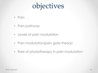
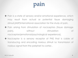
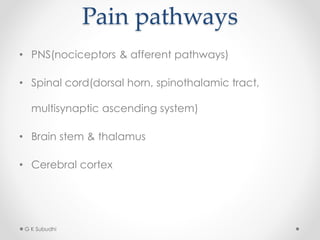
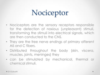
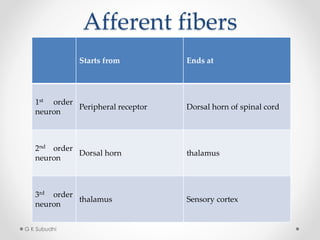
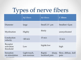
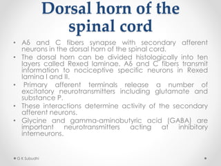
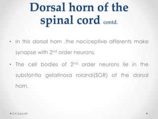
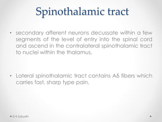
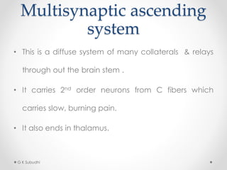
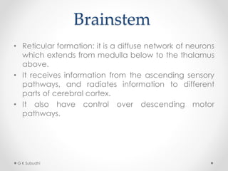
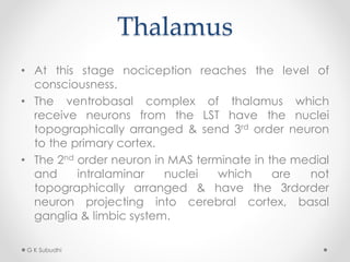
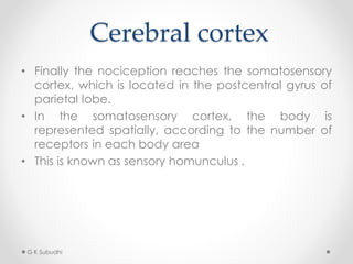
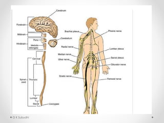
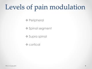
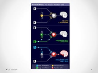
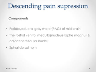
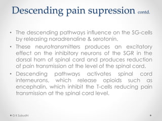
Gate control theory
of pain
Melzack & wall (1965)
Presented by:
Ganesh kumar subudhi
BPT, MPT(Neurology)
Asst. Prof. S J College of
Physiotherapy, Odisha
objectives
• Pain
• Pain pathway
• Levels of pain modulation
• Pain modulation(pain gate theory)
• Role of physiotherapy in pain modulation
G K Subudhi
pain
 Pain is a state of sensory and/or emotional experience, which
may result from actual or potential tissue damaging
stimuli.(IASP)international association for the study of pain.
 Pain arising from stimulation of nociceptors (tissue damage
pain), without stimulation of
nociceptors(emotional/psychological experience).
 Nociceptor is a sensory receptor of PNS that is cable of
transducing and encoding noxious stimuli i.e transmission of
noxious signal from the peripheri to cortex .
G K Subudhi
Pain pathways
• PNS(nociceptors & afferent pathways)
• Spinal cord(dorsal horn, spinothalamic tract,
multisynaptic ascending system)
• Brain stem & thalamus
• Cerebral cortex
G K Subudhi
Nociceptor
• Nociceptors are the sensory receptors responsible
for the detection of noxious (unpleasant) stimuli,
transforming the stimuli into electrical signals, which
are then conducted to the CNS.
• They are the free nerve endings of primary afferent
Aδ and C fibers.
• Distributed throughout the body (skin, viscera,
muscles, joints, meninges) they
• can be stimulated by mechanical, thermal or
chemical stimuli.
G K Subudhi
Afferent fibers
Starts from Ends at
1st order
neuron
Peripheral receptor Dorsal horn of spinal cord
2nd order
neuron
Dorsal horn thalamus
3rd order
neuron
thalamus Sensory cortex
G K Subudhi
Types of nerve fibers
Aβ fibers Aδ fibers C fibers
Diameter large Small 2-5 μm Smallest <2μm
Myelination Highly
thinly
unmyelinated
Conduction
velocity
>40 m/s
15 m/s
<2 m/s
Receptor
activation
threshold
Low
high& low
high
Sensation
on stimulation
Light touch,
non-noxious
Rapid, sharp,
localized pain
Slow, diffuse, dull
pain
G K Subudhi
Dorsal horn of the
spinal cord
• Aδ and C fibers synapse with secondary afferent
neurons in the dorsal horn of the spinal cord.
• The dorsal horn can be divided histologically into ten
layers called Rexed laminae. Aδ and C fibers transmit
information to nociceptive specific neurons in Rexed
lamina I and II.
• Primary afferent terminals release a number of
excitatory neurotransmitters including glutamate and
substance P.
• These interactions determine activity of the secondary
afferent neurons.
• Glycine and gamma-aminobutyric acid (GABA) are
important neurotransmitters acting at inhibitory
interneurons.
G K Subudhi
Dorsal horn of the
spinal cord contd.
• In this dorsal horn ,the nociceptive afferents make
synapse with 2nd order neurons.
• The cell bodies of 2nd order neurons lie in the
substantia gelatinosa rolandi(SGR) of the dorsal
horn.
G K Subudhi
Spinothalamic tract
• secondary afferent neurons decussate within a few
segments of the level of entry into the spinal cord
and ascend in the contralateral spinothalamic tract
to nuclei within the thalamus.
• Lateral spinothalamic tract contains Aδ fibers which
carries fast, sharp type pain.
G K Subudhi
Multisynaptic ascending
system
• This is a diffuse system of many collaterals & relays
through out the brain stem .
• It carries 2nd order neurons from C fibers which
carries slow, burning pain.
• It also ends in thalamus.
G K Subudhi
Brainstem
• Reticular formation: it is a diffuse network of neurons
which extends from medulla below to the thalamus
above.
• It receives information from the ascending sensory
pathways, and radiates information to different
parts of cerebral cortex.
• It also have control over descending motor
pathways.
G K Subudhi
Thalamus
• At this stage nociception reaches the level of
consciousness.
• The ventrobasal complex of thalamus which
receive neurons from the LST have the nuclei
topographically arranged & send 3rd order neuron
to the primary cortex.
• The 2nd order neuron in MAS terminate in the medial
and intralaminar nuclei which are not
topographically arranged & have the 3rdorder
neuron projecting into cerebral cortex, basal
ganglia & limbic system.
G K Subudhi
Cerebral cortex
• Finally the nociception reaches the somatosensory
cortex, which is located in the postcentral gyrus of
parietal lobe.
• In the somatosensory cortex, the body is
represented spatially, according to the number of
receptors in each body area
• This is known as sensory homunculus .
G K Subudhi
G K Subudhi
Levels of pain modulation
 Peripheral
 Spinal segment
 Supra spinal
 cortical
G K Subudhi
Pain modulation
 Gate control theory(presynaptic inhibition)
 Descending pain suppression system
G K Subudhi
Gate control theory
• This theory is suggested by Melzack & Wall in 1965.
• This theory suggest that pain perception is regulated by
a gate which may be opened or closed by means of
other inputs from the peripheral nerves or the CNS.
• The interneurons present in the SG-cells of the posterior
horn of spinal cord have an inhibitory influence on the T
cells.
• when the large diameter, low threshold
mechanoreceptors(Aβ-fibers) are stimulated by simple
mechanical stimulation of the receptors in the skin,
muscle or joints, the SG cells are stimulated via an
excitatory synapse.
G K Subudhi
G K Subudhi
Gate control theory contd.
• The SG cells receiving inputs from nociceptors (Aδ
&C-fibers) and the mechanoreceptors(Aβ-fibers),
integrates and modulates the pain fiber activity
and prevent transmission of nociceptive information
to higher centers by inhibitory influence on the T-
cells, resulting in closure of gate of pain transmission.
• It helps to explain why when we bang our head, it
feels better when we rub it.
• By activating Aβ fibers with tactile, non-noxious
stimuli inhibitory interneurons in the dorsal horn are
activated leading to inhibition of pain signals
transmitted via C fibers
G K Subudhi
G K Subudhi
Descending pain supression
Components
• Periaqueductal gray mater(PAG) of mid brain
• The rostral ventral medulla(nucleus raphe magnus &
adjacent reticular nuclei)
• Spinal dorsal horn
G K Subudhi
Descending pain supression contd.
• The descending pathways influence on the SG-cells
by releasing noradrenaline & serotonin.
• These neurotransmitters produces an excitatory
effect on the inhibitory neurons of the SGR in the
dorsal horn of spinal cord and produces reduction
of pain transmission at the level of the spinal cord.
• Descending pathways activates spinal cord
interneurons, which release opioids such as
encephalin, which inhibit the T-cells reducing pain
transmission at the spinal cord level.
G K Subudhi
Reference
• Electrotherapy simplified, B K Nanda
G K Subudhi
Thank u
G K Subudhi

Pain gait theory

  • 1.
    Gate control theory ofpain Melzack & wall (1965) Presented by: Ganesh kumar subudhi BPT, MPT(Neurology) Asst. Prof. S J College of Physiotherapy, Odisha
  • 2.
    objectives • Pain • Painpathway • Levels of pain modulation • Pain modulation(pain gate theory) • Role of physiotherapy in pain modulation G K Subudhi
  • 3.
    pain  Pain isa state of sensory and/or emotional experience, which may result from actual or potential tissue damaging stimuli.(IASP)international association for the study of pain.  Pain arising from stimulation of nociceptors (tissue damage pain), without stimulation of nociceptors(emotional/psychological experience).  Nociceptor is a sensory receptor of PNS that is cable of transducing and encoding noxious stimuli i.e transmission of noxious signal from the peripheri to cortex . G K Subudhi
  • 4.
    Pain pathways • PNS(nociceptors& afferent pathways) • Spinal cord(dorsal horn, spinothalamic tract, multisynaptic ascending system) • Brain stem & thalamus • Cerebral cortex G K Subudhi
  • 5.
    Nociceptor • Nociceptors arethe sensory receptors responsible for the detection of noxious (unpleasant) stimuli, transforming the stimuli into electrical signals, which are then conducted to the CNS. • They are the free nerve endings of primary afferent Aδ and C fibers. • Distributed throughout the body (skin, viscera, muscles, joints, meninges) they • can be stimulated by mechanical, thermal or chemical stimuli. G K Subudhi
  • 6.
    Afferent fibers Starts fromEnds at 1st order neuron Peripheral receptor Dorsal horn of spinal cord 2nd order neuron Dorsal horn thalamus 3rd order neuron thalamus Sensory cortex G K Subudhi
  • 7.
    Types of nervefibers Aβ fibers Aδ fibers C fibers Diameter large Small 2-5 μm Smallest <2μm Myelination Highly thinly unmyelinated Conduction velocity >40 m/s 15 m/s <2 m/s Receptor activation threshold Low high& low high Sensation on stimulation Light touch, non-noxious Rapid, sharp, localized pain Slow, diffuse, dull pain G K Subudhi
  • 8.
    Dorsal horn ofthe spinal cord • Aδ and C fibers synapse with secondary afferent neurons in the dorsal horn of the spinal cord. • The dorsal horn can be divided histologically into ten layers called Rexed laminae. Aδ and C fibers transmit information to nociceptive specific neurons in Rexed lamina I and II. • Primary afferent terminals release a number of excitatory neurotransmitters including glutamate and substance P. • These interactions determine activity of the secondary afferent neurons. • Glycine and gamma-aminobutyric acid (GABA) are important neurotransmitters acting at inhibitory interneurons. G K Subudhi
  • 9.
    Dorsal horn ofthe spinal cord contd. • In this dorsal horn ,the nociceptive afferents make synapse with 2nd order neurons. • The cell bodies of 2nd order neurons lie in the substantia gelatinosa rolandi(SGR) of the dorsal horn. G K Subudhi
  • 10.
    Spinothalamic tract • secondaryafferent neurons decussate within a few segments of the level of entry into the spinal cord and ascend in the contralateral spinothalamic tract to nuclei within the thalamus. • Lateral spinothalamic tract contains Aδ fibers which carries fast, sharp type pain. G K Subudhi
  • 11.
    Multisynaptic ascending system • Thisis a diffuse system of many collaterals & relays through out the brain stem . • It carries 2nd order neurons from C fibers which carries slow, burning pain. • It also ends in thalamus. G K Subudhi
  • 12.
    Brainstem • Reticular formation:it is a diffuse network of neurons which extends from medulla below to the thalamus above. • It receives information from the ascending sensory pathways, and radiates information to different parts of cerebral cortex. • It also have control over descending motor pathways. G K Subudhi
  • 13.
    Thalamus • At thisstage nociception reaches the level of consciousness. • The ventrobasal complex of thalamus which receive neurons from the LST have the nuclei topographically arranged & send 3rd order neuron to the primary cortex. • The 2nd order neuron in MAS terminate in the medial and intralaminar nuclei which are not topographically arranged & have the 3rdorder neuron projecting into cerebral cortex, basal ganglia & limbic system. G K Subudhi
  • 14.
    Cerebral cortex • Finallythe nociception reaches the somatosensory cortex, which is located in the postcentral gyrus of parietal lobe. • In the somatosensory cortex, the body is represented spatially, according to the number of receptors in each body area • This is known as sensory homunculus . G K Subudhi
  • 15.
  • 16.
    Levels of painmodulation  Peripheral  Spinal segment  Supra spinal  cortical G K Subudhi
  • 17.
    Pain modulation  Gatecontrol theory(presynaptic inhibition)  Descending pain suppression system G K Subudhi
  • 18.
    Gate control theory •This theory is suggested by Melzack & Wall in 1965. • This theory suggest that pain perception is regulated by a gate which may be opened or closed by means of other inputs from the peripheral nerves or the CNS. • The interneurons present in the SG-cells of the posterior horn of spinal cord have an inhibitory influence on the T cells. • when the large diameter, low threshold mechanoreceptors(Aβ-fibers) are stimulated by simple mechanical stimulation of the receptors in the skin, muscle or joints, the SG cells are stimulated via an excitatory synapse. G K Subudhi
  • 19.
  • 20.
    Gate control theorycontd. • The SG cells receiving inputs from nociceptors (Aδ &C-fibers) and the mechanoreceptors(Aβ-fibers), integrates and modulates the pain fiber activity and prevent transmission of nociceptive information to higher centers by inhibitory influence on the T- cells, resulting in closure of gate of pain transmission. • It helps to explain why when we bang our head, it feels better when we rub it. • By activating Aβ fibers with tactile, non-noxious stimuli inhibitory interneurons in the dorsal horn are activated leading to inhibition of pain signals transmitted via C fibers G K Subudhi
  • 21.
  • 22.
    Descending pain supression Components •Periaqueductal gray mater(PAG) of mid brain • The rostral ventral medulla(nucleus raphe magnus & adjacent reticular nuclei) • Spinal dorsal horn G K Subudhi
  • 23.
    Descending pain supressioncontd. • The descending pathways influence on the SG-cells by releasing noradrenaline & serotonin. • These neurotransmitters produces an excitatory effect on the inhibitory neurons of the SGR in the dorsal horn of spinal cord and produces reduction of pain transmission at the level of the spinal cord. • Descending pathways activates spinal cord interneurons, which release opioids such as encephalin, which inhibit the T-cells reducing pain transmission at the spinal cord level. G K Subudhi
  • 24.
  • 25.
    Thank u G KSubudhi