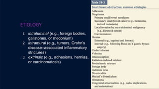
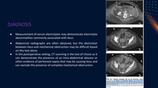
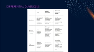
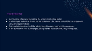
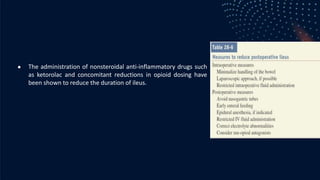
ILEUS OBSTRUCTIVE
PRECEPTOR:
NYENYEYEYE
PRESENTER:
INTAN
ANATOMY OF SMALL INTESTINE
● The small intestine is a tubular structure
that extends from the pylorus to the cecum.
● The estimated length varies depending on
whether radiologic, surgical, or autopsy
measurements are made. In the living, it is
thought to measure 4 to 6 meters.
● The small intestine consists of three
segments lying in series: the duodenum, the
jejunum, and the ileum.
ANATOMY OF SMALL
INTESTINE
● The duodenum, the most proximal segment,
lies in the retroperitoneum immediately
adjacent to the head and inferior border of the
body of the pancreas.
● The duodenum is demarcated from the
stomach by the pylorus and from the jejunum
by the ligament of Treitz.
● The jejunum and ileum lie within the
peritoneal cavity and are tethered to the
retroperitoneum by a broad-based mesentery.
● The ileum is demarcated from the cecum by
the ileocecal valve.
ANATOMY OF SMALL
INTESTINE
● The small intestine contains internal mucosal folds
known as plicae circulares or valvulae conniventes that
are visible upon gross inspection. These folds are also
visible radiographically and help in the distinction
between small intestine and colon. These folds are
more prominent in the proximal intestine than in the
distal small intestine.
● Other features evident on gross inspection that are
more characteristic of the proximal than distal small
intestine include larger circumference, thicker wall, less
fatty mesentery, and longer vasa recta.
ILEUS
OBSTRUCTIVE
DEFINITION
● Obstruksi usus adalah salah satu gangguan dalam saluran pencernaan yang
terjadi akibat adanya penyumbatan dalam usus, baik usus besar maupun
usus halus. Kondisi ini menyebabkan makanan dan cairan tidak bisa
melewati usus dengan baik dan menimbulkan tekanan pada usus
EPIDEMIOLOGY
● Mechanical small bowel obstruction is the most frequently
encountered surgical disorder of the small intestine.
● Small and large bowel obstructions are similar in incidence in
both males and females
● The overriding factor affecting incidence and distribution
depends on patient risk factors
● Intra-abdominal adhesions related to prior abdominal surgery
account for up to 75% of cases of small bowel obstruction.
ETIOLOGY
1. intraluminal (e.g., foreign bodies,
gallstones, or meconium)
2. intramural (e.g., tumors, Crohn’s
disease–associated inflammatory
strictures)
3. extrinsic (e.g., adhesions, hernias,
or carcinomatosis)
PATHOPHYSIOLOGY
● Partial/complete obstruction : onset of obstruction, gas and fluid
accumulate within the intestinal lumen proximal to the site of
obstruction -> distension -> increases intraluminal & intramural
pressure -> colicky pain, diarrhea
● Obstruction -> changes in luminal floral -> translocation of bacteria to
the regional lymph node
● Increase in intramural pressure -> decrease intestinal microvascular
perfusion -> ischemia -> necrosis (strangulated bowel obstruction)
● Closed loop obstruction : the accumulating gas and fluid cannot
escape either proximally or distally from the obstructed segment,
leading to a rapid rise in luminal pressure and a rapid progression to
strangulation.
CLINICAL
MANIFESTATION
● Colicky abdominal pain
● Nausea
● Vomiting -> proximal
● Obstipation
● Tachycardia
● Localized abdominal tenderness
● Fever
● Marked leukocitosis
● Acidosis
ANAMNESIS
● Riwayat operasi
● Riwayat konsumsi obat-obatan
● Nyeri kolik
● Nyeri meningkat setelah makan
● Mual & muntah
● BAB terakhir
● Flatus
PEMERIKSAAN FISIK
● Inspeksi : distensi abdomen, darm contour, darm steifung
● Palpasi : Nyeri tekan, massa,
● Perkusi : hipertympanic
● Auskultasi : Metallic sound, hiperperistaltik,
● Tanda dehidrasi (+)
● Rectal touche : ampula recti kolaps
DIAGNOSIS
● The abdominal series consists of (a) a radiograph of the abdomen with the
patient in a supine position, (b) a radiograph of the abdomen with the patient
in an upright position, and (c) a radiograph of the chest with the patient in an
upright position
● the triad of dilated small bowel loops (>3 cm in diameter), air-fluid levels seen
on upright films, and a paucity of air in the colon
DIAGNOSIS
● CT Scan with contrast
● discrete transition zone with dilation of bowel proximally, decompression of
bowel distally, intraluminal contrast that does not pass beyond the transition
zone, and a colon containing little gas or fluid
DIAGNOSIS
● Closed-loop obstruction is suggested by the
presence of a U-shaped or C-shaped dilated
bowel loop associated with a radial distribution of
mesenteric vessels converging toward a torsion
point.
● Strangulation is suggested by thickening of the
bowel wall, pneumatosis intestinalis (air in the
bowel wall), portal venous gas, mesenteric
haziness, and poor uptake of intravenous
contrast into the wall of the affected bowel
THERAPY
● NPO
● Fluid Resucitation
usually associated with a marked depletion of intravascular volume due to
decreased oral intake, vomiting, and sequestration of fluid in bowel lumen and
wall.
● NGT Decompression
The stomach should be continuously evacuated of air and fluid using a
nasogastric (NG) tube. Effective gastric decom- pression decreases nausea,
distention, and the risk of vomiting and aspiration
● The operative procedure performed for small bowel obstruction varies
according to the etiology of the obstruction. For example, adhesions are lysed,
tumors are resected, and hernias are reduced and repaired. Regardless of the
etiology, the affected intestine should be examined, and nonviable bowel
should be resected. Criteria suggesting viability are normal color, peristalsis,
DIFFERENTIAL
DIAGNOSIS
Viral or bacterial gastroenteritis
Paralytic ileus
Mesenteric ischemia
Acute pancreatitis
Intussusception
Constipation
DIFFERENTIAL
DIAGNOSIS
Bowel necrosis and perforation
Sepsis
Wound dehiscence
Intra-abdominal abscess
Aspiration
Inflammatory bowel syndrome
PROGNOSIS
If any strangulated bowel is left untreated, there is a mortality rate of close to
100%. However, if surgery is undertaken within 24-48 hours, the mortality rates
are less than 10%.
Factors that determine the morbidity include the age of patient, comorbidity,
and delay in treatment. Today, the overall mortality of bowel obstruction is still
about 5%-8%.
THANK YOU!
ETIOLOGY
● The most frequently encountered factors
are abdominal operations, infection and
inflammation, electrolyte abnormalities,
and drugs.
PATHOPHYSIOLOGY
● Following most abdominal operations or injuries, the motility of the gastrointestinal tract is
transiently impaired.
● Among the proposed mechanisms responsible for this dysmotility are surgical stress induced
sympathetic reflexes, inflammatory response mediator release, and anesthetic/analgesic side
effects; each of which can inhibit intestinal motility.
● Small-intestinal motility returning to normal within the first 24 hours after laparotomy and gastric
and colonic motility returning to normal by 48 hours and 2 to 5 days, respectively.
● Since small bowel motility is returned before colonic and gastric motility, listening for bowel
sounds is not a reliable indicator that ileus has fully resolved.
● Functional evidence of coordinated gastrointestinal motility in the form of passing flatus or bowel
movement is a more useful indicator.
CLINICAL MANIFESTATION
● Inability to tolerate liquids and solids by mouth, nausea, and lack of flatus or
bowel movements are the most common symptoms
● Vomiting
● Abdominal distension
● Although bowel sound characteristics are not diagnostic, they are usually
diminished or absent.
● The clinical manifestations of chronic intestinal pseudo-obstruction include
variable degrees of nausea and vomiting and abdominal pain and distention
DIAGNOSIS
● Definition of prolonged postoperative ileus has been varied but generally diagnosed if
ileus persists beyond 5 days postoperatively.
● Patient medication lists should be reviewed for the presence of drugs, especially opiates,
known to be associated with impaired intestinal motility.
● with prolonged postoperative ileus being defined as “two or more of nausea/vomiting,
inability to tolerate oral diet over 24 h, absence of flatus over 24 h, distension, radiologic
confirmation occurring on or after day 4 postoperatively without prior resolution of
postoperative ileus.
DIAGNOSIS
● Measurement of serum electrolytes may demonstrate electrolyte
abnormalities commonly associated with ileus.
● Abdominal radiographs are often obtained, but the distinction
between ileus and mechanical obstruction may be difficult based
on this test alone.
● In the postoperative setting, CT scanning is the test of choice as it
can demonstrate the presence of an intra-abdominal abscess or
other evidence of peritoneal sepsis that may be causing ileus and
can exclude the presence of complete mechanical obstruction.
DIFFERENTIAL DIAGNOSIS
TREATMENT
● Limiting oral intake and correcting the underlying inciting factor.
● If vomiting or abdominal distention are prominent, the stomach should be decompressed
using a nasogastric tube.
● Fluid and electrolytes should be administered intravenously until ileus resolves.
● If the duration of ileus is prolonged, total parental nutrition (TPN) may be required.
● The administration of nonsteroidal anti-inflammatory drugs such
as ketorolac and concomitant reductions in opioid dosing have
been shown to reduce the duration of ileus.
● Many studies have also suggested that limiting intra- and postoperative fluid
administration can also result in reduction of postoperative ileus and shortened
hospital stay. Furthermore, studies have shown that early postoperative feeding
after GI surgery is generally well tolerated and can lead to reduced postoperative
ileus and a shorter hospital stay.
● Although prokinetic agents have been tried to promote return of GI motility, they are
associated with efficacy- toxicity profiles that are too unfavorable to warrant routine use.
Recently, administration of alvimopan, a novel peripherally active mu opioid receptor
antagonist with limited oral absorp- tion, has been shown to reduce duration of
postoperative ileus, hospital stay.
COMPLICATION
● Most of the complications come from the prolonged hospital stay and the
possibility for subsequent procedures for a prolonged ileus (peripheral
inserted central catheter line, TPN, NG tube placement).
● There is the possibility of aspiration with increasing nausea and vomiting.
PROGNOSIS
● Having an ileus is only harmful in terms of the length of stay and decreased
nutrition.
● Longer hospital stays increase the risk of nosocomial infections, and a
prolonged ileus may lead to the need for TPN, which has its own risks and
benefit.
THANK YOU!
Any question?

Clinical Science Session - Ileus Obstruktif

  • 1.
  • 2.
    ANATOMY OF SMALLINTESTINE ● The small intestine is a tubular structure that extends from the pylorus to the cecum. ● The estimated length varies depending on whether radiologic, surgical, or autopsy measurements are made. In the living, it is thought to measure 4 to 6 meters. ● The small intestine consists of three segments lying in series: the duodenum, the jejunum, and the ileum.
  • 3.
    ANATOMY OF SMALL INTESTINE ●The duodenum, the most proximal segment, lies in the retroperitoneum immediately adjacent to the head and inferior border of the body of the pancreas. ● The duodenum is demarcated from the stomach by the pylorus and from the jejunum by the ligament of Treitz. ● The jejunum and ileum lie within the peritoneal cavity and are tethered to the retroperitoneum by a broad-based mesentery. ● The ileum is demarcated from the cecum by the ileocecal valve.
  • 4.
    ANATOMY OF SMALL INTESTINE ●The small intestine contains internal mucosal folds known as plicae circulares or valvulae conniventes that are visible upon gross inspection. These folds are also visible radiographically and help in the distinction between small intestine and colon. These folds are more prominent in the proximal intestine than in the distal small intestine. ● Other features evident on gross inspection that are more characteristic of the proximal than distal small intestine include larger circumference, thicker wall, less fatty mesentery, and longer vasa recta.
  • 5.
  • 6.
    DEFINITION ● Obstruksi ususadalah salah satu gangguan dalam saluran pencernaan yang terjadi akibat adanya penyumbatan dalam usus, baik usus besar maupun usus halus. Kondisi ini menyebabkan makanan dan cairan tidak bisa melewati usus dengan baik dan menimbulkan tekanan pada usus
  • 7.
    EPIDEMIOLOGY ● Mechanical smallbowel obstruction is the most frequently encountered surgical disorder of the small intestine. ● Small and large bowel obstructions are similar in incidence in both males and females ● The overriding factor affecting incidence and distribution depends on patient risk factors ● Intra-abdominal adhesions related to prior abdominal surgery account for up to 75% of cases of small bowel obstruction.
  • 8.
    ETIOLOGY 1. intraluminal (e.g.,foreign bodies, gallstones, or meconium) 2. intramural (e.g., tumors, Crohn’s disease–associated inflammatory strictures) 3. extrinsic (e.g., adhesions, hernias, or carcinomatosis)
  • 9.
    PATHOPHYSIOLOGY ● Partial/complete obstruction: onset of obstruction, gas and fluid accumulate within the intestinal lumen proximal to the site of obstruction -> distension -> increases intraluminal & intramural pressure -> colicky pain, diarrhea ● Obstruction -> changes in luminal floral -> translocation of bacteria to the regional lymph node ● Increase in intramural pressure -> decrease intestinal microvascular perfusion -> ischemia -> necrosis (strangulated bowel obstruction) ● Closed loop obstruction : the accumulating gas and fluid cannot escape either proximally or distally from the obstructed segment, leading to a rapid rise in luminal pressure and a rapid progression to strangulation.
  • 11.
    CLINICAL MANIFESTATION ● Colicky abdominalpain ● Nausea ● Vomiting -> proximal ● Obstipation ● Tachycardia ● Localized abdominal tenderness ● Fever ● Marked leukocitosis ● Acidosis
  • 12.
    ANAMNESIS ● Riwayat operasi ●Riwayat konsumsi obat-obatan ● Nyeri kolik ● Nyeri meningkat setelah makan ● Mual & muntah ● BAB terakhir ● Flatus
  • 13.
    PEMERIKSAAN FISIK ● Inspeksi: distensi abdomen, darm contour, darm steifung ● Palpasi : Nyeri tekan, massa, ● Perkusi : hipertympanic ● Auskultasi : Metallic sound, hiperperistaltik, ● Tanda dehidrasi (+) ● Rectal touche : ampula recti kolaps
  • 14.
    DIAGNOSIS ● The abdominalseries consists of (a) a radiograph of the abdomen with the patient in a supine position, (b) a radiograph of the abdomen with the patient in an upright position, and (c) a radiograph of the chest with the patient in an upright position ● the triad of dilated small bowel loops (>3 cm in diameter), air-fluid levels seen on upright films, and a paucity of air in the colon
  • 15.
    DIAGNOSIS ● CT Scanwith contrast ● discrete transition zone with dilation of bowel proximally, decompression of bowel distally, intraluminal contrast that does not pass beyond the transition zone, and a colon containing little gas or fluid
  • 16.
    DIAGNOSIS ● Closed-loop obstructionis suggested by the presence of a U-shaped or C-shaped dilated bowel loop associated with a radial distribution of mesenteric vessels converging toward a torsion point. ● Strangulation is suggested by thickening of the bowel wall, pneumatosis intestinalis (air in the bowel wall), portal venous gas, mesenteric haziness, and poor uptake of intravenous contrast into the wall of the affected bowel
  • 17.
    THERAPY ● NPO ● FluidResucitation usually associated with a marked depletion of intravascular volume due to decreased oral intake, vomiting, and sequestration of fluid in bowel lumen and wall. ● NGT Decompression The stomach should be continuously evacuated of air and fluid using a nasogastric (NG) tube. Effective gastric decom- pression decreases nausea, distention, and the risk of vomiting and aspiration ● The operative procedure performed for small bowel obstruction varies according to the etiology of the obstruction. For example, adhesions are lysed, tumors are resected, and hernias are reduced and repaired. Regardless of the etiology, the affected intestine should be examined, and nonviable bowel should be resected. Criteria suggesting viability are normal color, peristalsis,
  • 19.
    DIFFERENTIAL DIAGNOSIS Viral or bacterialgastroenteritis Paralytic ileus Mesenteric ischemia Acute pancreatitis Intussusception Constipation
  • 20.
    DIFFERENTIAL DIAGNOSIS Bowel necrosis andperforation Sepsis Wound dehiscence Intra-abdominal abscess Aspiration Inflammatory bowel syndrome
  • 21.
    PROGNOSIS If any strangulatedbowel is left untreated, there is a mortality rate of close to 100%. However, if surgery is undertaken within 24-48 hours, the mortality rates are less than 10%. Factors that determine the morbidity include the age of patient, comorbidity, and delay in treatment. Today, the overall mortality of bowel obstruction is still about 5%-8%.
  • 22.
  • 23.
    ETIOLOGY ● The mostfrequently encountered factors are abdominal operations, infection and inflammation, electrolyte abnormalities, and drugs.
  • 25.
    PATHOPHYSIOLOGY ● Following mostabdominal operations or injuries, the motility of the gastrointestinal tract is transiently impaired. ● Among the proposed mechanisms responsible for this dysmotility are surgical stress induced sympathetic reflexes, inflammatory response mediator release, and anesthetic/analgesic side effects; each of which can inhibit intestinal motility. ● Small-intestinal motility returning to normal within the first 24 hours after laparotomy and gastric and colonic motility returning to normal by 48 hours and 2 to 5 days, respectively. ● Since small bowel motility is returned before colonic and gastric motility, listening for bowel sounds is not a reliable indicator that ileus has fully resolved. ● Functional evidence of coordinated gastrointestinal motility in the form of passing flatus or bowel movement is a more useful indicator.
  • 26.
    CLINICAL MANIFESTATION ● Inabilityto tolerate liquids and solids by mouth, nausea, and lack of flatus or bowel movements are the most common symptoms ● Vomiting ● Abdominal distension ● Although bowel sound characteristics are not diagnostic, they are usually diminished or absent. ● The clinical manifestations of chronic intestinal pseudo-obstruction include variable degrees of nausea and vomiting and abdominal pain and distention
  • 27.
    DIAGNOSIS ● Definition ofprolonged postoperative ileus has been varied but generally diagnosed if ileus persists beyond 5 days postoperatively. ● Patient medication lists should be reviewed for the presence of drugs, especially opiates, known to be associated with impaired intestinal motility. ● with prolonged postoperative ileus being defined as “two or more of nausea/vomiting, inability to tolerate oral diet over 24 h, absence of flatus over 24 h, distension, radiologic confirmation occurring on or after day 4 postoperatively without prior resolution of postoperative ileus.
  • 28.
    DIAGNOSIS ● Measurement ofserum electrolytes may demonstrate electrolyte abnormalities commonly associated with ileus. ● Abdominal radiographs are often obtained, but the distinction between ileus and mechanical obstruction may be difficult based on this test alone. ● In the postoperative setting, CT scanning is the test of choice as it can demonstrate the presence of an intra-abdominal abscess or other evidence of peritoneal sepsis that may be causing ileus and can exclude the presence of complete mechanical obstruction.
  • 29.
  • 30.
    TREATMENT ● Limiting oralintake and correcting the underlying inciting factor. ● If vomiting or abdominal distention are prominent, the stomach should be decompressed using a nasogastric tube. ● Fluid and electrolytes should be administered intravenously until ileus resolves. ● If the duration of ileus is prolonged, total parental nutrition (TPN) may be required.
  • 31.
    ● The administrationof nonsteroidal anti-inflammatory drugs such as ketorolac and concomitant reductions in opioid dosing have been shown to reduce the duration of ileus.
  • 32.
    ● Many studieshave also suggested that limiting intra- and postoperative fluid administration can also result in reduction of postoperative ileus and shortened hospital stay. Furthermore, studies have shown that early postoperative feeding after GI surgery is generally well tolerated and can lead to reduced postoperative ileus and a shorter hospital stay. ● Although prokinetic agents have been tried to promote return of GI motility, they are associated with efficacy- toxicity profiles that are too unfavorable to warrant routine use. Recently, administration of alvimopan, a novel peripherally active mu opioid receptor antagonist with limited oral absorp- tion, has been shown to reduce duration of postoperative ileus, hospital stay.
  • 33.
    COMPLICATION ● Most ofthe complications come from the prolonged hospital stay and the possibility for subsequent procedures for a prolonged ileus (peripheral inserted central catheter line, TPN, NG tube placement). ● There is the possibility of aspiration with increasing nausea and vomiting.
  • 34.
    PROGNOSIS ● Having anileus is only harmful in terms of the length of stay and decreased nutrition. ● Longer hospital stays increase the risk of nosocomial infections, and a prolonged ileus may lead to the need for TPN, which has its own risks and benefit.
  • 35.