The internal carotid artery begins at the bifurcation of the common carotid artery and ascends through the neck embedded in the carotid sheath. It passes through the carotid canal in the skull.
The internal jugular vein receives blood from the brain, face, and neck. It starts in the skull and descends through the neck lateral to nerves and arteries before joining the subclavian vein.
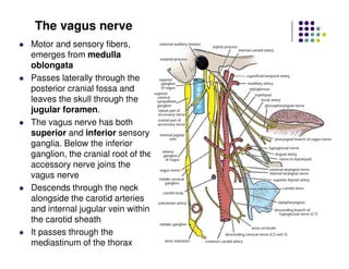
The vagus nerve emerges from the medulla and passes through the neck giving off branches including the recurrent laryngeal nerve before descending into the thorax. It provides both motor and sensory functions.