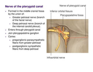
Nerve of the pterygoid canal
Formed in the middle cranial fossa
by the union of:
Greater petrosal nerve (branch
of the facial nerve)
Deep petrosal nerve (branch of
the internal carotid plexus).
Enters through pterygoid canal
Join pterygopalatine ganglion
Caries:
preganglionic parasympathetic
fibers from greater petrosal
postganglionic sympathetic
fibers from deep petrosal
Branch from
facial in
middle ear
cavity
Medial wall
of the
tympanic
cavity from
geniculate
ganglion
Leave to
Middle
cranial fossa
through the
greater
petrosal
foramen
Greater petrosal nerve
Passes over
Foramen
lacerum, where it
joins deep
petrosal nerve to
form the nerve to
pterygoid canal
Pterygoid canal
Pterygopalatine
gangilion
Maxillary nerve
Greater petrosal nerve
Posterior margin of
the middle cranial
fossa
Under the internal
carotid artery
Superior surface of
the cartilage filling
the foramen lacerum
Joined by the deep
petrosal nerve to
form the nerve of the
pterygoid canal
Greater petrosal nerve
Postganglionic
sympathetic
fibers
originate in the
superior
cervical
sympathetic
ganglion in the
neck
leave the
ganglion
(suoerior
cervical) as the
internal carotid
nerve
Deep petrosal nerve
largest of the four
parasympathetic ganglia in the
head
postganglionic parasympathetic
fibers originate in the
pterygopalatine ganglion +
postganglionic sympathetic
fibers passing through the
ganglion
Distribute with
orbital, palatine, nasal, and
pharyngeal branches
Supplies:
Mucous glands in the nasal
cavity
Salivary glands in the upper
half of the oral cavity
lacrimal gland in the orbit.
Pterygopalatine ganglion
Fibers from
pterygopalatine
ganglion join
main trunk of
the maxillary
nerve and
distributed
with:
Zygomatic
Posterior
superior
alveolar
Infra-orbital
Deep petrosal nerve
Fibers leave the zygomaticotemporal branch of the zygomatic nerve travels up the
lateral orbital wall to join the lacrimal nerve
Nerve supply of lacrimal gland
The lacrimal nerve is a major general sensory branch of the ophthalmic nerve
Parasympathetic innervation: originally from great petrosal nerve branch of facial
Sympathatic innervation : originally from deep petrosal nerve from carotid plexus,
superior cervical ganglia
through the infra-orbital foramen
Anterior superior alveolar arteries: incisor and canine teeth
Maxillary artery
Branches from 3rd
part:
(pterygopalatine
fossa)
Anterior to
pterygopalatine
ganglion
Posterior superior
alveolar artery
molar and
premolar
Infra-orbital artery
through the inferior
orbital fissure
Greater palatine artery
Into the palatine canal
Gives lesser palatine
branch (soft palate)
Then superiorly
through the incisive
canal
supply the anterior
aspect of the septal
wall of the nasal
cavity
Maxillary artery
Sphenopalatine artery: (largest) terminal branch of the maxillary artery
Branches
Posterior lateral nasal branches
Posterior septal branches
Maxillary
artery
Maxillary artery
Pharyngeal branch
Through the
palatovaginal canal
Supplies:
posterior aspect of
the roof of the
nasal cavity
Sphenoidal sinus
Pharyngotympanic
tube
Artery of pterygoid
canal
Passes into the
pterygoid canal
Foramen lacerum
Terminates in
nasopharynx
Type: Synovial (hinge and sliding)
Articulation: Articular tubercle and the anterior portion of the mandibular
fossa above and the head (condyloid process) of the mandible below
The articular surfaces are covered by fibrocartilage
Articular disc: divides the joint into upper and lower cavities
attached to the capsule and tendon of the lateral pterygoid
TMJ
TMJ
Capsule: attached above to the
articular tubercle and the margins
of the mandibular fossa and below
to the neck of the mandible.
Synovial membrane: lines both
compartments of the joint and
attached to the margins of the
articular disc
Ligaments:
Lateral ligament
Prevents lateral and posterior
displacement of the condyle
Sphenomandibular ligament
Keeps same amount of tension
during both opening and closing of
the mouth
Stylomandibular ligament
Limit anterior protrusion of the
mandible
TMJ
Nerve supply: Auriculotemporal
and masseteric
Blood supply: Deep auricular,
Anterior tympanic, Superficial
temporal
Relations:
Anteriorly: Mandibular notch,
masseteric nerve and artery
Posteriorly: external auditory
meatus, glenoid process of the
parotid gland
Laterally: parotid gland, fascia, and
skin
Medially: maxillary artery and vein
and the auriculotemporal nerve

pterygopalatine_fosssa.pdf

  • 1.
    Nerve of thepterygoid canal Formed in the middle cranial fossa by the union of: Greater petrosal nerve (branch of the facial nerve) Deep petrosal nerve (branch of the internal carotid plexus). Enters through pterygoid canal Join pterygopalatine ganglion Caries: preganglionic parasympathetic fibers from greater petrosal postganglionic sympathetic fibers from deep petrosal
  • 2.
    Branch from facial in middleear cavity Medial wall of the tympanic cavity from geniculate ganglion Leave to Middle cranial fossa through the greater petrosal foramen Greater petrosal nerve
  • 3.
    Passes over Foramen lacerum, whereit joins deep petrosal nerve to form the nerve to pterygoid canal Pterygoid canal Pterygopalatine gangilion Maxillary nerve Greater petrosal nerve
  • 4.
    Posterior margin of themiddle cranial fossa Under the internal carotid artery Superior surface of the cartilage filling the foramen lacerum Joined by the deep petrosal nerve to form the nerve of the pterygoid canal Greater petrosal nerve
  • 5.
    Postganglionic sympathetic fibers originate in the superior cervical sympathetic ganglionin the neck leave the ganglion (suoerior cervical) as the internal carotid nerve Deep petrosal nerve
  • 6.
    largest of thefour parasympathetic ganglia in the head postganglionic parasympathetic fibers originate in the pterygopalatine ganglion + postganglionic sympathetic fibers passing through the ganglion Distribute with orbital, palatine, nasal, and pharyngeal branches Supplies: Mucous glands in the nasal cavity Salivary glands in the upper half of the oral cavity lacrimal gland in the orbit. Pterygopalatine ganglion
  • 7.
    Fibers from pterygopalatine ganglion join maintrunk of the maxillary nerve and distributed with: Zygomatic Posterior superior alveolar Infra-orbital Deep petrosal nerve Fibers leave the zygomaticotemporal branch of the zygomatic nerve travels up the lateral orbital wall to join the lacrimal nerve
  • 8.
    Nerve supply oflacrimal gland The lacrimal nerve is a major general sensory branch of the ophthalmic nerve Parasympathetic innervation: originally from great petrosal nerve branch of facial Sympathatic innervation : originally from deep petrosal nerve from carotid plexus, superior cervical ganglia
  • 10.
    through the infra-orbitalforamen Anterior superior alveolar arteries: incisor and canine teeth Maxillary artery Branches from 3rd part: (pterygopalatine fossa) Anterior to pterygopalatine ganglion Posterior superior alveolar artery molar and premolar Infra-orbital artery through the inferior orbital fissure
  • 11.
    Greater palatine artery Intothe palatine canal Gives lesser palatine branch (soft palate) Then superiorly through the incisive canal supply the anterior aspect of the septal wall of the nasal cavity Maxillary artery
  • 12.
    Sphenopalatine artery: (largest)terminal branch of the maxillary artery Branches Posterior lateral nasal branches Posterior septal branches Maxillary artery
  • 13.
    Maxillary artery Pharyngeal branch Throughthe palatovaginal canal Supplies: posterior aspect of the roof of the nasal cavity Sphenoidal sinus Pharyngotympanic tube Artery of pterygoid canal Passes into the pterygoid canal Foramen lacerum Terminates in nasopharynx
  • 16.
    Type: Synovial (hingeand sliding) Articulation: Articular tubercle and the anterior portion of the mandibular fossa above and the head (condyloid process) of the mandible below The articular surfaces are covered by fibrocartilage Articular disc: divides the joint into upper and lower cavities attached to the capsule and tendon of the lateral pterygoid TMJ
  • 17.
    TMJ Capsule: attached aboveto the articular tubercle and the margins of the mandibular fossa and below to the neck of the mandible. Synovial membrane: lines both compartments of the joint and attached to the margins of the articular disc Ligaments: Lateral ligament Prevents lateral and posterior displacement of the condyle Sphenomandibular ligament Keeps same amount of tension during both opening and closing of the mouth Stylomandibular ligament Limit anterior protrusion of the mandible
  • 18.
    TMJ Nerve supply: Auriculotemporal andmasseteric Blood supply: Deep auricular, Anterior tympanic, Superficial temporal Relations: Anteriorly: Mandibular notch, masseteric nerve and artery Posteriorly: external auditory meatus, glenoid process of the parotid gland Laterally: parotid gland, fascia, and skin Medially: maxillary artery and vein and the auriculotemporal nerve