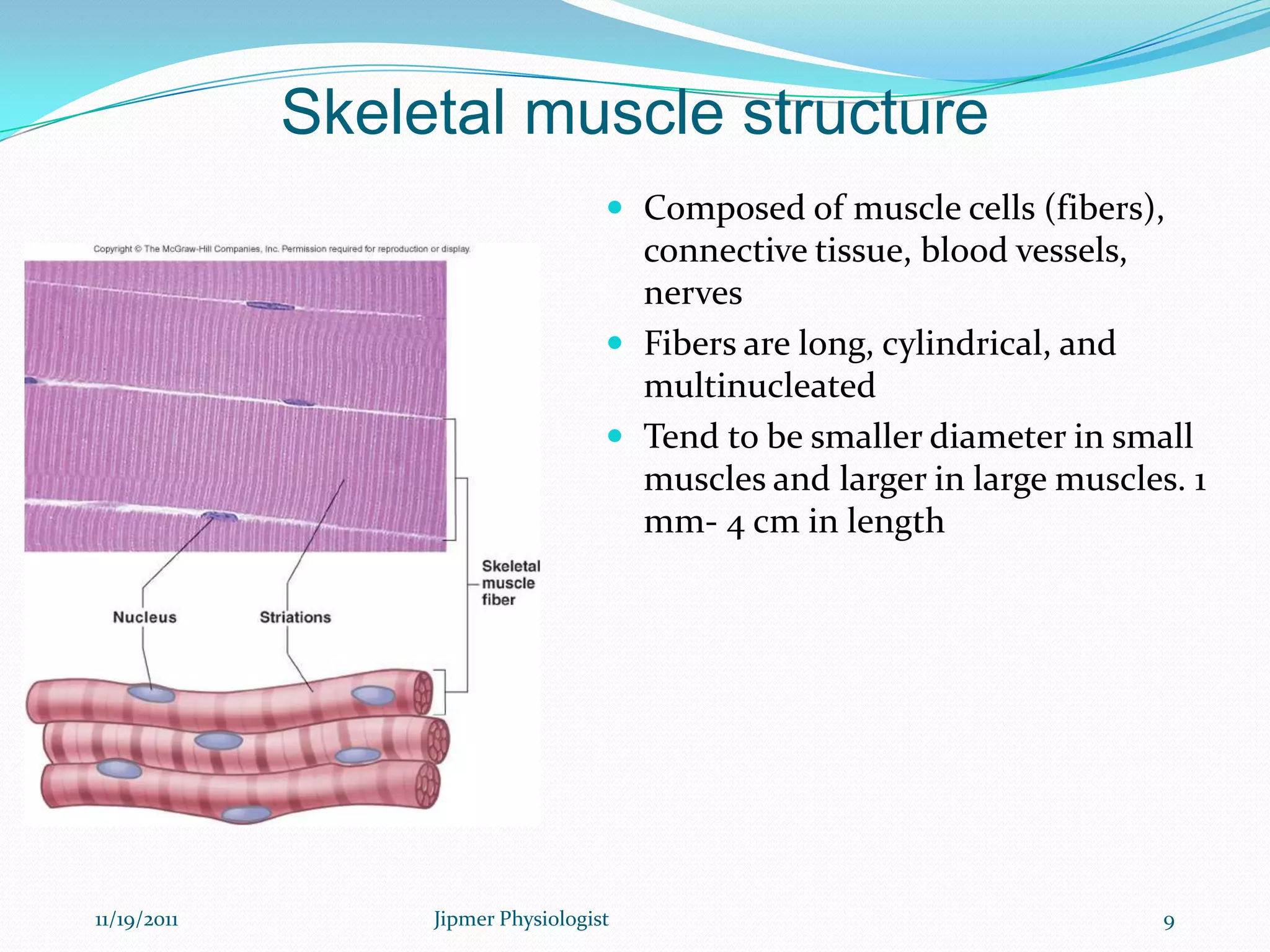
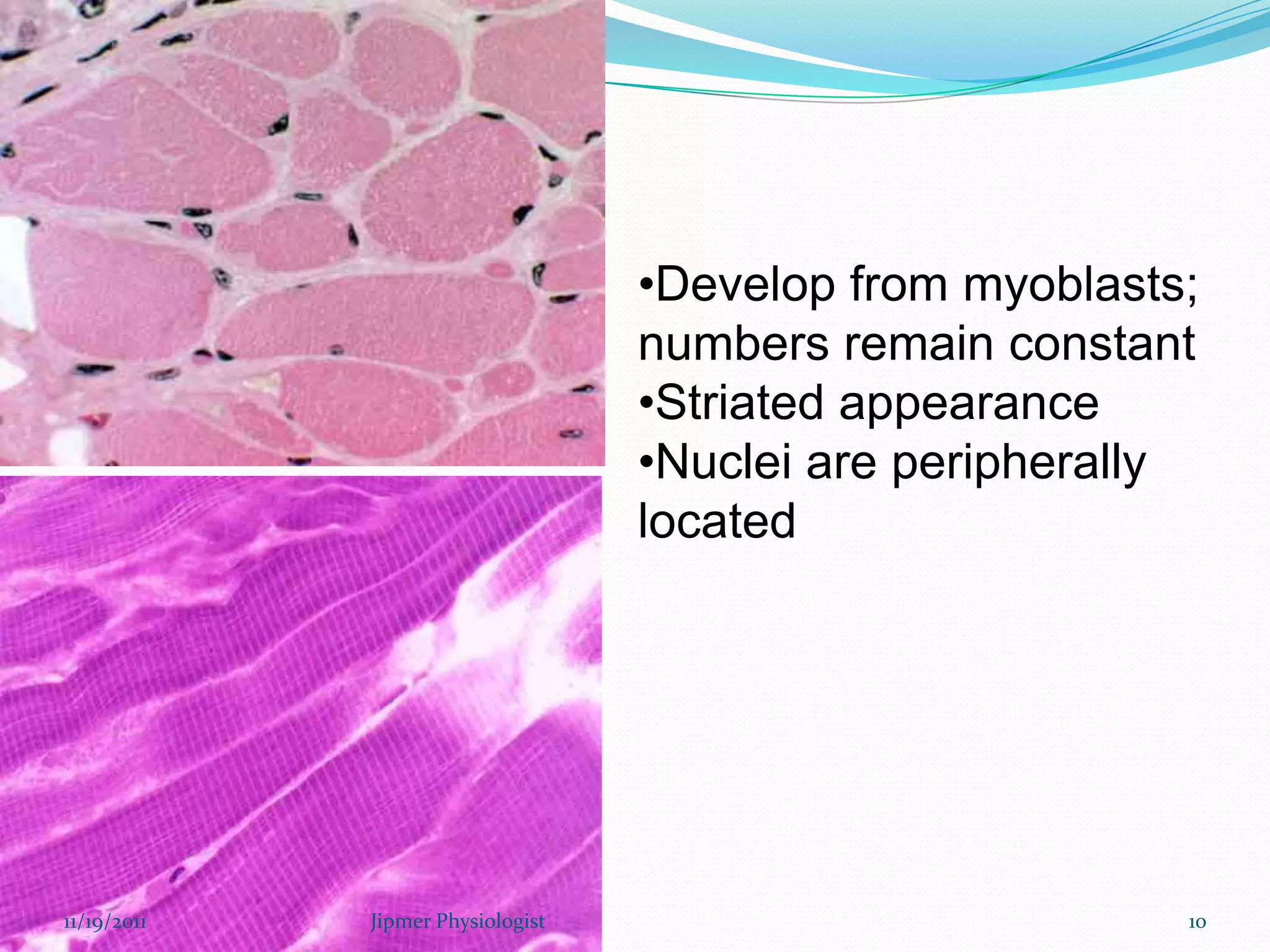
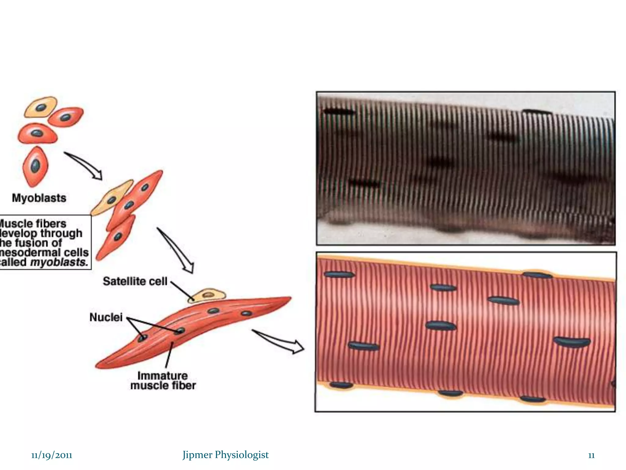
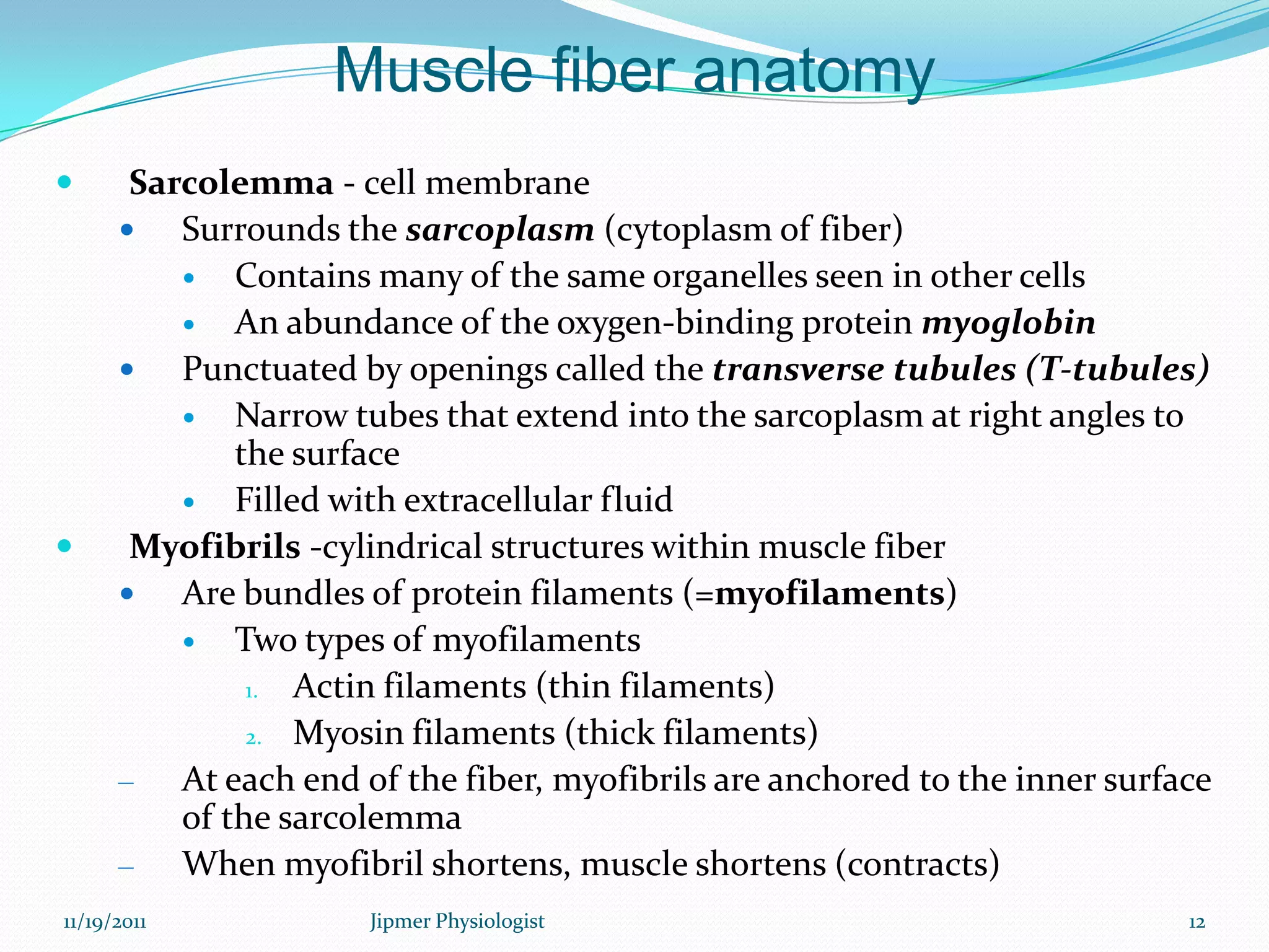
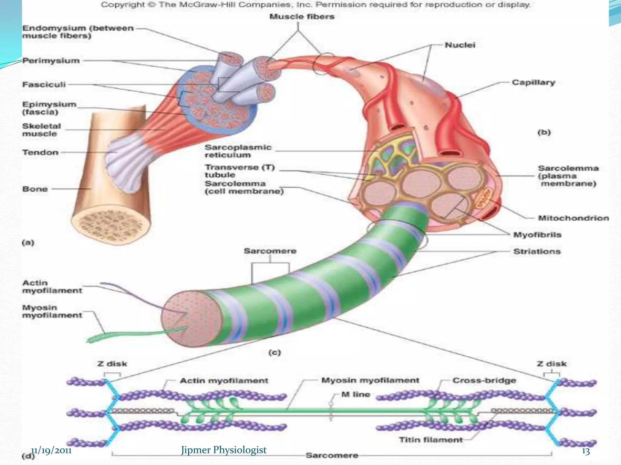
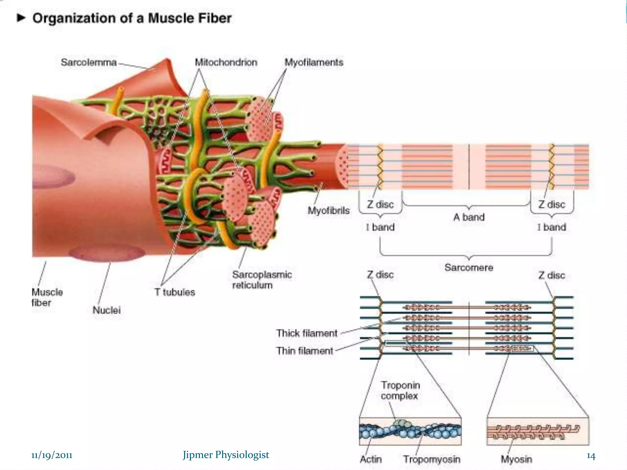
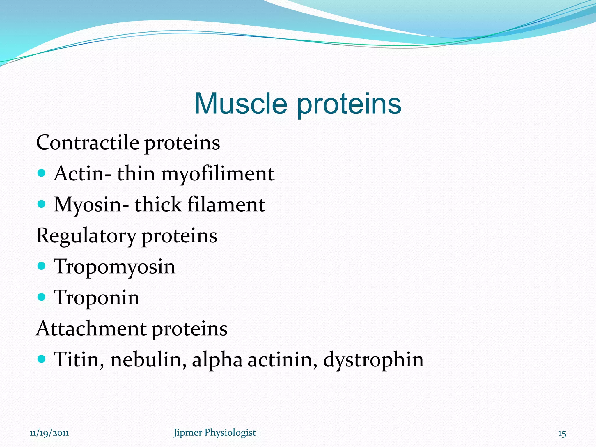
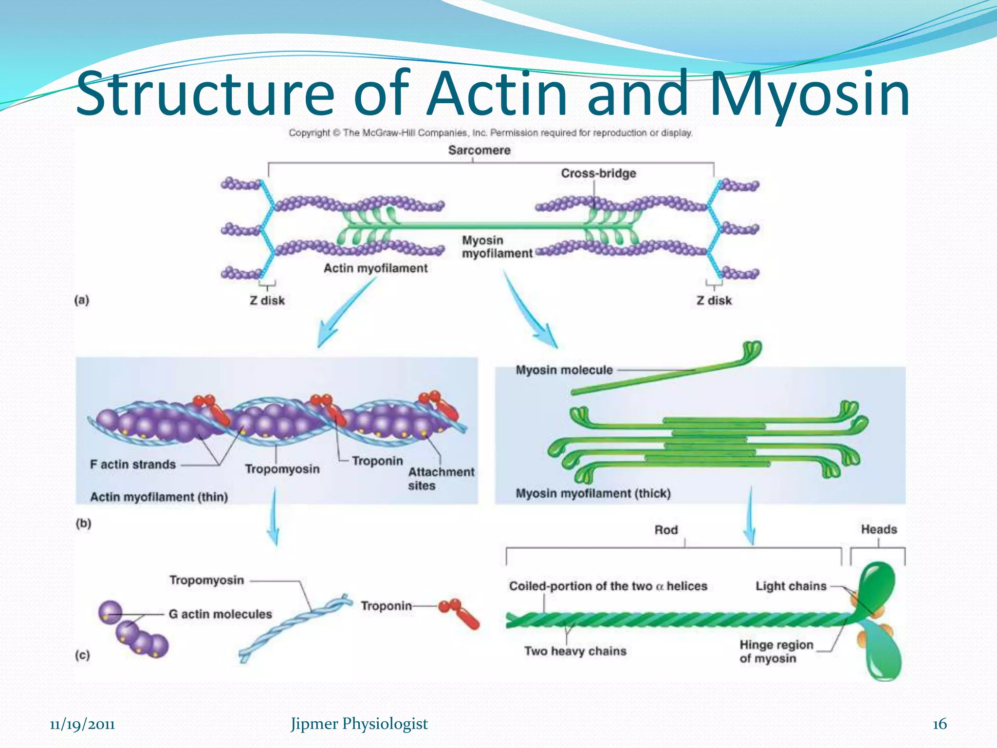
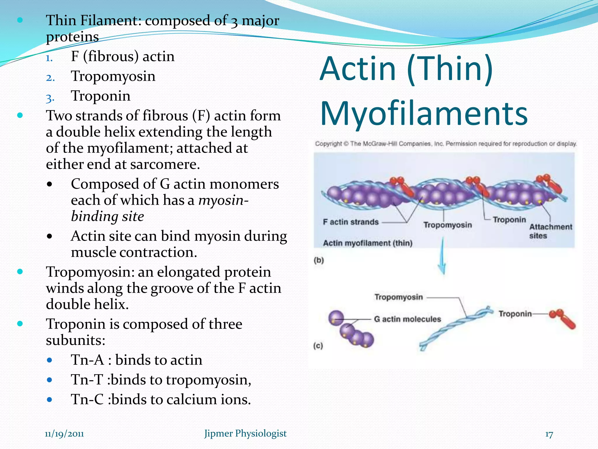
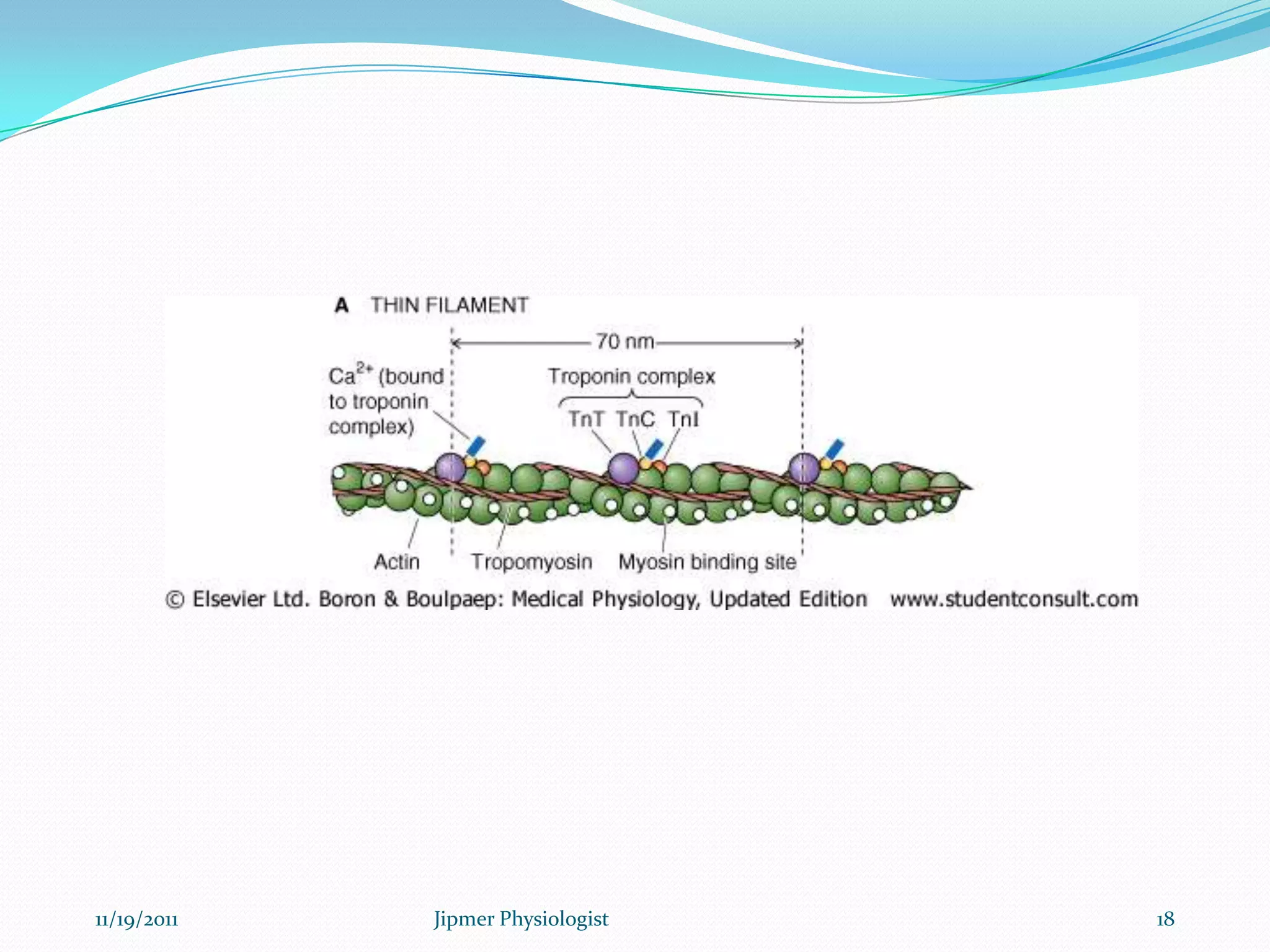
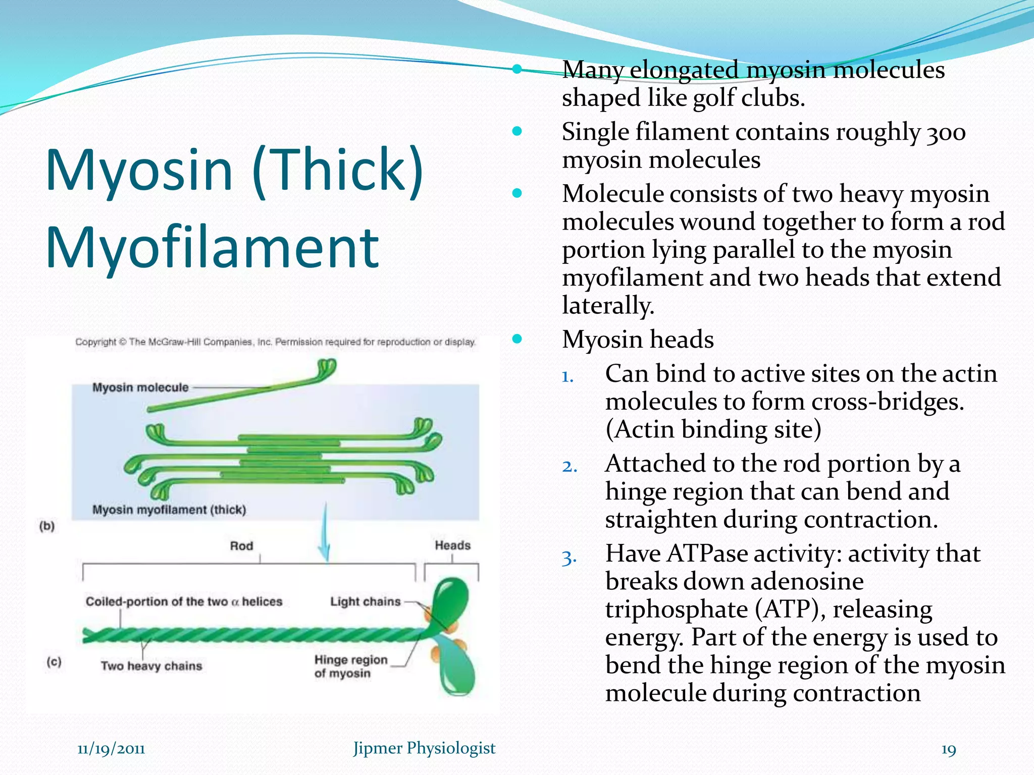
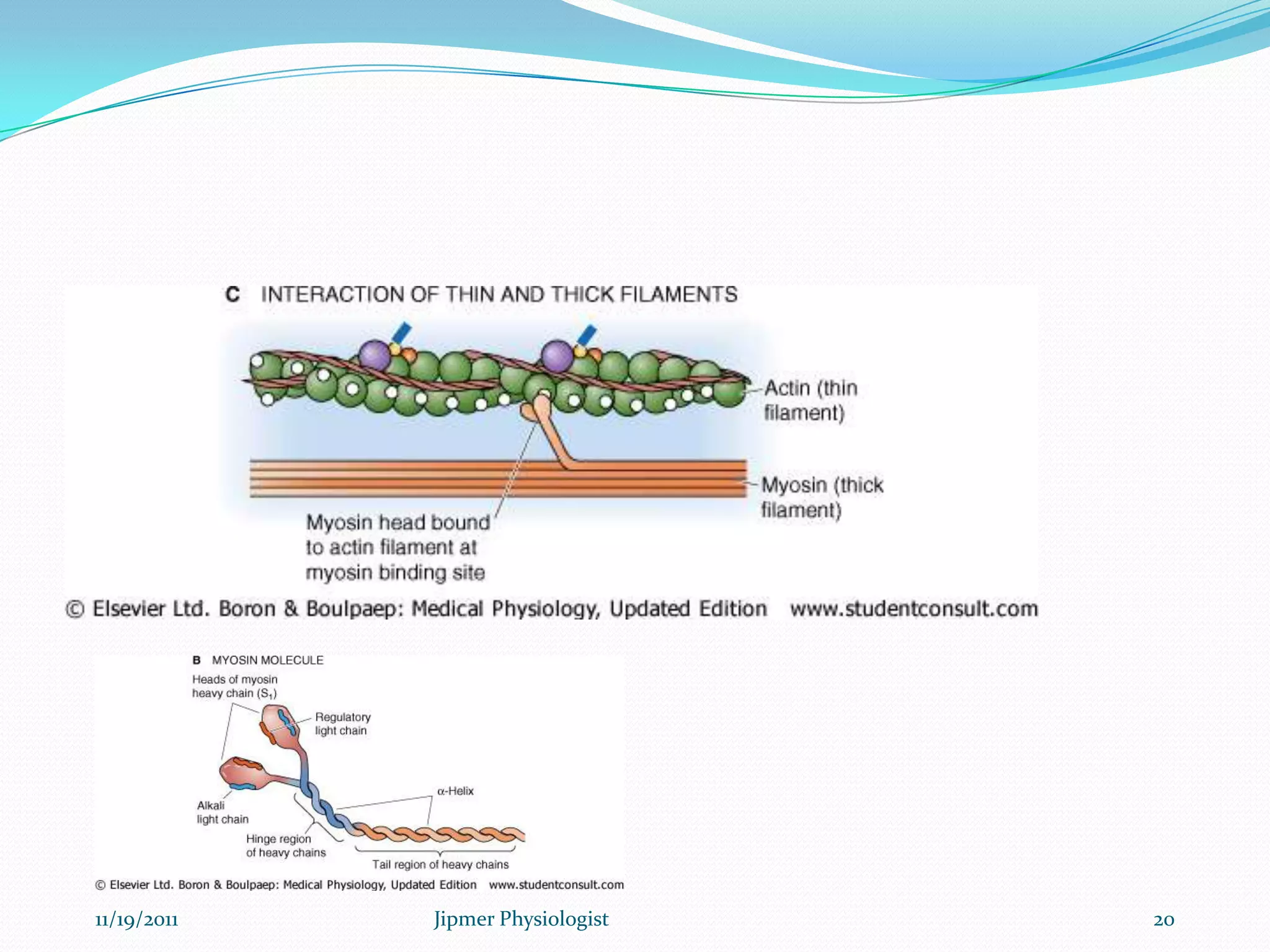
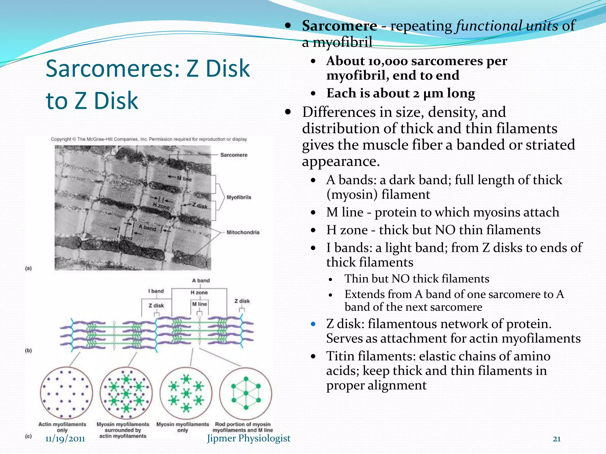
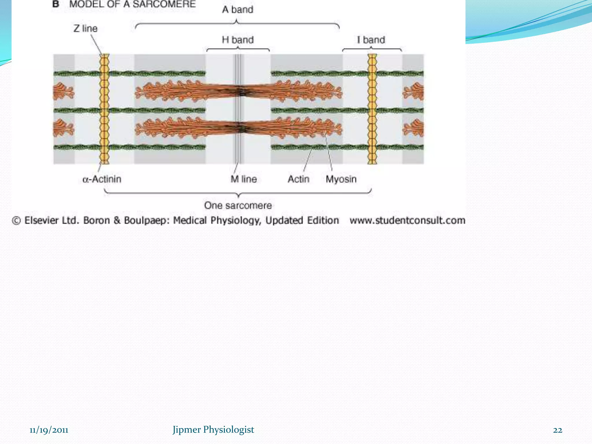
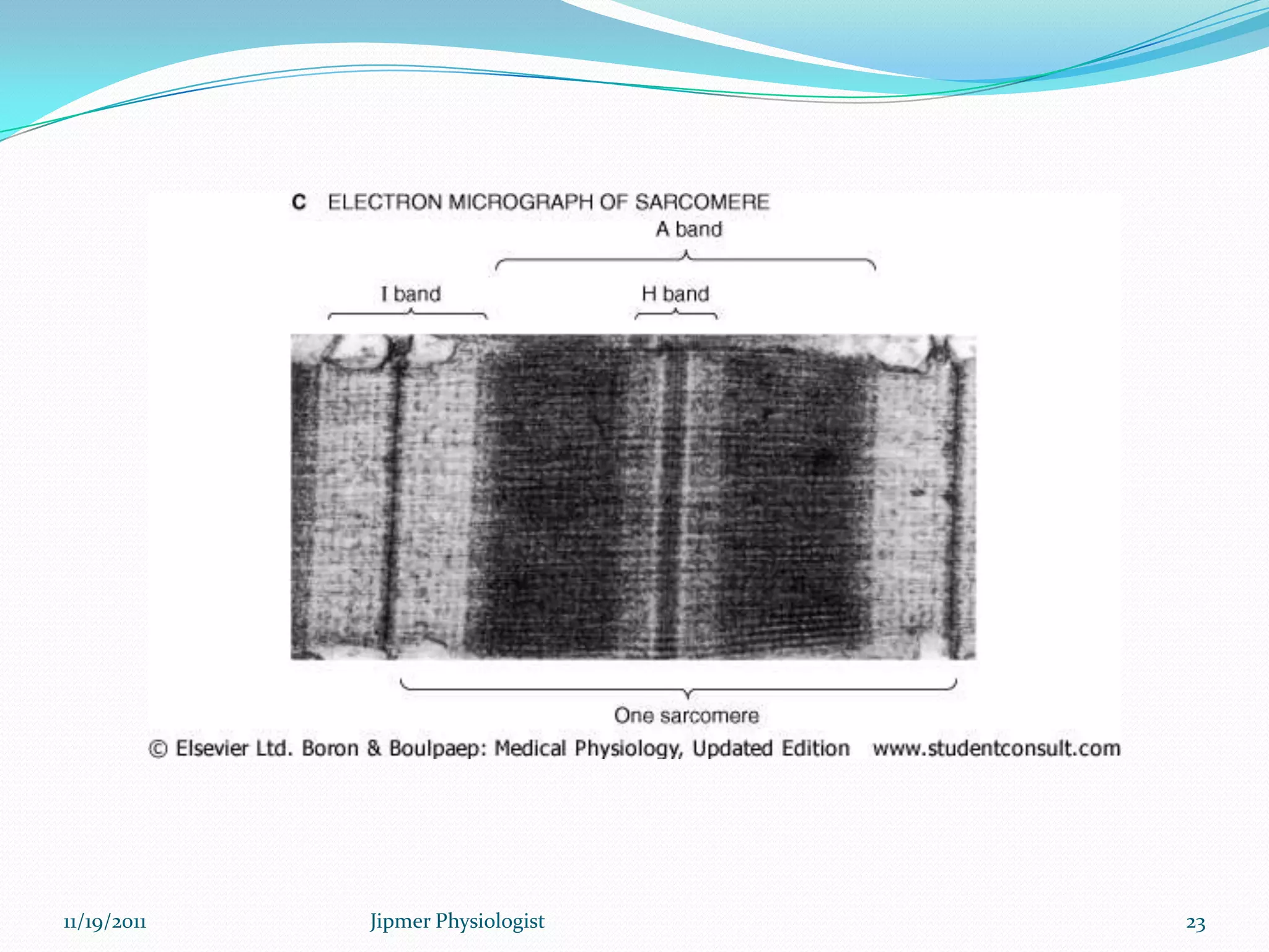
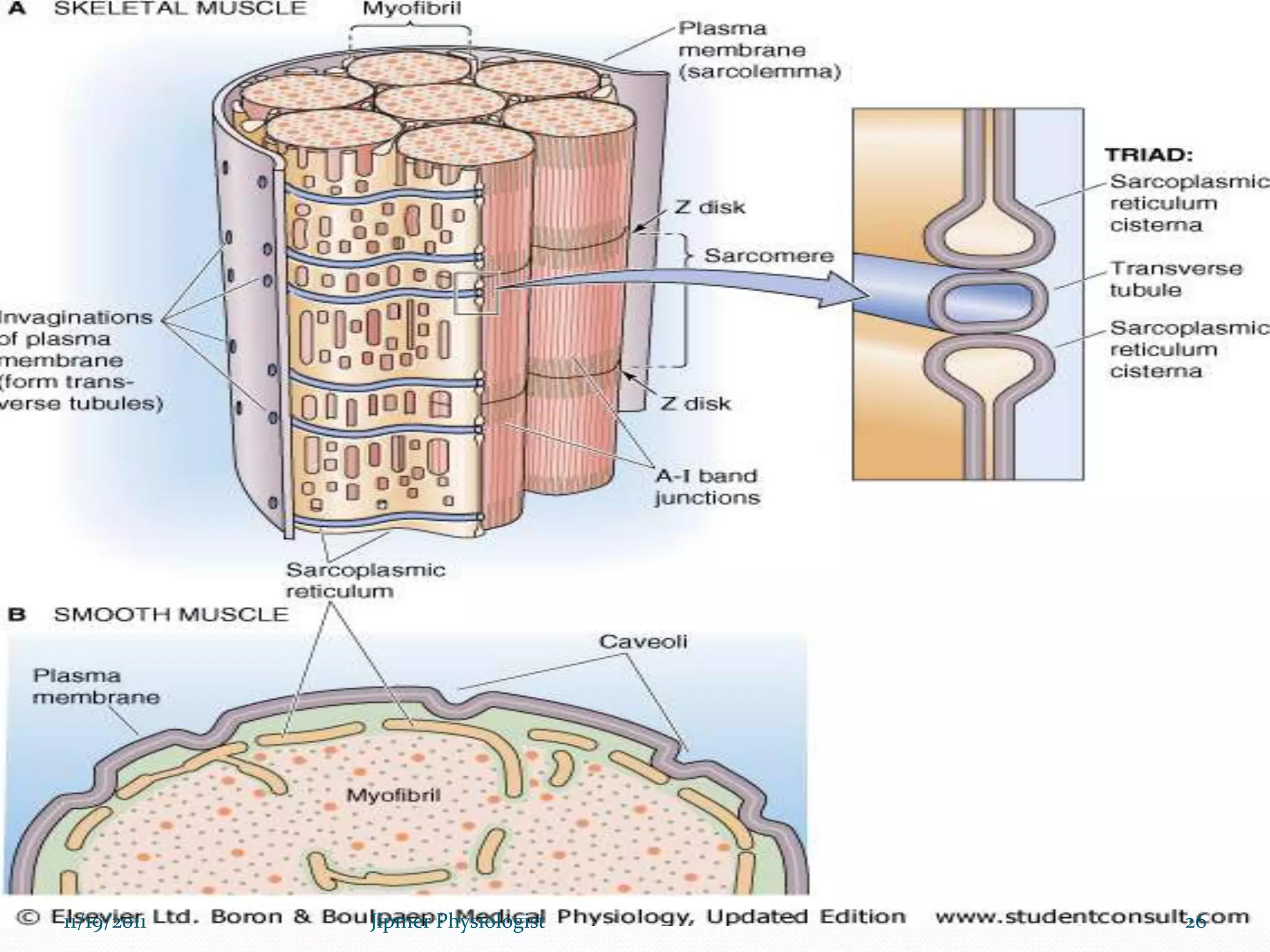
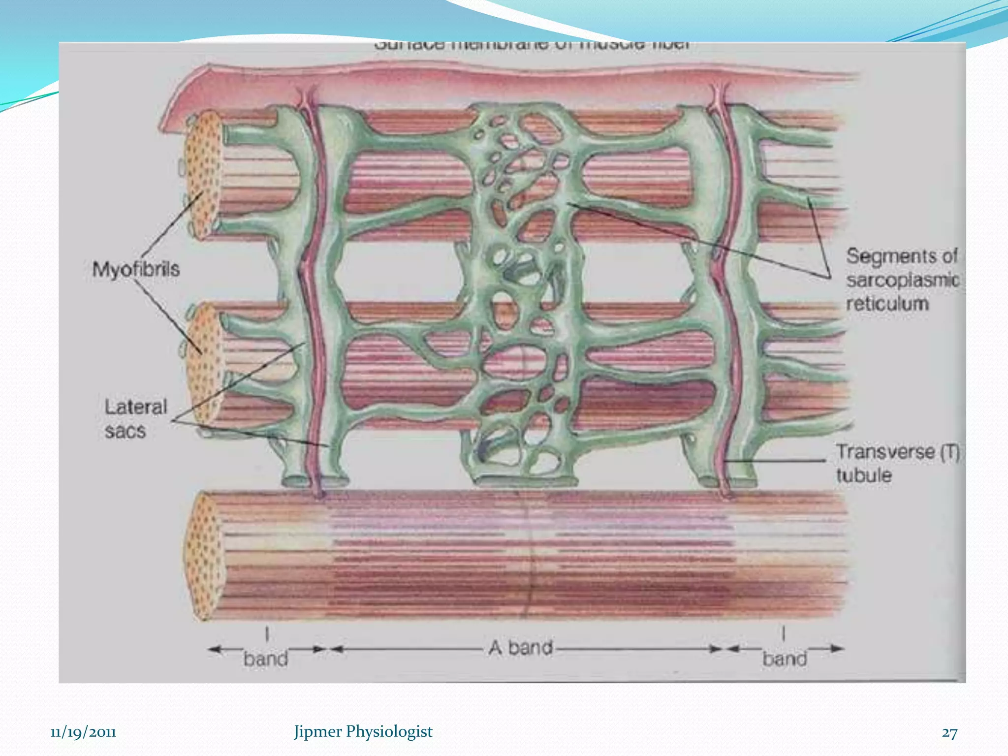
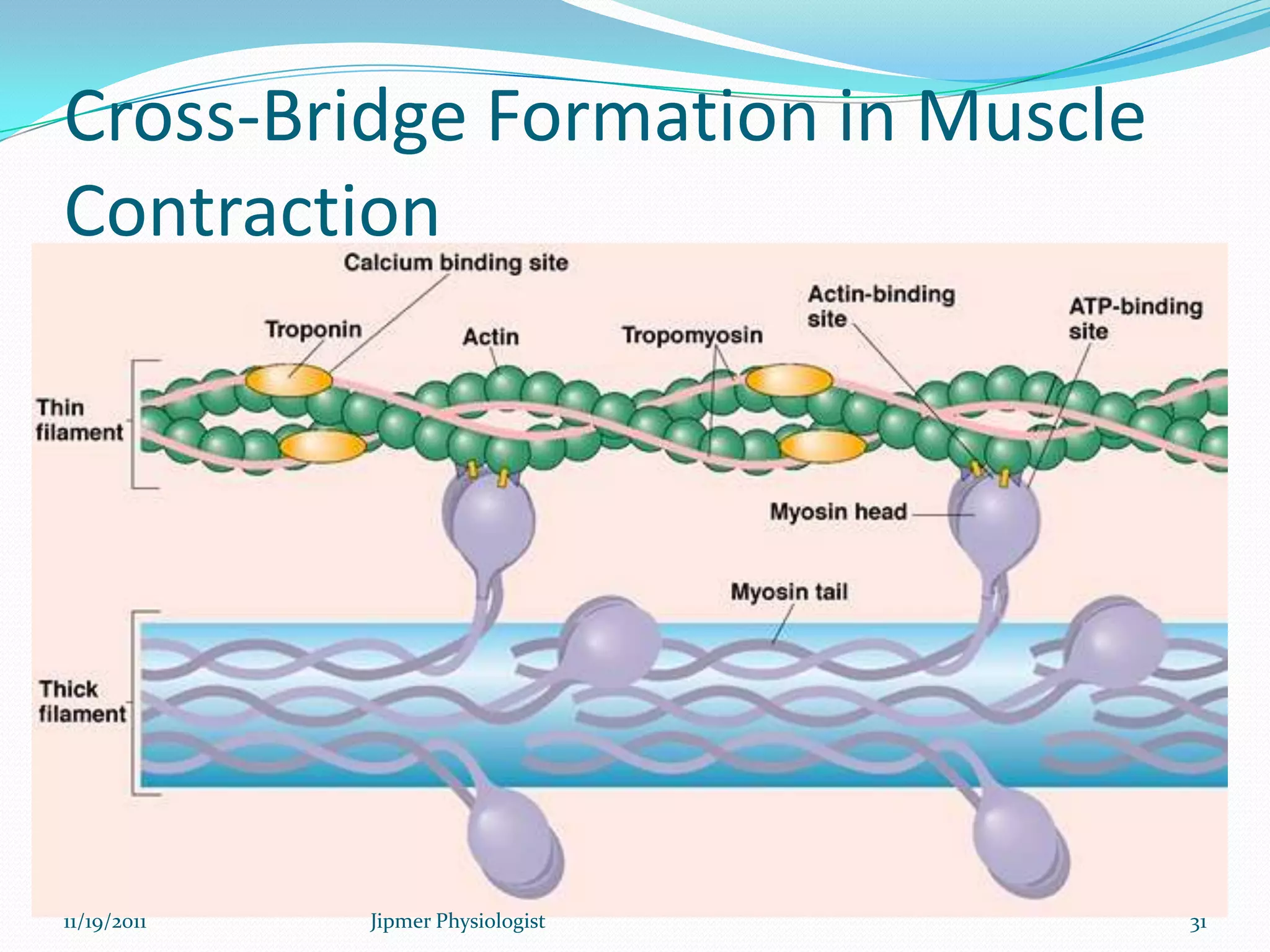
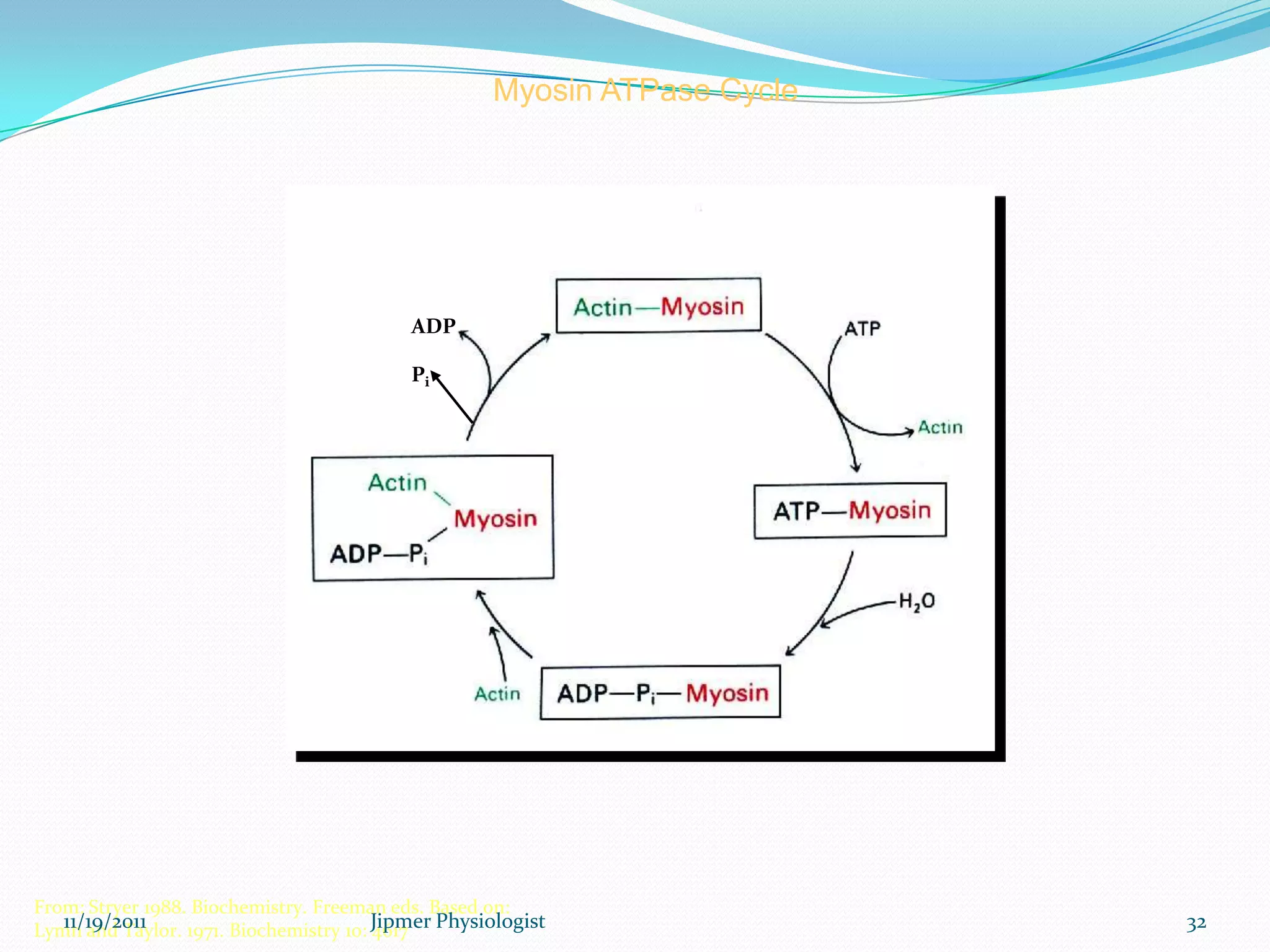
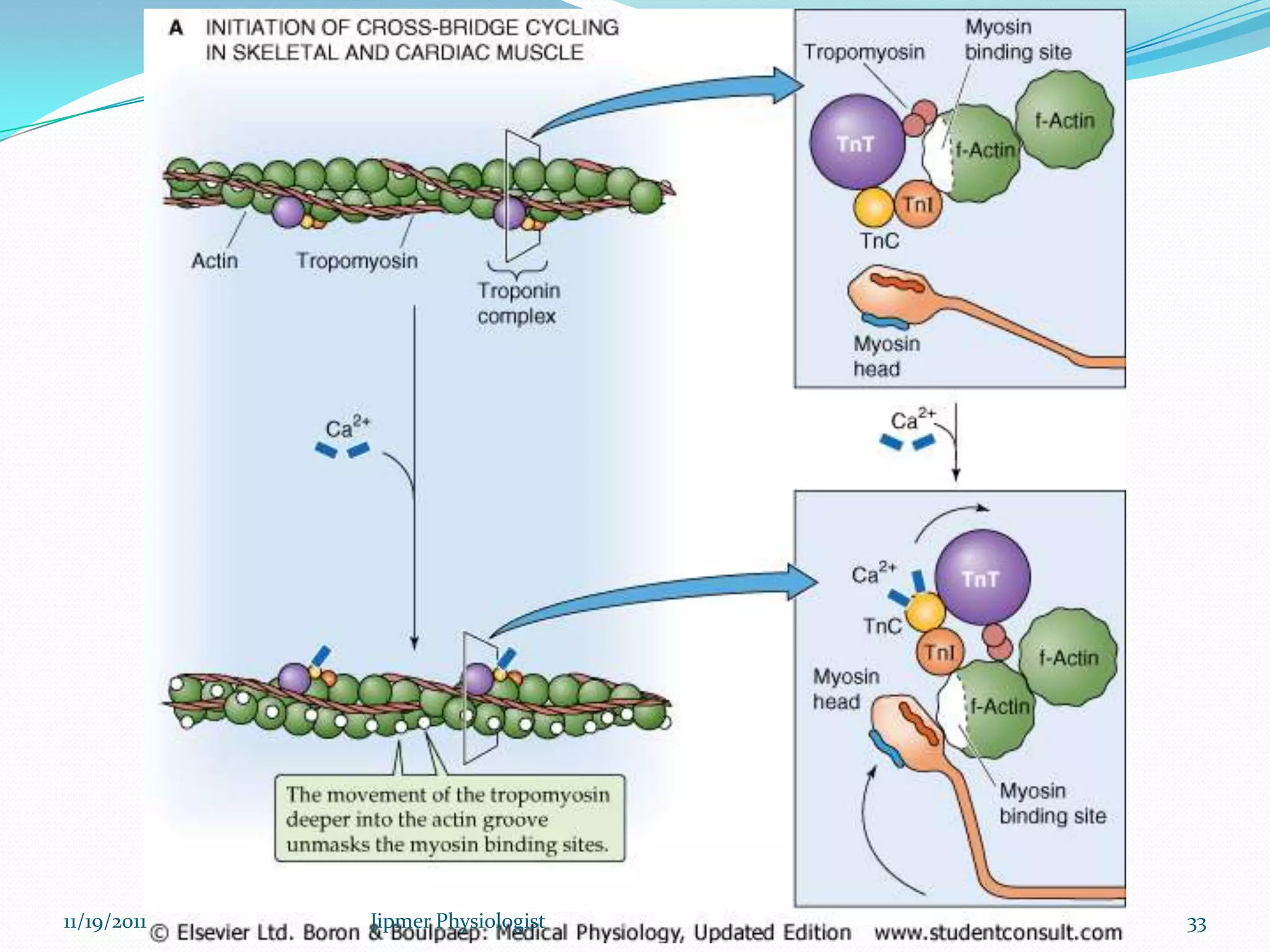
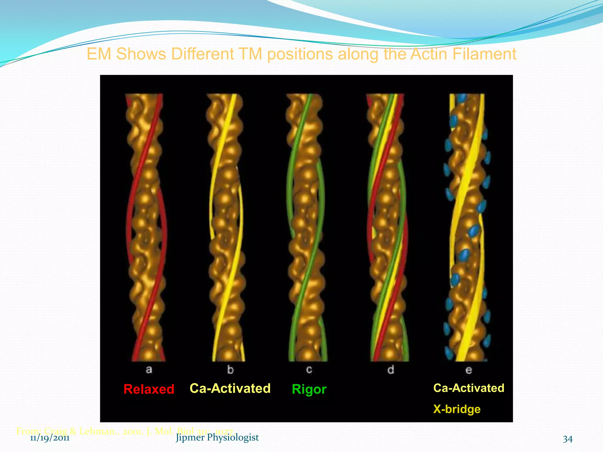
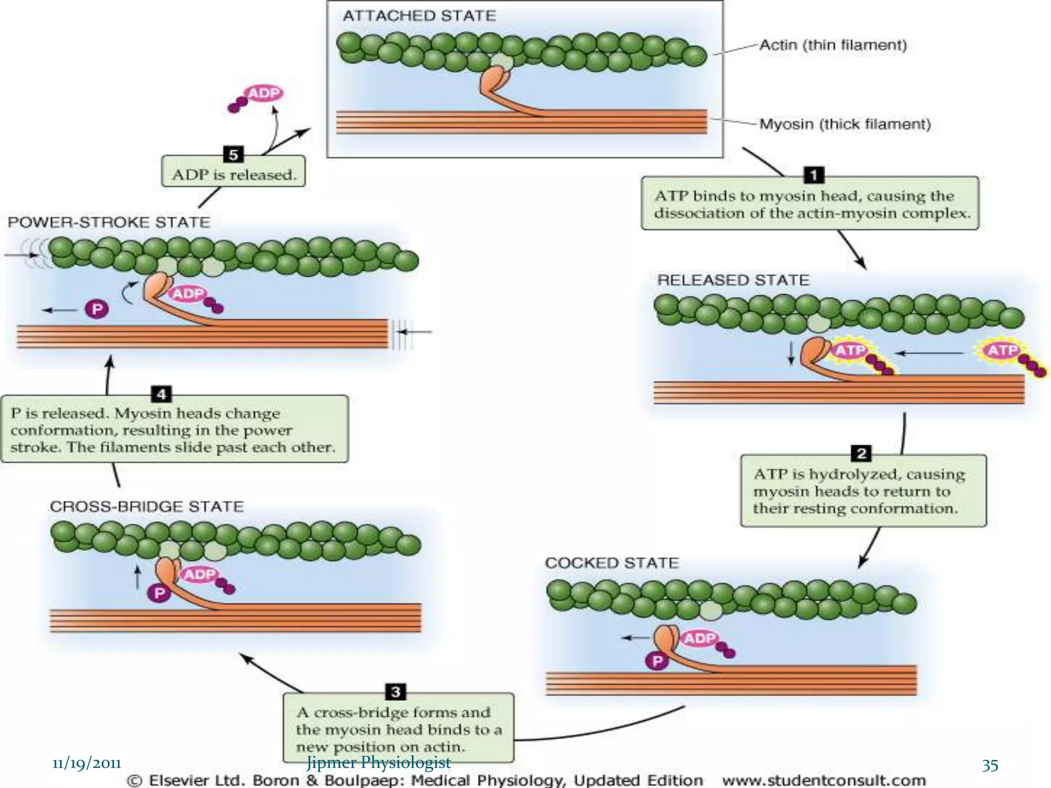
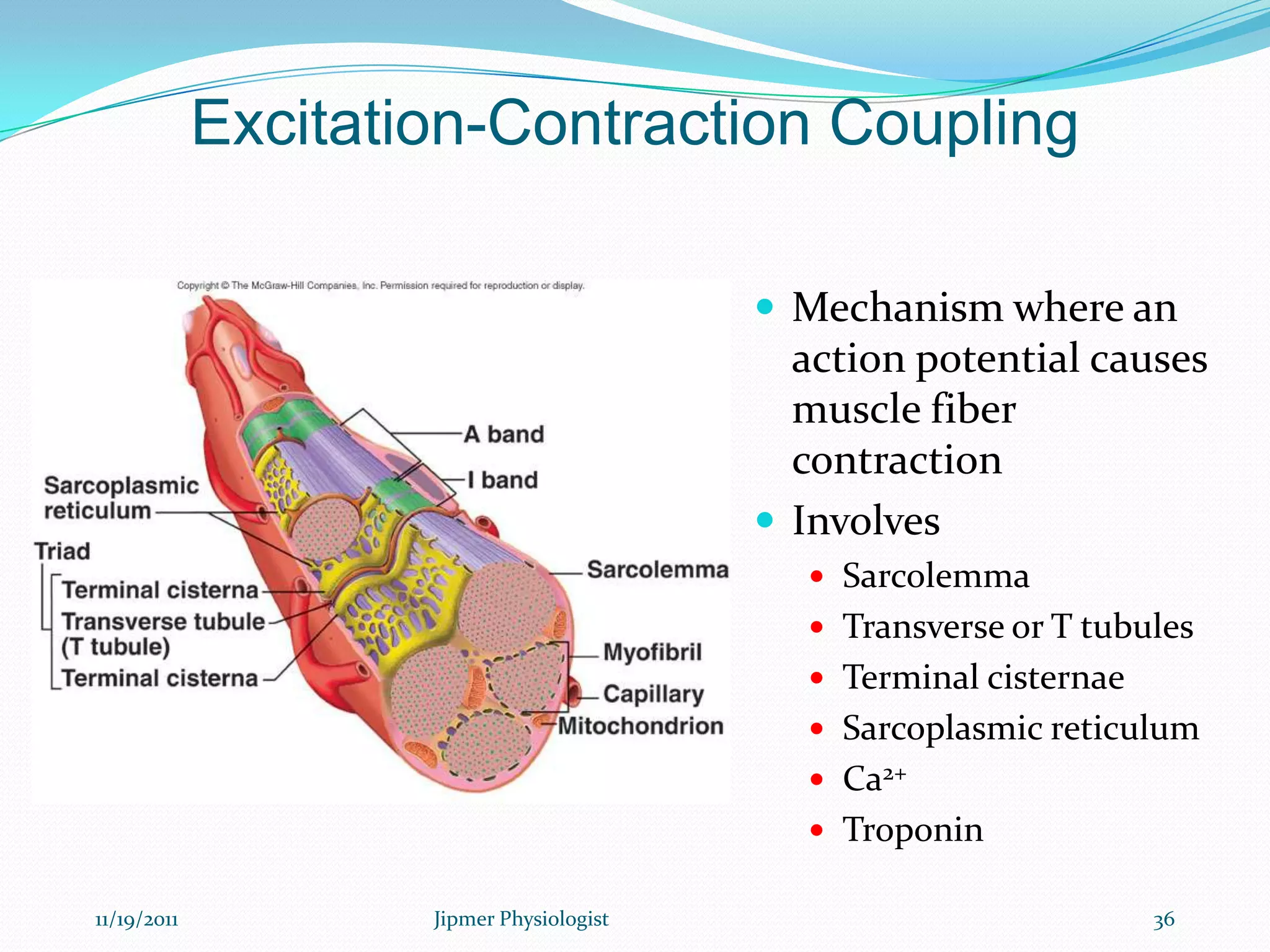
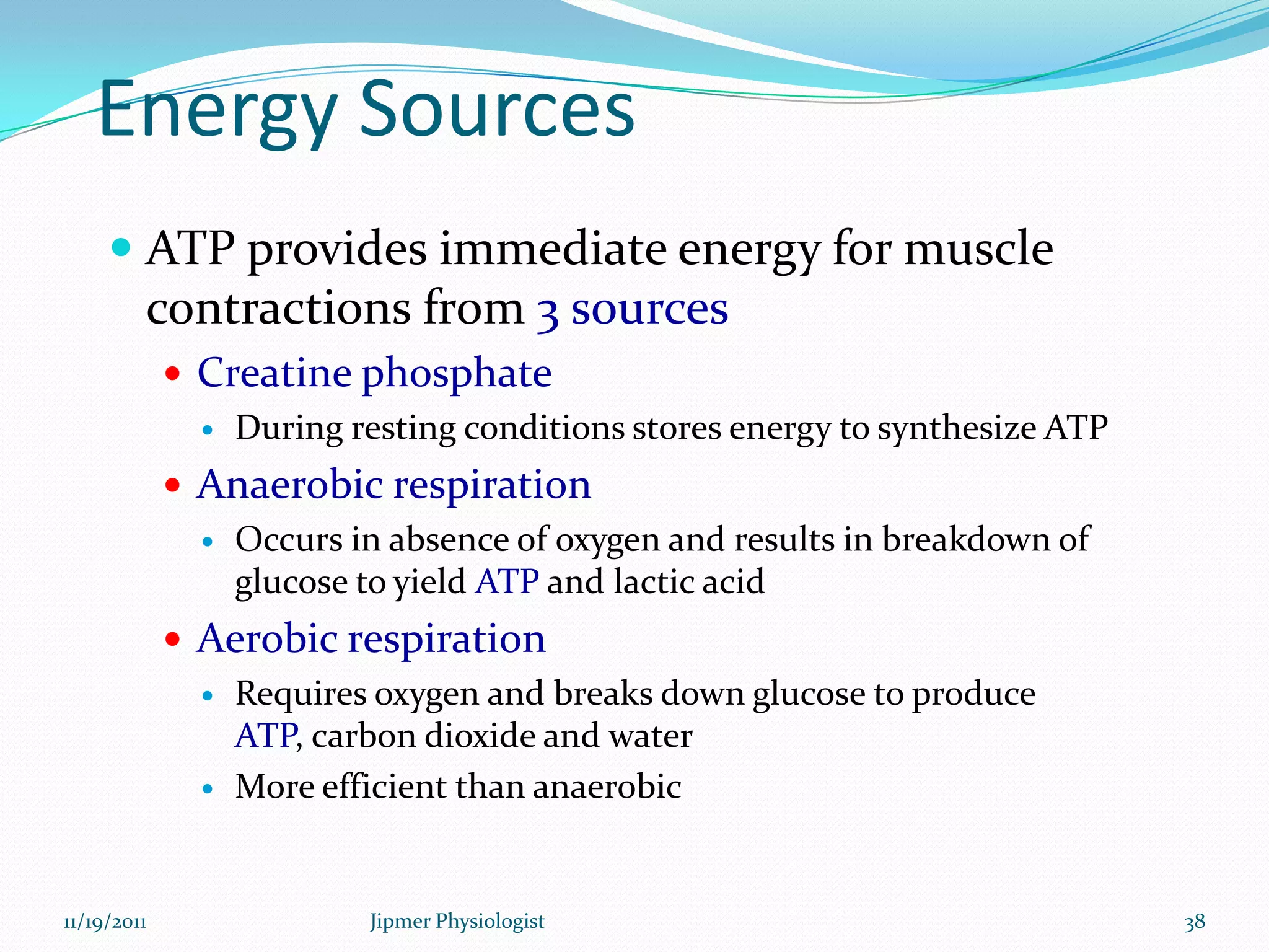
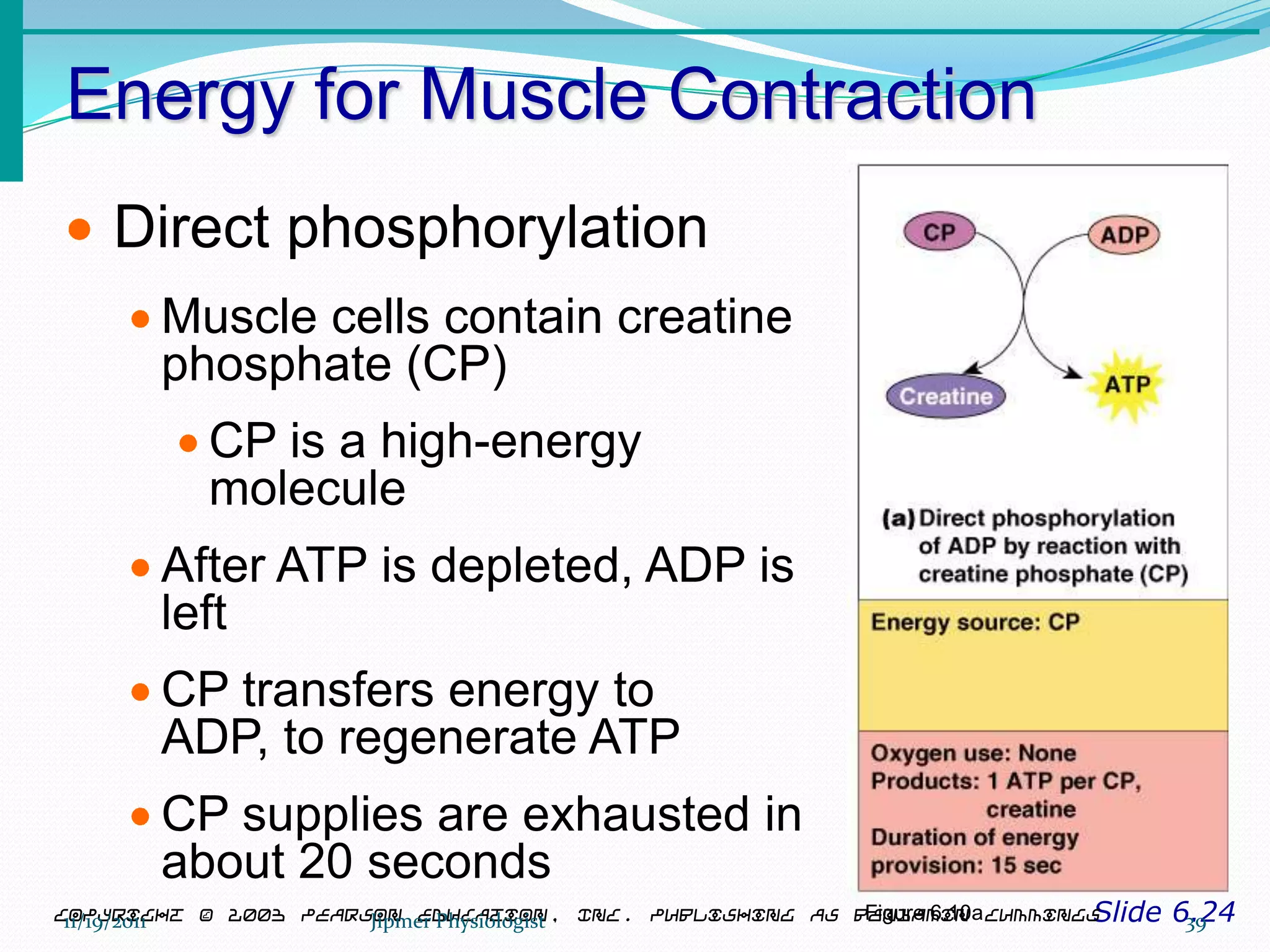
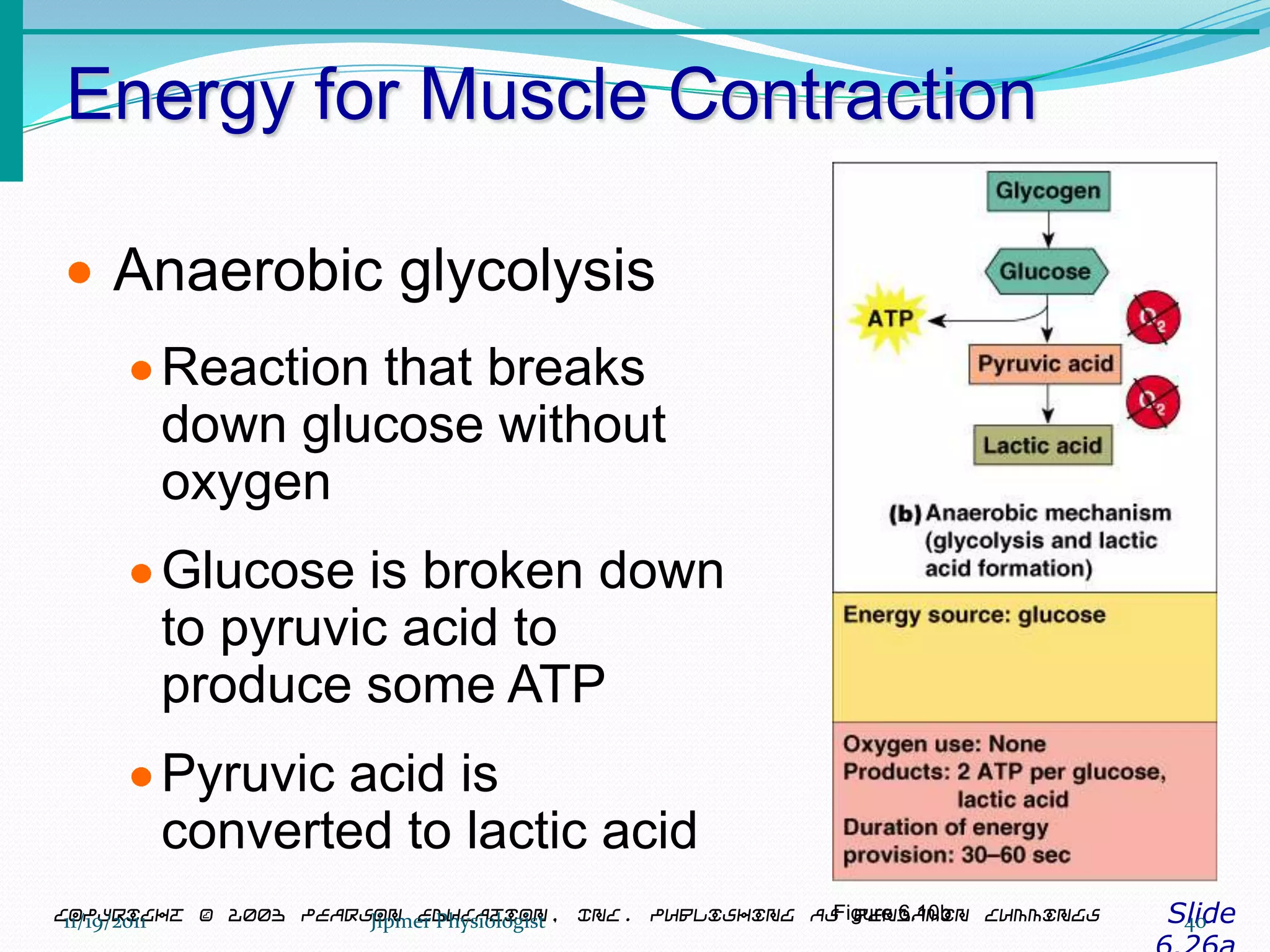
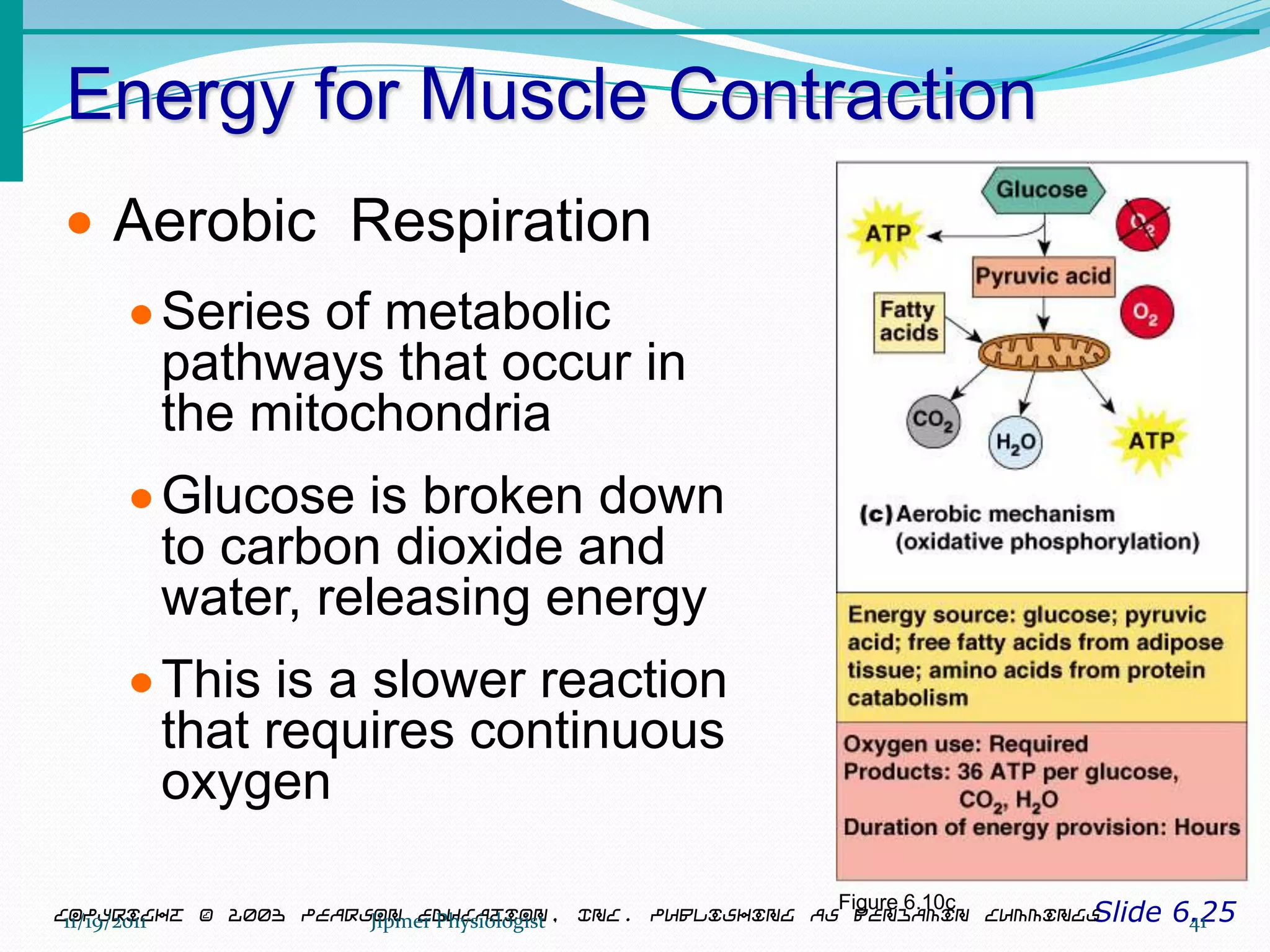
Dr. Dinesh T,
Junior resident.
11/19/2011 1Jipmer Physiologist
Discussion topics
 Introduction
 Myocyte- structure, development
 Muscle proteins
 Sarcomere
 Sarcotubular system
 Excitation- contraction coupling
 Molecular basis of muscle contraction
 Summary
 Muscle relaxation
 Conclusion
11/19/2011 2Jipmer Physiologist
Introduction
 Human body contains over 400 skeletal muscles
 40-50% of total body weight
Functions of skeletal muscle
 Body movement (Locomotion)
 Maintenance of posture
 Respiration
 Diaphragm and intercostal contractions
 Communication (Verbal and Facial)
 Constriction of organs and vessels
 Peristalsis of intestinal tract
 Vasoconstriction of b.v. and other structures (pupils)
 Production of body heat (Thermogenesis)
11/19/2011 3Jipmer Physiologist
Skeletal Muscle Characteristics
Slide 6.3Copyright © 2003 Pearson Education, Inc. publishing as Benjamin Cummings
Most are attached by tendons to bones
Cells are multinucleate
Striated – have visible banding
Voluntary – subject to conscious control
Cells are surrounded and bundled by
connective tissue = great force, but tires
easily
11/19/2011 4Jipmer Physiologist
Terms
Sarcolemma = Cell membrane
Sarcoplasm = Cytoplasm
Sarcoplasmic Reticulum =
Endoplasmic Reticulum
Sarcosomes = Mitochontria
11/19/2011 5Jipmer Physiologist
Structure of skeletal muscle:
connective tissue covering
 Epimysium
 Surrounds entire muscle
 Perimysium
 Surrounds bundles of muscle fibers
 Endomysium
 Surrounds individual muscle fibers
11/19/2011 6Jipmer Physiologist
11/19/2011 7Jipmer Physiologist
11/19/2011 8Jipmer Physiologist
Skeletal muscle structure
 Composed of muscle cells (fibers),
connective tissue, blood vessels,
nerves
 Fibers are long, cylindrical, and
multinucleated
 Tend to be smaller diameter in small
muscles and larger in large muscles. 1
mm- 4 cm in length
11/19/2011 9Jipmer Physiologist
•Develop from myoblasts;
numbers remain constant
•Striated appearance
•Nuclei are peripherally
located
11/19/2011 10Jipmer Physiologist
Embryologic origin:
11/19/2011 11Jipmer Physiologist
Muscle fiber anatomy
 Sarcolemma - cell membrane
 Surrounds the sarcoplasm (cytoplasm of fiber)
 Contains many of the same organelles seen in other cells
 An abundance of the oxygen-binding protein myoglobin
 Punctuated by openings called the transverse tubules (T-tubules)
 Narrow tubes that extend into the sarcoplasm at right angles to
the surface
 Filled with extracellular fluid
 Myofibrils -cylindrical structures within muscle fiber
 Are bundles of protein filaments (=myofilaments)
 Two types of myofilaments
1. Actin filaments (thin filaments)
2. Myosin filaments (thick filaments)
– At each end of the fiber, myofibrils are anchored to the inner surface
of the sarcolemma
– When myofibril shortens, muscle shortens (contracts)
11/19/2011 12Jipmer Physiologist
11/19/2011 13Jipmer Physiologist
11/19/2011 14Jipmer Physiologist
Muscle proteins
Contractile proteins
 Actin- thin myofiliment
 Myosin- thick filament
Regulatory proteins
 Tropomyosin
 Troponin
Attachment proteins
 Titin, nebulin, alpha actinin, dystrophin
11/19/2011 15Jipmer Physiologist
Structure of Actin and Myosin
11/19/2011 16Jipmer Physiologist
Actin (Thin)
Myofilaments
 Thin Filament: composed of 3 major
proteins
1. F (fibrous) actin
2. Tropomyosin
3. Troponin
 Two strands of fibrous (F) actin form
a double helix extending the length
of the myofilament; attached at
either end at sarcomere.
 Composed of G actin monomers
each of which has a myosin-
binding site
 Actin site can bind myosin during
muscle contraction.
 Tropomyosin: an elongated protein
winds along the groove of the F actin
double helix.
 Troponin is composed of three
subunits:
 Tn-A : binds to actin
 Tn-T :binds to tropomyosin,
 Tn-C :binds to calcium ions.
11/19/2011 17Jipmer Physiologist
11/19/2011 18Jipmer Physiologist
Myosin (Thick)
Myofilament
 Many elongated myosin molecules
shaped like golf clubs.
 Single filament contains roughly 300
myosin molecules
 Molecule consists of two heavy myosin
molecules wound together to form a rod
portion lying parallel to the myosin
myofilament and two heads that extend
laterally.
 Myosin heads
1. Can bind to active sites on the actin
molecules to form cross-bridges.
(Actin binding site)
2. Attached to the rod portion by a
hinge region that can bend and
straighten during contraction.
3. Have ATPase activity: activity that
breaks down adenosine
triphosphate (ATP), releasing
energy. Part of the energy is used to
bend the hinge region of the myosin
molecule during contraction
11/19/2011 19Jipmer Physiologist
11/19/2011 20Jipmer Physiologist
Sarcomeres: Z Disk
to Z Disk
 Sarcomere - repeating functional units of
a myofibril
 About 10,000 sarcomeres per
myofibril, end to end
 Each is about 2 µm long
 Differences in size, density, and
distribution of thick and thin filaments
gives the muscle fiber a banded or striated
appearance.
 A bands: a dark band; full length of thick
(myosin) filament
 M line - protein to which myosins attach
 H zone - thick but NO thin filaments
 I bands: a light band; from Z disks to ends of
thick filaments
 Thin but NO thick filaments
 Extends from A band of one sarcomere to A
band of the next sarcomere
 Z disk: filamentous network of protein.
Serves as attachment for actin myofilaments
 Titin filaments: elastic chains of amino
acids; keep thick and thin filaments in
proper alignment
11/19/2011 21Jipmer Physiologist
11/19/2011 22Jipmer Physiologist
11/19/2011 23Jipmer Physiologist
Sarcoplasmic Reticulum (SR)
 SR is an elaborate, smooth endoplasmic
reticulum
 runs longitudinally and surrounds each myofibril
 Form chambers called terminal cisternae on either
side of the T-tubules
 A single T-tubule and the 2 terminal cisternae
form a triad
 SR stores Ca++ when muscle not contracting
 When stimulated, calcium released into sarcoplasm
 SR membrane has Ca++ pumps that function to pump
Ca++ out of the sarcoplasm back into the SR after
contraction
11/19/2011 24Jipmer Physiologist
Sarcoplasmic Reticulum (SR)
Figure 9.5
11/19/2011 25Jipmer Physiologist
11/19/2011 26Jipmer Physiologist
11/19/2011 27Jipmer Physiologist
Muscular Contraction
 The sliding filament model
 Muscle shortening occurs due to the movement of the
actin filament over the myosin filament
 Formation of cross-bridges between actin and myosin
filaments
 Reduction in the distance between Z-lines of the
sarcomere
11/19/2011 28Jipmer Physiologist
Sliding Filament Theory
 Rest – uncharged ATP cross-bridge complex
 Excitation-coupling – charged ATP cross-bridge
complex, “turned on”
 Contraction – actomyosin – ATP > ADP & Pi +
energy
 Recharging – reload cross-bridge with ATP
 Relaxation – cross-bridges “turned off”
11/19/2011 29Jipmer Physiologist
Sliding Filament Model of
Contraction
 Thin filaments slide past the thick ones so that the
actin and myosin filaments overlap to a greater degree
 In the relaxed state, thin and thick filaments overlap
only slightly
 Upon stimulation, myosin heads bind to actin and
sliding begins
11/19/2011 30Jipmer Physiologist
Cross-Bridge Formation in Muscle
Contraction
11/19/2011 31Jipmer Physiologist
Myosin ATPase Cycle
From: Stryer 1988. Biochemistry. Freeman eds. Based on:
Lymn and Taylor. 1971. Biochemistry 10: 4617
Pi
ADP
11/19/2011 32Jipmer Physiologist
11/19/2011 33Jipmer Physiologist
EM Shows Different TM positions along the Actin Filament
Relaxed Ca-Activated Rigor Ca-Activated
X-bridge
From: Craig & Lehman.. 2001. J. Mol. Biol 311: 1027.
11/19/2011 34Jipmer Physiologist
11/19/2011 35Jipmer Physiologist
Excitation-Contraction Coupling
 Mechanism where an
action potential causes
muscle fiber
contraction
 Involves
 Sarcolemma
 Transverse or T tubules
 Terminal cisternae
 Sarcoplasmic reticulum
 Ca2+
 Troponin
11/19/2011 36Jipmer Physiologist
Sources of ATP for Muscle
Contraction
11/19/2011 37Jipmer Physiologist
Energy Sources
 ATP provides immediate energy for muscle
contractions from 3 sources
 Creatine phosphate
 During resting conditions stores energy to synthesize ATP
 Anaerobic respiration
 Occurs in absence of oxygen and results in breakdown of
glucose to yield ATP and lactic acid
 Aerobic respiration
 Requires oxygen and breaks down glucose to produce
ATP, carbon dioxide and water
 More efficient than anaerobic
11/19/2011 38Jipmer Physiologist
Energy for Muscle Contraction
Slide 6.24Copyright © 2003 Pearson Education, Inc. publishing as Benjamin Cummings
Direct phosphorylation
Muscle cells contain creatine
phosphate (CP)
CP is a high-energy
molecule
After ATP is depleted, ADP is
left
CP transfers energy to
ADP, to regenerate ATP
CP supplies are exhausted in
about 20 seconds
Figure 6.10a11/19/2011 39Jipmer Physiologist
Energy for Muscle Contraction
SlideCopyright © 2003 Pearson Education, Inc. publishing as Benjamin Cummings
Anaerobic glycolysis
Reaction that breaks
down glucose without
oxygen
Glucose is broken down
to pyruvic acid to
produce some ATP
Pyruvic acid is
converted to lactic acid
Figure 6.10b11/19/2011 40Jipmer Physiologist
Energy for Muscle Contraction
Slide 6.25Copyright © 2003 Pearson Education, Inc. publishing as Benjamin Cummings
Aerobic Respiration
Series of metabolic
pathways that occur in
the mitochondria
Glucose is broken down
to carbon dioxide and
water, releasing energy
This is a slower reaction
that requires continuous
oxygen
Figure 6.10c
11/19/2011 41Jipmer Physiologist
11/19/2011 Jipmer Physiologist ‹#›
11/19/2011 43Jipmer Physiologist

Skeletalmusclestructurefunction09 11-2011-111120005142-phpapp01

  • 1.
    Dr. Dinesh T, Juniorresident. 11/19/2011 1Jipmer Physiologist
  • 2.
    Discussion topics  Introduction Myocyte- structure, development  Muscle proteins  Sarcomere  Sarcotubular system  Excitation- contraction coupling  Molecular basis of muscle contraction  Summary  Muscle relaxation  Conclusion 11/19/2011 2Jipmer Physiologist
  • 3.
    Introduction  Human bodycontains over 400 skeletal muscles  40-50% of total body weight Functions of skeletal muscle  Body movement (Locomotion)  Maintenance of posture  Respiration  Diaphragm and intercostal contractions  Communication (Verbal and Facial)  Constriction of organs and vessels  Peristalsis of intestinal tract  Vasoconstriction of b.v. and other structures (pupils)  Production of body heat (Thermogenesis) 11/19/2011 3Jipmer Physiologist
  • 4.
    Skeletal Muscle Characteristics Slide6.3Copyright © 2003 Pearson Education, Inc. publishing as Benjamin Cummings Most are attached by tendons to bones Cells are multinucleate Striated – have visible banding Voluntary – subject to conscious control Cells are surrounded and bundled by connective tissue = great force, but tires easily 11/19/2011 4Jipmer Physiologist
  • 5.
    Terms Sarcolemma = Cellmembrane Sarcoplasm = Cytoplasm Sarcoplasmic Reticulum = Endoplasmic Reticulum Sarcosomes = Mitochontria 11/19/2011 5Jipmer Physiologist
  • 6.
    Structure of skeletalmuscle: connective tissue covering  Epimysium  Surrounds entire muscle  Perimysium  Surrounds bundles of muscle fibers  Endomysium  Surrounds individual muscle fibers 11/19/2011 6Jipmer Physiologist
  • 7.
  • 8.
  • 9.
    Skeletal muscle structure Composed of muscle cells (fibers), connective tissue, blood vessels, nerves  Fibers are long, cylindrical, and multinucleated  Tend to be smaller diameter in small muscles and larger in large muscles. 1 mm- 4 cm in length 11/19/2011 9Jipmer Physiologist
  • 10.
    •Develop from myoblasts; numbersremain constant •Striated appearance •Nuclei are peripherally located 11/19/2011 10Jipmer Physiologist
  • 11.
  • 12.
    Muscle fiber anatomy Sarcolemma - cell membrane  Surrounds the sarcoplasm (cytoplasm of fiber)  Contains many of the same organelles seen in other cells  An abundance of the oxygen-binding protein myoglobin  Punctuated by openings called the transverse tubules (T-tubules)  Narrow tubes that extend into the sarcoplasm at right angles to the surface  Filled with extracellular fluid  Myofibrils -cylindrical structures within muscle fiber  Are bundles of protein filaments (=myofilaments)  Two types of myofilaments 1. Actin filaments (thin filaments) 2. Myosin filaments (thick filaments) – At each end of the fiber, myofibrils are anchored to the inner surface of the sarcolemma – When myofibril shortens, muscle shortens (contracts) 11/19/2011 12Jipmer Physiologist
  • 13.
  • 14.
  • 15.
    Muscle proteins Contractile proteins Actin- thin myofiliment  Myosin- thick filament Regulatory proteins  Tropomyosin  Troponin Attachment proteins  Titin, nebulin, alpha actinin, dystrophin 11/19/2011 15Jipmer Physiologist
  • 16.
    Structure of Actinand Myosin 11/19/2011 16Jipmer Physiologist
  • 17.
    Actin (Thin) Myofilaments  ThinFilament: composed of 3 major proteins 1. F (fibrous) actin 2. Tropomyosin 3. Troponin  Two strands of fibrous (F) actin form a double helix extending the length of the myofilament; attached at either end at sarcomere.  Composed of G actin monomers each of which has a myosin- binding site  Actin site can bind myosin during muscle contraction.  Tropomyosin: an elongated protein winds along the groove of the F actin double helix.  Troponin is composed of three subunits:  Tn-A : binds to actin  Tn-T :binds to tropomyosin,  Tn-C :binds to calcium ions. 11/19/2011 17Jipmer Physiologist
  • 18.
  • 19.
    Myosin (Thick) Myofilament  Manyelongated myosin molecules shaped like golf clubs.  Single filament contains roughly 300 myosin molecules  Molecule consists of two heavy myosin molecules wound together to form a rod portion lying parallel to the myosin myofilament and two heads that extend laterally.  Myosin heads 1. Can bind to active sites on the actin molecules to form cross-bridges. (Actin binding site) 2. Attached to the rod portion by a hinge region that can bend and straighten during contraction. 3. Have ATPase activity: activity that breaks down adenosine triphosphate (ATP), releasing energy. Part of the energy is used to bend the hinge region of the myosin molecule during contraction 11/19/2011 19Jipmer Physiologist
  • 20.
  • 21.
    Sarcomeres: Z Disk toZ Disk  Sarcomere - repeating functional units of a myofibril  About 10,000 sarcomeres per myofibril, end to end  Each is about 2 µm long  Differences in size, density, and distribution of thick and thin filaments gives the muscle fiber a banded or striated appearance.  A bands: a dark band; full length of thick (myosin) filament  M line - protein to which myosins attach  H zone - thick but NO thin filaments  I bands: a light band; from Z disks to ends of thick filaments  Thin but NO thick filaments  Extends from A band of one sarcomere to A band of the next sarcomere  Z disk: filamentous network of protein. Serves as attachment for actin myofilaments  Titin filaments: elastic chains of amino acids; keep thick and thin filaments in proper alignment 11/19/2011 21Jipmer Physiologist
  • 22.
  • 23.
  • 24.
    Sarcoplasmic Reticulum (SR) SR is an elaborate, smooth endoplasmic reticulum  runs longitudinally and surrounds each myofibril  Form chambers called terminal cisternae on either side of the T-tubules  A single T-tubule and the 2 terminal cisternae form a triad  SR stores Ca++ when muscle not contracting  When stimulated, calcium released into sarcoplasm  SR membrane has Ca++ pumps that function to pump Ca++ out of the sarcoplasm back into the SR after contraction 11/19/2011 24Jipmer Physiologist
  • 25.
    Sarcoplasmic Reticulum (SR) Figure9.5 11/19/2011 25Jipmer Physiologist
  • 26.
  • 27.
  • 28.
    Muscular Contraction  Thesliding filament model  Muscle shortening occurs due to the movement of the actin filament over the myosin filament  Formation of cross-bridges between actin and myosin filaments  Reduction in the distance between Z-lines of the sarcomere 11/19/2011 28Jipmer Physiologist
  • 29.
    Sliding Filament Theory Rest – uncharged ATP cross-bridge complex  Excitation-coupling – charged ATP cross-bridge complex, “turned on”  Contraction – actomyosin – ATP > ADP & Pi + energy  Recharging – reload cross-bridge with ATP  Relaxation – cross-bridges “turned off” 11/19/2011 29Jipmer Physiologist
  • 30.
    Sliding Filament Modelof Contraction  Thin filaments slide past the thick ones so that the actin and myosin filaments overlap to a greater degree  In the relaxed state, thin and thick filaments overlap only slightly  Upon stimulation, myosin heads bind to actin and sliding begins 11/19/2011 30Jipmer Physiologist
  • 31.
    Cross-Bridge Formation inMuscle Contraction 11/19/2011 31Jipmer Physiologist
  • 32.
    Myosin ATPase Cycle From:Stryer 1988. Biochemistry. Freeman eds. Based on: Lymn and Taylor. 1971. Biochemistry 10: 4617 Pi ADP 11/19/2011 32Jipmer Physiologist
  • 33.
  • 34.
    EM Shows DifferentTM positions along the Actin Filament Relaxed Ca-Activated Rigor Ca-Activated X-bridge From: Craig & Lehman.. 2001. J. Mol. Biol 311: 1027. 11/19/2011 34Jipmer Physiologist
  • 35.
  • 36.
    Excitation-Contraction Coupling  Mechanismwhere an action potential causes muscle fiber contraction  Involves  Sarcolemma  Transverse or T tubules  Terminal cisternae  Sarcoplasmic reticulum  Ca2+  Troponin 11/19/2011 36Jipmer Physiologist
  • 37.
    Sources of ATPfor Muscle Contraction 11/19/2011 37Jipmer Physiologist
  • 38.
    Energy Sources  ATPprovides immediate energy for muscle contractions from 3 sources  Creatine phosphate  During resting conditions stores energy to synthesize ATP  Anaerobic respiration  Occurs in absence of oxygen and results in breakdown of glucose to yield ATP and lactic acid  Aerobic respiration  Requires oxygen and breaks down glucose to produce ATP, carbon dioxide and water  More efficient than anaerobic 11/19/2011 38Jipmer Physiologist
  • 39.
    Energy for MuscleContraction Slide 6.24Copyright © 2003 Pearson Education, Inc. publishing as Benjamin Cummings Direct phosphorylation Muscle cells contain creatine phosphate (CP) CP is a high-energy molecule After ATP is depleted, ADP is left CP transfers energy to ADP, to regenerate ATP CP supplies are exhausted in about 20 seconds Figure 6.10a11/19/2011 39Jipmer Physiologist
  • 40.
    Energy for MuscleContraction SlideCopyright © 2003 Pearson Education, Inc. publishing as Benjamin Cummings Anaerobic glycolysis Reaction that breaks down glucose without oxygen Glucose is broken down to pyruvic acid to produce some ATP Pyruvic acid is converted to lactic acid Figure 6.10b11/19/2011 40Jipmer Physiologist
  • 41.
    Energy for MuscleContraction Slide 6.25Copyright © 2003 Pearson Education, Inc. publishing as Benjamin Cummings Aerobic Respiration Series of metabolic pathways that occur in the mitochondria Glucose is broken down to carbon dioxide and water, releasing energy This is a slower reaction that requires continuous oxygen Figure 6.10c 11/19/2011 41Jipmer Physiologist
  • 42.
  • 43.