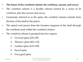
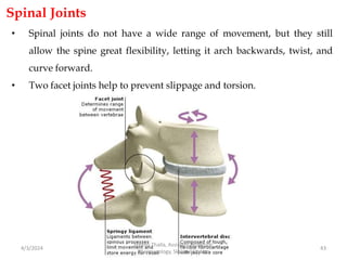
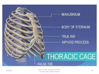
Download to read offline
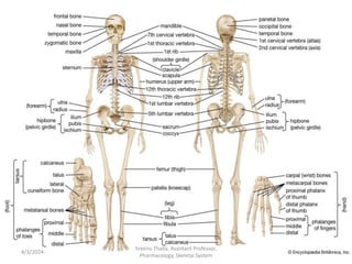

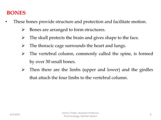
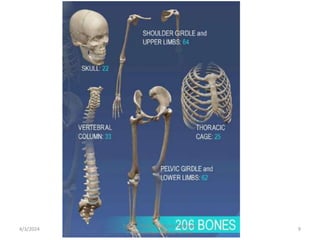
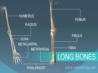
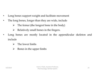
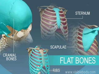
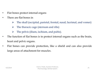
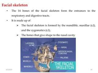
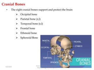
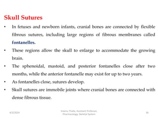
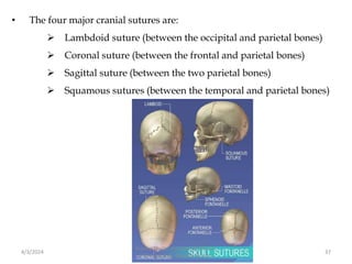
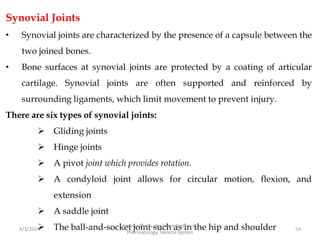
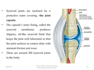
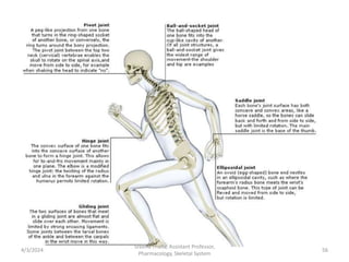
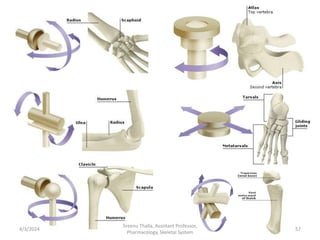


The document discusses the human skeletal system. It describes that the skeleton provides structure, protects organs, and facilitates movement. It is made up of bones, cartilage, and ligaments. The skeleton includes the axial skeleton which protects organs, and the appendicular skeleton which facilitates limb movement. Bones are categorized by shape into long, short, flat, irregular, and sesamoid types. Joints also vary in range of motion. The document outlines the components and functions of the skull, vertebrae, ribs, and other bones that make up the human skeletal system.


