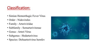
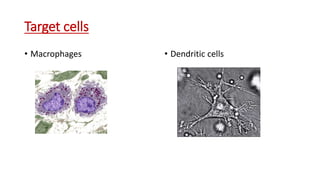
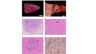
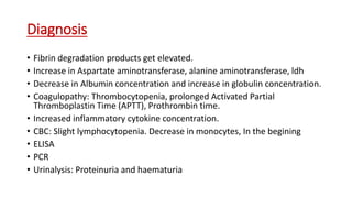
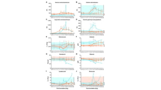
Simian Hemorrhagic Fever
Dr Zaid wani
• Viral hemorrhagic fever disease
characterized by the release of pro-
inflammatory cytokines from infected
macrophages (MΦs) and dendritic cells
(DCs) of liver and spleen that induces
tissue factor production and subsequent
disseminated intravascular coagulopathy.
Simian hemorrhagic fever virus (SHFV)
causes highly lethal disease in Asian
macaques resembling human illness
caused by Ebola or Lassa virus.
History
• 1964, two simultaneous outbreaks of lethal viral hemorrhagic fever
among captive Asian macaques at primate-holding facilities at the
Institute of Experimental Pathology and Therapy in Sukhumi, Soviet
Union; and at the National Institutes of Health, Primate Quarantine
Unit in Bethesda, MD.
Classification:
• Simian Hemorrhagic Fever Virus
• Order : Nidovirales
• Family : Arteriviridae
• Subfamily : Semarterivirenae
• Genus : Arteri Virus
• Subgenus : Hedarterivirus
• Species: Deltaarterivirus hemfev
Hosts
• Origin from African Monkeys
(Patas and Baboons) –
Subclinical
• Highly virulent in rhesus
monkeys, crab-eating macaques,
stump-tailed macaques (Macaca
arctoides), and Japanese
macaques
Morphology
• Spherical to pleomorphic
• 40 to 55 nm in diameter
• Enveloped, contains small
surface protrusions.
• Non-segmented, linear, single-
stranded RNA genome of
positive polarity.
• Highly contagious
• Mortality : 100%
Target cells
• Macrophages • Dendritic cells
Transmission
• Blood-to-blood transmission
• Use of the same needle for tattooing or tuberculosis testing
• During fighting
• Spread efficiently among macaques by both direct and indirect
contact
Cultivation and isolation
• In vitro culture on primary macaque macrophage cultures and on MA
– 104, African green monkey foetal kidney continuous cell line.
• Plaque assay of plasma on MA-104.
• Rt PCR of Peripherial blood Mononuclear cells (PBMC) detects
Intracellular viral RNA
Pathogenesis
• Understudied
• Host cell receptors not identified
• Entry into the cells by low pH
dependent Clathrin mediated
endocytosis.
• CD163 is a crucial SHFV cell
entry factor.
Clinical signs
• African NHPs; low viremia, persistent infection.
• Bacterial sepsis in 75% of cases.
• Highly fatal in Asian macaques; Fever, facial edema, anorexia,
dehydration, depression, retrobulbar hemorrhages and subcutaneous
hematomas. Coagulation defects indicated by skin petechiae
Death occurs 7 to 13 days after infection
3-7 days IP
Bleeding diathesis
Alternatively sudden death. No signs
Gross lesions
Necropsy:
• Oedema
• Splenomegaly : Pale, smooth capsule, rounded margins, white pulp
not visible grossly.
• Local extensive congestion
• Haemorrhage: Skin, subcutis, sclera, base of tongue, spleen, LN, liver,
kidney, lung, myocardium, pancreas, trachea, GIT.
• Disseminated intravascular coagulopathy (Hallmark)
Microscopic findings
• Extensive lymphoid necrosis and congestion in spleen, LNs, GALT.
• Sinuses distended by plasma and fibrin.
• Complete cortical thymic necrosis with sparing of medulla - unique to
SHFV
• Thrombi within glomeruli, hepatic sinusoids and lung.
• Intestinal epithelial cell necrosis.
• Fetal loss
• Lymphohistocytic meningioencephalitis
Diagnosis
• Fibrin degradation products get elevated.
• Increase in Aspartate aminotransferase, alanine aminotransferase, ldh
• Decrease in Albumin concentration and increase in globulin concentration.
• Coagulopathy: Thrombocytopenia, prolonged Activated Partial
Thromboplastin Time (APTT), Prothrombin time.
• Increased inflammatory cytokine concentration.
• CBC: Slight lymphocytopenia. Decrease in monocytes, In the begining
• ELISA
• PCR
• Urinalysis: Proteinuria and haematuria
Treatment and prevention
• No specific treatment
• Symptomatic
• Treatments with drugs that disrupt cellular pH
• Antibody targeting CD163
• Elimination of clinically ill and exposed animals
• Quarentine
• Disinfection
• Notifying the concerned authorities
• Monitoring human contacts
• Exposure to African primates to be prevented
Zoonotic importance
• No evidence of zoonosis
References
• Divergent Simian Arteriviruses Cause Simian Hemorrhagic Fever of Differing
Severities in Macaques (DOI: 10.1128/mBio.02009-15)
• Clinical Characterization of Host Response to Simian Hemorrhagic Fever Virus
Infection in Permissive and Refractory Hosts: A Model for Determining
Mechanisms of VHF Pathogenesis (https://doi.org/10.3390/v11010067)
• Simian Hemorrhagic Fever Virus Cell Entry Is Dependent on CD163 and Uses a
Clathrin-Mediated Endocytosis-Like Pathway (DOI: 10.1128/JVI.02697-14)
• Characterization of the Simian Hemorrhagic Fever Virus Cell Entry Pathway (DOI:
10.13140/2.1.3913.2487 )
• A simian hemorrhagic fever virus isolate from persistently infected baboons
efficiently induces hemorrhagic fever disease in Japanese macaques
(https://doi.org/10.1016/j.virol.2014.10.018)
THANKYOU

Simian haemorrhagic fever - SHF

  • 1.
  • 2.
    • Viral hemorrhagicfever disease characterized by the release of pro- inflammatory cytokines from infected macrophages (MΦs) and dendritic cells (DCs) of liver and spleen that induces tissue factor production and subsequent disseminated intravascular coagulopathy. Simian hemorrhagic fever virus (SHFV) causes highly lethal disease in Asian macaques resembling human illness caused by Ebola or Lassa virus.
  • 3.
    History • 1964, twosimultaneous outbreaks of lethal viral hemorrhagic fever among captive Asian macaques at primate-holding facilities at the Institute of Experimental Pathology and Therapy in Sukhumi, Soviet Union; and at the National Institutes of Health, Primate Quarantine Unit in Bethesda, MD.
  • 4.
    Classification: • Simian HemorrhagicFever Virus • Order : Nidovirales • Family : Arteriviridae • Subfamily : Semarterivirenae • Genus : Arteri Virus • Subgenus : Hedarterivirus • Species: Deltaarterivirus hemfev
  • 5.
    Hosts • Origin fromAfrican Monkeys (Patas and Baboons) – Subclinical • Highly virulent in rhesus monkeys, crab-eating macaques, stump-tailed macaques (Macaca arctoides), and Japanese macaques
  • 6.
    Morphology • Spherical topleomorphic • 40 to 55 nm in diameter • Enveloped, contains small surface protrusions. • Non-segmented, linear, single- stranded RNA genome of positive polarity. • Highly contagious • Mortality : 100%
  • 7.
    Target cells • Macrophages• Dendritic cells
  • 8.
    Transmission • Blood-to-blood transmission •Use of the same needle for tattooing or tuberculosis testing • During fighting • Spread efficiently among macaques by both direct and indirect contact
  • 9.
    Cultivation and isolation •In vitro culture on primary macaque macrophage cultures and on MA – 104, African green monkey foetal kidney continuous cell line. • Plaque assay of plasma on MA-104. • Rt PCR of Peripherial blood Mononuclear cells (PBMC) detects Intracellular viral RNA
  • 10.
    Pathogenesis • Understudied • Hostcell receptors not identified • Entry into the cells by low pH dependent Clathrin mediated endocytosis. • CD163 is a crucial SHFV cell entry factor.
  • 11.
    Clinical signs • AfricanNHPs; low viremia, persistent infection. • Bacterial sepsis in 75% of cases. • Highly fatal in Asian macaques; Fever, facial edema, anorexia, dehydration, depression, retrobulbar hemorrhages and subcutaneous hematomas. Coagulation defects indicated by skin petechiae Death occurs 7 to 13 days after infection 3-7 days IP Bleeding diathesis Alternatively sudden death. No signs
  • 12.
    Gross lesions Necropsy: • Oedema •Splenomegaly : Pale, smooth capsule, rounded margins, white pulp not visible grossly. • Local extensive congestion • Haemorrhage: Skin, subcutis, sclera, base of tongue, spleen, LN, liver, kidney, lung, myocardium, pancreas, trachea, GIT. • Disseminated intravascular coagulopathy (Hallmark)
  • 14.
    Microscopic findings • Extensivelymphoid necrosis and congestion in spleen, LNs, GALT. • Sinuses distended by plasma and fibrin. • Complete cortical thymic necrosis with sparing of medulla - unique to SHFV • Thrombi within glomeruli, hepatic sinusoids and lung. • Intestinal epithelial cell necrosis. • Fetal loss • Lymphohistocytic meningioencephalitis
  • 17.
    Diagnosis • Fibrin degradationproducts get elevated. • Increase in Aspartate aminotransferase, alanine aminotransferase, ldh • Decrease in Albumin concentration and increase in globulin concentration. • Coagulopathy: Thrombocytopenia, prolonged Activated Partial Thromboplastin Time (APTT), Prothrombin time. • Increased inflammatory cytokine concentration. • CBC: Slight lymphocytopenia. Decrease in monocytes, In the begining • ELISA • PCR • Urinalysis: Proteinuria and haematuria
  • 19.
    Treatment and prevention •No specific treatment • Symptomatic • Treatments with drugs that disrupt cellular pH • Antibody targeting CD163 • Elimination of clinically ill and exposed animals • Quarentine • Disinfection • Notifying the concerned authorities • Monitoring human contacts • Exposure to African primates to be prevented
  • 20.
    Zoonotic importance • Noevidence of zoonosis
  • 21.
    References • Divergent SimianArteriviruses Cause Simian Hemorrhagic Fever of Differing Severities in Macaques (DOI: 10.1128/mBio.02009-15) • Clinical Characterization of Host Response to Simian Hemorrhagic Fever Virus Infection in Permissive and Refractory Hosts: A Model for Determining Mechanisms of VHF Pathogenesis (https://doi.org/10.3390/v11010067) • Simian Hemorrhagic Fever Virus Cell Entry Is Dependent on CD163 and Uses a Clathrin-Mediated Endocytosis-Like Pathway (DOI: 10.1128/JVI.02697-14) • Characterization of the Simian Hemorrhagic Fever Virus Cell Entry Pathway (DOI: 10.13140/2.1.3913.2487 ) • A simian hemorrhagic fever virus isolate from persistently infected baboons efficiently induces hemorrhagic fever disease in Japanese macaques (https://doi.org/10.1016/j.virol.2014.10.018)
  • 22.