
Recommended
More Related Content
What's hot
What's hot (20)
Similar to The Pelvis.pptx
Similar to The Pelvis.pptx (20)
Recently uploaded
Recently uploaded (20)
The Pelvis.pptx
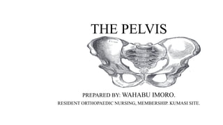
- 1. THE PELVIS PREPARED BY: WAHABU IMORO. RESIDENT ORTHOPAEDIC NURSING, MEMBERSHIP. KUMASI SITE.
- 2. OUTLINE OF PRESENTATION i. Describe the pelvis ii. Structure of the pelvis iii.Parts of the pelvis iv.Types of pelvis v. Functions of the pelvis vi.Common conditions affecting the pelvis vii.References
- 3. DESCRIPTION OF THE PELVIS • The pelvis is made up of two hipbones connected anteriorly at the pubic symphysis and posteriorly to the sacrum and the coccyx, each is made up of 3 bones-the blade shaped ILIUM-on each side- the ISCHIUM posterior and inferior on which the weight falls when sitting and the PUBIS anteriorly. @teachmeseries
- 4. STRUCTURE OF THE PELVIS • Anterior view of the pelvis Anterior view of the pelvic girdle. Adapted from [Van de Graaff, 2001, Ch. 7]
- 5. STRUCTURE OF THE PELVIS cont… • Posterior view
- 6. PARTS OF THE PELVIS AND THEIR FUNCTIONS • Base of the sacrum: It is a large oval articular surface, the upper surface of the body of the first sacral vertebra which is connected with the under surface of the last lumbar vertebra by an intervertebral fibrocartilage( intervertebral disc) • The pelvic surface of the sacrum: It is concave from top and curved slightly from side to the side. • Iliac crest: The top border of the ilium.
- 7. Cont… • Ilium: It is the widest and largest of the three parts of the hip bones • Sacroiliac articulation: it connects the hip bones to the sacrum and the coccyx. It serves as a shock absorber between the upper body and the legs. • Anterior sacral foramina: They are four in number and their size diminishes from top to bottom, they give exit to the anterior sacral nerves and entrance to the lateral sacral arteries. • Spine of ischium: it is a thin pointed triangular eminence at the posterior border of the ischium bone, it supports and resist intra- abdominal pressure that is exerted from above.
- 8. Cont… • Acetabulum: The cup-shaped socket that forms the hip joint with the head of the femur. • Obturator foramen: it the largest foramen in the body, it allows passage of the obturator artery, vein and nerve, it is situated between the ischium and the pubis. • Superior and inferior rami: they are what we refer to as the pubic bones. • Pubic tubercle: It serve as a point of attachment for the inguinal ligament, it is clinically used to locate the rings of the inguinal canal.
- 9. Cont…. • Symphysis pubis: It is a cartilaginous joint between the two pubic bones, it keeps the two pelvic bones together and steady during activities, it absorb shock and allow delivery of a baby. • Acetabular notch: it is an anterioinferior depression of the margin of the acetabulum, the margins serves as attachment of ligaments of the head of the femur. • Coccyx: triangular bony structure located at the end of the vertebral column, also known as tailbone, its made up of 3- 5 bones fused together.
- 10. Cont…… iliac spine: Serves as attachment for ligaments and muscle, it is an important landmark for measurement of the true length of the leg. Iliac fossa: it is a large, smooth, concave area on the internal surface of the ilium. Sacral canal: Is a continuation of the spinal canal and runs throughout the greater part of the sacrum. The canal lodges the sacral nerves via the anterior and posterior sacral foramina. Sacral hiatus: Located at the distal part of the sacrum, it's the area in which epidural medication are administered.
- 12. TYPES OF PELVIS • Gynecoid. This is the most common type of pelvis in females and is generally considered to be the typical female pelvis. Its overall shape is round, shallow, and open. • Android. This type of pelvis bears more resemblance to the male pelvis. It’s narrower than the gynecoid pelvis and is shaped more like a heart or a wedge. • Anthropoid. An anthropoid pelvis is narrow and deep. Its shape is similar to an upright egg or oval. • Platypelloid. The platypelloid pelvis is also called a flat pelvis. This is the least common type. It’s wide but shallow, and it resembles an egg or oval lying on its side.
- 13. FUNCTIONS OF THE PELVIS • For locomotion: The body weight is transmitted through the pelvis to the lower limbs. • Childbirth: The birth canal lies within the pelvic girdle, it serves as a passage for the neonate. • Support: It supports the organs of the pelvic cavity.
- 14. BLOOD PASSAGE
- 15. BLOOD SUPPLY
- 16. Cont…
- 17. LIGAMENTS OF THE PELVIS 1. ILIOLUMBAR LIGAMENT 2.SACROILIAC LIGAMENT 3.SACROSPNOUS LIGAMENT 4.SACROTUBEROUS LIGAMENT 5.INGUINAL LIGAMENT
- 18. Iliolumbar Ligament • It spans between the tip of the 4 and 5th transverse process of the spine to the waist bone (iliac crest). It functions to restrain movement in the lumbosacral and sacroiliac joints. In doing so, the iliolumbar ligament is a major stabilizer of the low back and sacroiliac joint
- 20. Sacroiliac (SI) Joint Ligaments: • These are the massive ligaments at the back of the sacroiliac joints (the joints between the tailbone and back of the pelvis/hip). Women have more issues here than men, and these can be stretched during childbirth, falls onto the butt, and trauma. The pain is often in this area at the back of the hip and can refer down the leg.
- 22. Sacrospinous Ligament • It is a thin triangular ligament that stretches between the sacrum and boney outcropping on the ischium. It functions to prevent posterior rotation of the ilium. Stress to this ligament occurs most often when leaning forward or getting out of a chair
- 24. Sacrotuberous Ligament • It stretches between the sacrum and the Sitz bone (ischial spine). It functions to prevent rotation of the ilium. The sacrotuberous ligament also contributes to the strength of the pelvis, prevents rotation of the ilium, and provides an attachment point for buttock and thigh muscles.
- 27. CONT.. • The inguinal ligament (also ligamentum inguinale, arcus inguinalis or Pouparts’s ligament) is a band of connective tissue that extends from the anterior superior iliac spine of the ilium to the pubic tubercle on the pubic bone
- 30. Cont..
- 31. NERVE PASSAGE
- 32. NERVE SUPPLY
- 33. Cont..
- 34. Cont..
- 35. Cont..
- 36. COMMON DISORDERS OF THE PELVIS • Osteitis pubis: Inflammation of the pubis symphysis, characterized by pain in the groin and tenderness over the front of the pelvis. Common with footballers due to repeated kicking. • Fractures: Common with motorcycle users, acetabulum and sacroiliac dislocation, open book and vertical shears, • Contracted pelvis: due to developmental and severe nutritional deficiencies. e.g. Naegeles pelvis, Roberts pelvis, Osteomalacic pelvis etc. • Osteoarthritis of the hip
- 37. Osteitis pubis
- 39. Classification of Acetabular fractures
- 40. Vertical Shears
- 42. Cont..
- 43. Cont.…
- 45. Cont.. • Osteoarthritis of the hip joint
- 46. Cont..
- 47. References • Clinical Application of Neuromuscular Techniques, Volume 2(second edition)2011. • https/www.physio-pedia.com • https://www.britanica.com>science • https://radiopedia.org>articles • https://teachmeanatomy.info
- 48. END