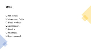
SEPSIS AND SEPTIC SHOCK
To: Dr.Dejene
By: Biniam Mekonen
Outline
DEFINITIONS
ETIOLOGY
EPIDEMIOLOGY
RISK FACTORS
PATHOPHYSIOLOGY
DIAGNOSIS
TREATMENT
Definition
❑Sepsis is a life threating condition that arise when body response to infection cause
injury to its own tissue and organ
❑SIRS may have an infectious or a noninfectious etiology. If infection is suspected or
proven, a patient with SIRS is said to have sepsis.
❑When sepsis is associated with dysfunction of organs distant from the site of infection,
the patient has severe sepsis. Severe sepsis may be accompanied by hypotension or
evidence of hypoperfusion.
❑When hypotension cannot be corrected by infusing fluids, the diagnosis is septic
shock.
❑ As sepsis progresses to septic shock, the risk of dying increases substantially. Sepsis is
usually reversible, whereas patients with septic shock often succumb despite aggressive
therapy.
Etiology
❑Sepsis and Septic shock has a mortality rate of over 50% ranking the first among the
causes of death in intensive care units.
❑It results from the spread & expansion of an initially localized infection like
pneumonia into the blood stream.
❑Most causes of septic shock (~70%) are caused by endotoxin-producing gram-
negative bacilli, hence the term endotoxic shock.
❑Endotoxins are bacterial wall lipopolyschardes(LPS) released when cell walls are
degraded.
Cont.
❑Analogues molecules in the walls of grampositive bacteria & fungi can also elicit
septic shock. LPS bind with CD14 molecule on leucocytes, especially monocytes &
macrophages, endothelial cells & others. Depending on
❑the dosage of LPS – protein complex, initiation of a cascade of cytokine-mediated
events
❑Several medical conditions increase a person's susceptibility to infection and
developing sepsis. Common sepsis risk factors include age (especially the very young
and old); conditions that weaken the immune system such as cancer, diabetes, or the
absence of a spleen; and major trauma and burns
Cont.
Epidemology
• Sepsis and septic shock causes millions of deaths globally each year and is the most
common cause of death in people who have been hospitalized.The number of new
cases worldwide of sepsis is estimated to be 18 million cases per year.
• In the United States sepsis affects approximately 3 in 1,000 people and severe
sepsis contributes to more than 200,000 deaths per year.
• Due to it rarely being reported as a primary diagnosis (often being a complication of
cancer or other illness), the incidence, mortality, and morbidity rates of sepsis are
likely underestimated.
Pathophysiology
sepsis and septic shock
Cont.
❑Initially, arteries and arterioles dilate, decreasing peripheral arterial resistance;
cardiac output typically increases.
❑This stage has been referred to as warm shock. Later, cardiac output may decrease,
blood pressure falls (with or without an increase in peripheral resistance), and typical
features of shock appear.
❑Even in the stage of increased cardiac output, vasoactive mediators cause blood flow
to bypass capillary exchange vessels (a distributive defect).
❑Poor capillary flow resulting from this shunting, along with capillary obstruction by
microthrombi, decreases delivery of oxygen and impairs removal of carbon dioxide
and waste products.
❑Decreased perfusion causes dysfunction and sometimes failure of one or more
organs, including the kidneys, lungs, liver, brain, and heart.
CLINICAL
MANIFESTATIONS
Cont.
The rate at which signs and symptoms develop may differ from patient to patient, and
there are striking individual variations in presentation.
For example, some patients with sepsis are normo- or hypothermic; the absence of
fever is most common in neonates, in elderly patients, and in persons with uremia or
alcoholism.
Hyperventilation is often an early sign. Disorientation, confusion, and other
manifestations of encephalopathy may also develop early in the septic response,
particularly in the elderly and in individuals with pre-existing
neurologic impairment.
Focal neurologic signs are uncommon, although pre-existing focal deficits may
become more prominent
Gastrointestinal manifestations such as nausea, vomiting, diarrhea, and ileus may
suggest acute gastroenteritis. Stress ulceration can lead to upper gastrointestinal
bleeding. Cholestatic jaundice, with elevated levels of serum bilirubin (mostly
conjugated) and alkaline phosphatase, may precede other signs of sepsis.
Hepatocellular or canalicular dysfunction appears to underlie most cases, and the
results of hepatic function tests return to normal with resolution of the infection.
Prolonged or severe hypotension may induce acute hepatic injury or ischemic bowel
necrosis.
Cont..
❑Many tissues may be unable to extract oxygen normally from the blood, so that
anaerobic metabolism occurs despite near-normal mixed venous oxygen saturation.
Blood lactate levels rise early, in part because of increased glycolysis with impaired
clearance of the resulting lactate and pyruvate by the liver and kidneys.
❑As hypoperfusion develops, tissue hypoxia generates more lactic acid, contributing to
metabolic acidosis.
Cont.
❑The blood glucose concentration often increases, particularly in patients with
diabetes, although impaired gluconeogenesis and excessive insulin release on
occasion produce hypoglycemia.
❑The cytokine-driven acute-phase response inhibits the synthesis of transthyretin
while enhancing the production of C-reactive protein, fibrinogen, and complement
components. Protein catabolism is often markedly accelerated.
Complication
Cardiopulmonary Complications
ARDS develops in 50% of patients with severe sepsis or septic shock. The failure of the
respiratory muscles can exacerbate hypoxemia and hypercapnia.
An elevated pulmonary capillary wedge pressure (18 mmHg) suggests fluid volume
overload or cardiac failure rather than ARDS.
Pneumonia caused by viruses or by Pneumocystis may be clinically indistinguishable
from ARDS.
Cont.
❑Sepsis-induced hypotension (see “Septic Shock,” above) usually results from a
generalized maldistribution of blood flow and blood volume and from hypovolemia
that is due, at least in part, to diffuse capillary leakage of intravascular fluid.
❑Other factors that may decrease effective intravascular volume include dehydration
from antecedent disease or insensible fluid losses, vomiting or diarrhea, and polyuria.
❑During early septic shock, systemic vascular resistance is usually elevated and
cardiac output may be low. After fluid repletion, in contrast, cardiac output typically
increases and systemic vascular resistance falls.
❑Indeed, normal or increased cardiac output and decreased systemic vascular
resistance distinguish septic shock from cardiogenic, extracardiac obstructive, and
hypovolemic shock; other processes that can produce this combination include
anaphylaxis, beriberi, cirrhosis, and overdoses of nitroprusside or narcotics.
Cont.
❑Depression of myocardial function, manifested as increased end diastolic and systolic
ventricular volumes with a decreased ejection fraction, develops within 24 h in most
patients with severe sepsis.
❑Cardiac output is maintained despite the low ejection fraction because ventricular
dilatation permits a normal stroke volume. In survivors, myocardial function returns
to normal over several days.
❑Although myocardial dysfunction may contribute to hypotension, refractory
hypotension is usually due to a low systemic vascular resistance, and death results
from refractory shock or the failure of multiple organs rather than from cardiac
dysfunction per s
Renal Complications
Oliguria, azotemia, proteinuria, and nonspecific urinary casts are frequently found.
Many patients are inappropriately polyuric; hyperglycemia may exacerbate this
tendency.
Most renal failure is due to acute tubular necrosis induced by hypotension or
capillary injury, although some patients also have glomerulonephritis, renal cortical
necrosis, or interstitial nephritis.
Drug-induced renal damage may complicate therapy, particularly when hypotensive
patients are given aminoglycoside antibiotics
Coagulation
Although thrombocytopenia occurs in 10 to 30% of patients, the underlying
mechanisms are not understood.
Platelet counts are usually very low (50,000/L) in patients with DIC; these low
counts may reflect diffuse endothelial injury or microvascular thrombosis.
Neurologic Complications
❑When the septic illness lasts for weeks to months, “critical-illness” polyneuropathy
may prevent weaning from ventilatory support and produce distal motor weakness.
Electrophysiologic studies are diagnostic. Guillain-Barre´ syndrome, metabolic
disturbances, and toxin activity must be ruled out.
Diagnosis
cont.
❑Early diagnosis is necessary to properly manage sepsis, as the initiation of rapid
therapy is key to reducing deaths from severe sepsis.
❑Some hospitals use alerts generated from electronic health records to bring attention
to potential cases as early as possible.
❑Within the first three hours of suspected sepsis, diagnostic studies should include
white blood cell counts, measuring serum lactate, and obtaining appropriate
cultures before starting antibiotics, so long as this does not delay their use by more
than 45 minutes.
cont.
❑To identify the causative organism(s), at least two sets of blood cultures using bottles
with media for aerobic and anaerobic organisms are necessary.
❑At least one should be drawn through the skin and one through each vascular access
device (such as an IV catheter) that has been in place more than 48 hours. Bacteria
are present in the blood in only about 30% of cases.
❑Another possible method of detection is by polymerase chain reaction. If other
sources of infection are suspected, cultures of these sources, such as urine,
cerebrospinal fluid, wounds, or respiratory secretions, also should be obtained, as
long as this does not delay the use of antibiotics.
cont.
❑Within six hours, if blood pressure remains low despite initial fluid resuscitation of
30 mL/kg, or if initial lactate is ≥ four mmol/L (36 mg/dL), central venous pressure
and central venous oxygen saturation should be measured.
❑Lactate should be re-measured if the initial lactate was elevated. Evidence for point
of care lactate measurement over usual methods of measurement, however, is poor
cont.
❑Within twelve hours, it is essential to diagnose or exclude any source of infection that
would require emergent source control, such as a necrotizing soft tissue infection, an
infection causing inflammation of the abdominal cavity lining, an infection of the bile
duct, or an intestinal infarction.
❑A pierced internal organ (free air on an abdominal X-ray or CT scan), an abnormal
chest X-ray consistent with pneumonia (with focal opacification), or petechiae,
purpura, or purpura fulminans may indicate the presence of an infection.
Treatment
Cont..
❑Early recognition and focused management may improve the outcomes in sepsis.
Current professional recommendations include a number of actions ("bundles") to be
followed as soon as possible after diagnosis.
❑Within the first three hours, someone with sepsis should have received antibiotics
and, intravenous fluids if there is evidence of either low blood pressure or other
evidence for inadequate blood supply to organs (as evidenced by a raised level of
lactate); blood cultures also should be obtained within this time period.
❑After six hours the blood pressure should be adequate, close monitoring of blood
pressure and blood supply to organs should be in place, and the lactate should be
measured again if initially it was raised
Cont.
❑A related bundle, the "Sepsis Six", is in widespread use in the United Kingdom; this
requires the administration Antibiotics within an hour of recognition, Blood cultures,
Lactate, andhemoglobin determination, Urine output monitoring, High-flow oxygen,
and Intravenous fluids.
Cont.
❑Apart from the timely administration of fluids and antibiotics, the management of
sepsis also involves surgical drainage of infected fluid collections and appropriate
support for organ dysfunction.
❑This may include hemodialysis in kidney failure, mechanical ventilation in lung
dysfunction, transfusion of blood products, and drug and fluid therapy for circulatory
failure.
❑Ensuring adequate nutrition—preferably by enteral feeding, but if necessary, by
parenteral nutrition—is important during prolonged illness.Medication to prevent
deep vein thrombosis and gastric ulcers also may be use
Cont.
❑A related bundle, the "Sepsis Six", is in widespread use in the United Kingdom; this
requires the administration Antibiotics within an hour of recognition, Blood cultures,
Lactate, andhemoglobin determination, Urine output monitoring, High-flow oxygen,
and Intravenous fluids.
cont
❑Apart from the timely administration of fluids and antibiotics, the management of
sepsis also involves surgical drainage of infected fluid collections and appropriate
support for organ dysfunction.
❑This may include hemodialysis in kidney failure, mechanical ventilation in lung
dysfunction, transfusion of blood products, and drug and fluid therapy for circulatory
failure.
❑Ensuring adequate nutrition—preferably by enteral feeding, but if necessary, by
parenteral nutrition—is important during prolonged illness.
❑ Medication to prevent deep vein thrombosis and gastric ulcers also may be used.
new born
❑Neonatal sepsis can be difficult to diagnose as newborns may be asymptomatic.
❑If a newborn shows signs and symptoms suggestive of sepsis, antibiotics are
immediately started and are either changed to target a specific organism identified by
diagnostic testing or discontinued after an infectious cause for the symptoms has been
ruled out.
❑Despite early intervention, death occurs in 13% of children who develop septic
shock, with the risk partly based on other health problems.
❑Those without multiple organ system failures or who require only one inotropic agent
mortality is low.
cont
❑Antibiotics
❑Intravenous fluids
❑Blood products
❑Vasopressors
❑Steroids
❑Anesthesia
❑Source control
THANK YOU

sepsis and septic shock

  • 1.
    SEPSIS AND SEPTICSHOCK To: Dr.Dejene By: Biniam Mekonen
  • 2.
  • 3.
    Definition ❑Sepsis is alife threating condition that arise when body response to infection cause injury to its own tissue and organ ❑SIRS may have an infectious or a noninfectious etiology. If infection is suspected or proven, a patient with SIRS is said to have sepsis. ❑When sepsis is associated with dysfunction of organs distant from the site of infection, the patient has severe sepsis. Severe sepsis may be accompanied by hypotension or evidence of hypoperfusion. ❑When hypotension cannot be corrected by infusing fluids, the diagnosis is septic shock. ❑ As sepsis progresses to septic shock, the risk of dying increases substantially. Sepsis is usually reversible, whereas patients with septic shock often succumb despite aggressive therapy.
  • 5.
    Etiology ❑Sepsis and Septicshock has a mortality rate of over 50% ranking the first among the causes of death in intensive care units. ❑It results from the spread & expansion of an initially localized infection like pneumonia into the blood stream. ❑Most causes of septic shock (~70%) are caused by endotoxin-producing gram- negative bacilli, hence the term endotoxic shock. ❑Endotoxins are bacterial wall lipopolyschardes(LPS) released when cell walls are degraded.
  • 6.
    Cont. ❑Analogues molecules inthe walls of grampositive bacteria & fungi can also elicit septic shock. LPS bind with CD14 molecule on leucocytes, especially monocytes & macrophages, endothelial cells & others. Depending on ❑the dosage of LPS – protein complex, initiation of a cascade of cytokine-mediated events ❑Several medical conditions increase a person's susceptibility to infection and developing sepsis. Common sepsis risk factors include age (especially the very young and old); conditions that weaken the immune system such as cancer, diabetes, or the absence of a spleen; and major trauma and burns
  • 7.
  • 8.
    Epidemology • Sepsis andseptic shock causes millions of deaths globally each year and is the most common cause of death in people who have been hospitalized.The number of new cases worldwide of sepsis is estimated to be 18 million cases per year. • In the United States sepsis affects approximately 3 in 1,000 people and severe sepsis contributes to more than 200,000 deaths per year. • Due to it rarely being reported as a primary diagnosis (often being a complication of cancer or other illness), the incidence, mortality, and morbidity rates of sepsis are likely underestimated.
  • 9.
  • 11.
    Cont. ❑Initially, arteries andarterioles dilate, decreasing peripheral arterial resistance; cardiac output typically increases. ❑This stage has been referred to as warm shock. Later, cardiac output may decrease, blood pressure falls (with or without an increase in peripheral resistance), and typical features of shock appear. ❑Even in the stage of increased cardiac output, vasoactive mediators cause blood flow to bypass capillary exchange vessels (a distributive defect). ❑Poor capillary flow resulting from this shunting, along with capillary obstruction by microthrombi, decreases delivery of oxygen and impairs removal of carbon dioxide and waste products. ❑Decreased perfusion causes dysfunction and sometimes failure of one or more organs, including the kidneys, lungs, liver, brain, and heart.
  • 12.
  • 13.
    Cont. The rate atwhich signs and symptoms develop may differ from patient to patient, and there are striking individual variations in presentation. For example, some patients with sepsis are normo- or hypothermic; the absence of fever is most common in neonates, in elderly patients, and in persons with uremia or alcoholism. Hyperventilation is often an early sign. Disorientation, confusion, and other manifestations of encephalopathy may also develop early in the septic response, particularly in the elderly and in individuals with pre-existing neurologic impairment.
  • 14.
    Focal neurologic signsare uncommon, although pre-existing focal deficits may become more prominent Gastrointestinal manifestations such as nausea, vomiting, diarrhea, and ileus may suggest acute gastroenteritis. Stress ulceration can lead to upper gastrointestinal bleeding. Cholestatic jaundice, with elevated levels of serum bilirubin (mostly conjugated) and alkaline phosphatase, may precede other signs of sepsis. Hepatocellular or canalicular dysfunction appears to underlie most cases, and the results of hepatic function tests return to normal with resolution of the infection. Prolonged or severe hypotension may induce acute hepatic injury or ischemic bowel necrosis.
  • 15.
    Cont.. ❑Many tissues maybe unable to extract oxygen normally from the blood, so that anaerobic metabolism occurs despite near-normal mixed venous oxygen saturation. Blood lactate levels rise early, in part because of increased glycolysis with impaired clearance of the resulting lactate and pyruvate by the liver and kidneys. ❑As hypoperfusion develops, tissue hypoxia generates more lactic acid, contributing to metabolic acidosis.
  • 16.
    Cont. ❑The blood glucoseconcentration often increases, particularly in patients with diabetes, although impaired gluconeogenesis and excessive insulin release on occasion produce hypoglycemia. ❑The cytokine-driven acute-phase response inhibits the synthesis of transthyretin while enhancing the production of C-reactive protein, fibrinogen, and complement components. Protein catabolism is often markedly accelerated.
  • 17.
  • 19.
    Cardiopulmonary Complications ARDS developsin 50% of patients with severe sepsis or septic shock. The failure of the respiratory muscles can exacerbate hypoxemia and hypercapnia. An elevated pulmonary capillary wedge pressure (18 mmHg) suggests fluid volume overload or cardiac failure rather than ARDS. Pneumonia caused by viruses or by Pneumocystis may be clinically indistinguishable from ARDS.
  • 20.
    Cont. ❑Sepsis-induced hypotension (see“Septic Shock,” above) usually results from a generalized maldistribution of blood flow and blood volume and from hypovolemia that is due, at least in part, to diffuse capillary leakage of intravascular fluid. ❑Other factors that may decrease effective intravascular volume include dehydration from antecedent disease or insensible fluid losses, vomiting or diarrhea, and polyuria. ❑During early septic shock, systemic vascular resistance is usually elevated and cardiac output may be low. After fluid repletion, in contrast, cardiac output typically increases and systemic vascular resistance falls. ❑Indeed, normal or increased cardiac output and decreased systemic vascular resistance distinguish septic shock from cardiogenic, extracardiac obstructive, and hypovolemic shock; other processes that can produce this combination include anaphylaxis, beriberi, cirrhosis, and overdoses of nitroprusside or narcotics.
  • 21.
    Cont. ❑Depression of myocardialfunction, manifested as increased end diastolic and systolic ventricular volumes with a decreased ejection fraction, develops within 24 h in most patients with severe sepsis. ❑Cardiac output is maintained despite the low ejection fraction because ventricular dilatation permits a normal stroke volume. In survivors, myocardial function returns to normal over several days. ❑Although myocardial dysfunction may contribute to hypotension, refractory hypotension is usually due to a low systemic vascular resistance, and death results from refractory shock or the failure of multiple organs rather than from cardiac dysfunction per s
  • 22.
    Renal Complications Oliguria, azotemia,proteinuria, and nonspecific urinary casts are frequently found. Many patients are inappropriately polyuric; hyperglycemia may exacerbate this tendency. Most renal failure is due to acute tubular necrosis induced by hypotension or capillary injury, although some patients also have glomerulonephritis, renal cortical necrosis, or interstitial nephritis. Drug-induced renal damage may complicate therapy, particularly when hypotensive patients are given aminoglycoside antibiotics
  • 23.
    Coagulation Although thrombocytopenia occursin 10 to 30% of patients, the underlying mechanisms are not understood. Platelet counts are usually very low (50,000/L) in patients with DIC; these low counts may reflect diffuse endothelial injury or microvascular thrombosis.
  • 24.
    Neurologic Complications ❑When theseptic illness lasts for weeks to months, “critical-illness” polyneuropathy may prevent weaning from ventilatory support and produce distal motor weakness. Electrophysiologic studies are diagnostic. Guillain-Barre´ syndrome, metabolic disturbances, and toxin activity must be ruled out.
  • 25.
  • 26.
    cont. ❑Early diagnosis isnecessary to properly manage sepsis, as the initiation of rapid therapy is key to reducing deaths from severe sepsis. ❑Some hospitals use alerts generated from electronic health records to bring attention to potential cases as early as possible. ❑Within the first three hours of suspected sepsis, diagnostic studies should include white blood cell counts, measuring serum lactate, and obtaining appropriate cultures before starting antibiotics, so long as this does not delay their use by more than 45 minutes.
  • 27.
    cont. ❑To identify thecausative organism(s), at least two sets of blood cultures using bottles with media for aerobic and anaerobic organisms are necessary. ❑At least one should be drawn through the skin and one through each vascular access device (such as an IV catheter) that has been in place more than 48 hours. Bacteria are present in the blood in only about 30% of cases. ❑Another possible method of detection is by polymerase chain reaction. If other sources of infection are suspected, cultures of these sources, such as urine, cerebrospinal fluid, wounds, or respiratory secretions, also should be obtained, as long as this does not delay the use of antibiotics.
  • 28.
    cont. ❑Within six hours,if blood pressure remains low despite initial fluid resuscitation of 30 mL/kg, or if initial lactate is ≥ four mmol/L (36 mg/dL), central venous pressure and central venous oxygen saturation should be measured. ❑Lactate should be re-measured if the initial lactate was elevated. Evidence for point of care lactate measurement over usual methods of measurement, however, is poor
  • 29.
    cont. ❑Within twelve hours,it is essential to diagnose or exclude any source of infection that would require emergent source control, such as a necrotizing soft tissue infection, an infection causing inflammation of the abdominal cavity lining, an infection of the bile duct, or an intestinal infarction. ❑A pierced internal organ (free air on an abdominal X-ray or CT scan), an abnormal chest X-ray consistent with pneumonia (with focal opacification), or petechiae, purpura, or purpura fulminans may indicate the presence of an infection.
  • 30.
  • 31.
    Cont.. ❑Early recognition andfocused management may improve the outcomes in sepsis. Current professional recommendations include a number of actions ("bundles") to be followed as soon as possible after diagnosis. ❑Within the first three hours, someone with sepsis should have received antibiotics and, intravenous fluids if there is evidence of either low blood pressure or other evidence for inadequate blood supply to organs (as evidenced by a raised level of lactate); blood cultures also should be obtained within this time period. ❑After six hours the blood pressure should be adequate, close monitoring of blood pressure and blood supply to organs should be in place, and the lactate should be measured again if initially it was raised
  • 32.
    Cont. ❑A related bundle,the "Sepsis Six", is in widespread use in the United Kingdom; this requires the administration Antibiotics within an hour of recognition, Blood cultures, Lactate, andhemoglobin determination, Urine output monitoring, High-flow oxygen, and Intravenous fluids.
  • 33.
    Cont. ❑Apart from thetimely administration of fluids and antibiotics, the management of sepsis also involves surgical drainage of infected fluid collections and appropriate support for organ dysfunction. ❑This may include hemodialysis in kidney failure, mechanical ventilation in lung dysfunction, transfusion of blood products, and drug and fluid therapy for circulatory failure. ❑Ensuring adequate nutrition—preferably by enteral feeding, but if necessary, by parenteral nutrition—is important during prolonged illness.Medication to prevent deep vein thrombosis and gastric ulcers also may be use
  • 34.
    Cont. ❑A related bundle,the "Sepsis Six", is in widespread use in the United Kingdom; this requires the administration Antibiotics within an hour of recognition, Blood cultures, Lactate, andhemoglobin determination, Urine output monitoring, High-flow oxygen, and Intravenous fluids.
  • 35.
    cont ❑Apart from thetimely administration of fluids and antibiotics, the management of sepsis also involves surgical drainage of infected fluid collections and appropriate support for organ dysfunction. ❑This may include hemodialysis in kidney failure, mechanical ventilation in lung dysfunction, transfusion of blood products, and drug and fluid therapy for circulatory failure. ❑Ensuring adequate nutrition—preferably by enteral feeding, but if necessary, by parenteral nutrition—is important during prolonged illness. ❑ Medication to prevent deep vein thrombosis and gastric ulcers also may be used.
  • 36.
    new born ❑Neonatal sepsiscan be difficult to diagnose as newborns may be asymptomatic. ❑If a newborn shows signs and symptoms suggestive of sepsis, antibiotics are immediately started and are either changed to target a specific organism identified by diagnostic testing or discontinued after an infectious cause for the symptoms has been ruled out. ❑Despite early intervention, death occurs in 13% of children who develop septic shock, with the risk partly based on other health problems. ❑Those without multiple organ system failures or who require only one inotropic agent mortality is low.
  • 37.
  • 38.