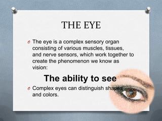
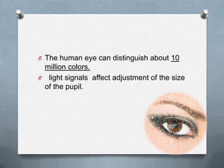
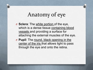
PRINCIPLE SENSORY
     SYSTEM
       GROUP 1
   AYESHA MEHBOOB
     SUNDAS GILL
    MARYAM YAQUB
      FIZZA RAJA
    SUMERA MALIK
What is a Sensory System?
O Window to physical energies. Give rise to
  sensory perceptions.

O General plan of sensory pathways:
  receptor, thalamus (diencephalon), cortex
  (telencephalon).
Different sensory systems
O Vision
O Audition
O Somatosensory
O Taste
O Smell
Why do we have different SYSTEMS
when the neural activity is the same : action
                potential


O Theory of specific sensory energies by
  Johannes Muller, 1826. Labeled
  pathways.
Vision
Introduction and structure of an eye
                by
         Ayesha Mehboob
      Function of human eye
                By
         Sundas Saba Gill
THE EYE
O The eye is a complex sensory organ
 consisting of various muscles, tissues,
 and nerve sensors, which work together to
 create the phenomenon we know as
 vision:

      The ability to see
O Complex eyes can distinguish shapes
 and colors.
TYPES OF EYE
Eye types can be categorized into
1. Simple eyes: with one concave
   photoreceptive surface, and
2. Compound eyes: which comprise a
   number of individual lenses laid out on a
   convex surface
O  Any eye type can be adapted for almost
  any behavior or environment.
O The only limitations specific to eye types
  are that of resolution e.g.
 The compound eyes achieving a resolution
better than 1°. Also, superposition eyes can
achieve greater sensitivity and apposition
eyes are better suited to dark-dwelling
creature
HUMAN EYES

O Human eye is an organ which reacts to
  light for several purposes.
O As a conscious sense organ,
  the eye allows vision.
O Rod and cone cells in the retina allow
  conscious light perception and vision
  including color differentiation and the
  perception of depth.
O The human eye can distinguish about 10
 million colors.
O light signals affect adjustment of the size
 of the pupil.
STRUCTURE OF AN EYE
OUTER STRUCTURE
INNER STRUCTURE
Anatomy of eye
O Sclera: The white portion of the eye,
  which is a dense tissue containing blood
  vessels and providing a surface for
  attaching the external muscles of the eye.
O Pupil: The round, black opening in the
  center of the iris that allows light to pass
  through the eye and onto the retina.
O Iris: The colored part of the outer eye.
  This thin muscle constricts or dilates to
  adjust the diameter of the pupil, thus
  controlling the amount of light that enters
  the eye.
O Cornea: The transparent tissue that
  covers the front of the eye including the
  pupil and iris.
O responsible for most of the eye’s focusing
  abilities.
Aqueous humor : A liquid substance found
in the eye chambers, made mostly of water.
O delivers vital nutrients to the eyes
O maintain correct pressure balance in the
  eye chamber
O Help in maintain its shape
O Ciliary muscle: is a muscle in an area of
  the eye which helps people focus.
O It assist the lens of the eye to be flattened
  or rounded to allow people to focus on
  distant and near objects and maintain the
  proper fluid pressure in the eye.
O Lens: After light enters through the pupil,
  it passes through the lens, which focuses
  the light and projects it onto the surface of
  the retina in the back of the eye.
O vitreous humor: helps keep the retina in
  place.
O transparent jelly-like mass located behind
  the lens.
O helps to maintain the shape of the
  posterior chamber of the eyeball.
O Retina: A light sensitive tissue that lines
  the inner surface of the back of the eye.
O It transforms light that enters through the
  pupil and passes through the lens into
  nerve signals that are converted into an
  image by the visual cortex of the brain
VISUAL FIELD
O All of these components function
  simultaneously to allow us to see our
  surroundings.
O The resulting picture, called the visual
  field:
Combination of two primary types of vision
with distinct functions and characteristics
TYPES OF VISIONS
1. FOVEAL VISION (smallest portion of our
    visual field)
O Objects within the scope of foveal vision
  are clear and colorful.
2. PERIPHERAL VISION(The majority of
the scene we see)
O to detect movements and color and shape
  contrasts.
TYPES OF LIGHT RECPTORS
O These two different types of vision are a result
  of the two kinds of light receptor cells found in
  the retina.
1. Cones: Foveal vision is created by cone
    cells.
O that are tightly packed in a small area in the
  center of the retina.
O only account for 6% of the total retinal light
  receptors.
O Cone cells require the most light for creating a
  clear, detailed image.
2. Rods: The rod cells account for the other
94% of light receptors in the retina.
O They require less light.
O create the blurry, less colorful qualities of
  peripheral vision.
BINOCULAR &
   MONOCULAR VISION
O The visual fields of many organisms like
  human , involve large areas of binocular
  vision to improve depth perception.(3
  dimension)
O vision using two eyes,
O In other organisms, eyes are located so
  as to maximize the field of view, such as
  in rabbits and horses, which
  have monocular vision.
SUNDAS SABA GILL
ANATOMICAL PROCESS OF
       VISION
1. The eye is a complex optical system
   which collects light from the surrounding
   environment,
2. regulates its intensity through
   a diaphragm,
3. focuses it through an adjustable
   assembly of lenses to form an image,
4. converts this image into a set of
   electrical signals
5. and transmits these signals to
   the brain through complex neural
   pathways that connect the eye via
   the optic nerve to the visual cortex and
   other areas of the brain.
In simple words:
O clear image of the visual world produce on
  a sheet of photoreceptors called the
  retina, which is part of the central nervous
  system but located at the back of the eye.
O Photoreceptors gather visual information
  and sending electrical signals to other
  retinal neurons for initial processing
O The signals are then sent via the optic
  nerve to other parts of brain, which
  ultimately processes the image and allows
  us to see.
O The image on the retina is reversed:
Objects to the right of center project images
to the left part of the retina and vice versa.

Vision

  • 1.
    PRINCIPLE SENSORY SYSTEM GROUP 1 AYESHA MEHBOOB SUNDAS GILL MARYAM YAQUB FIZZA RAJA SUMERA MALIK
  • 2.
    What is aSensory System? O Window to physical energies. Give rise to sensory perceptions. O General plan of sensory pathways: receptor, thalamus (diencephalon), cortex (telencephalon).
  • 3.
    Different sensory systems OVision O Audition O Somatosensory O Taste O Smell
  • 4.
    Why do wehave different SYSTEMS when the neural activity is the same : action potential O Theory of specific sensory energies by Johannes Muller, 1826. Labeled pathways.
  • 5.
    Vision Introduction and structureof an eye by Ayesha Mehboob Function of human eye By Sundas Saba Gill
  • 7.
    THE EYE O Theeye is a complex sensory organ consisting of various muscles, tissues, and nerve sensors, which work together to create the phenomenon we know as vision: The ability to see O Complex eyes can distinguish shapes and colors.
  • 8.
    TYPES OF EYE Eyetypes can be categorized into 1. Simple eyes: with one concave photoreceptive surface, and 2. Compound eyes: which comprise a number of individual lenses laid out on a convex surface
  • 9.
    O Anyeye type can be adapted for almost any behavior or environment. O The only limitations specific to eye types are that of resolution e.g. The compound eyes achieving a resolution better than 1°. Also, superposition eyes can achieve greater sensitivity and apposition eyes are better suited to dark-dwelling creature
  • 10.
    HUMAN EYES O Humaneye is an organ which reacts to light for several purposes. O As a conscious sense organ, the eye allows vision. O Rod and cone cells in the retina allow conscious light perception and vision including color differentiation and the perception of depth.
  • 11.
    O The humaneye can distinguish about 10 million colors. O light signals affect adjustment of the size of the pupil.
  • 12.
  • 13.
  • 14.
  • 15.
    Anatomy of eye OSclera: The white portion of the eye, which is a dense tissue containing blood vessels and providing a surface for attaching the external muscles of the eye. O Pupil: The round, black opening in the center of the iris that allows light to pass through the eye and onto the retina.
  • 16.
    O Iris: Thecolored part of the outer eye. This thin muscle constricts or dilates to adjust the diameter of the pupil, thus controlling the amount of light that enters the eye. O Cornea: The transparent tissue that covers the front of the eye including the pupil and iris. O responsible for most of the eye’s focusing abilities.
  • 17.
    Aqueous humor :A liquid substance found in the eye chambers, made mostly of water. O delivers vital nutrients to the eyes O maintain correct pressure balance in the eye chamber O Help in maintain its shape
  • 18.
    O Ciliary muscle:is a muscle in an area of the eye which helps people focus. O It assist the lens of the eye to be flattened or rounded to allow people to focus on distant and near objects and maintain the proper fluid pressure in the eye.
  • 19.
    O Lens: Afterlight enters through the pupil, it passes through the lens, which focuses the light and projects it onto the surface of the retina in the back of the eye. O vitreous humor: helps keep the retina in place. O transparent jelly-like mass located behind the lens. O helps to maintain the shape of the posterior chamber of the eyeball.
  • 20.
    O Retina: Alight sensitive tissue that lines the inner surface of the back of the eye. O It transforms light that enters through the pupil and passes through the lens into nerve signals that are converted into an image by the visual cortex of the brain
  • 22.
    VISUAL FIELD O Allof these components function simultaneously to allow us to see our surroundings. O The resulting picture, called the visual field: Combination of two primary types of vision with distinct functions and characteristics
  • 23.
    TYPES OF VISIONS 1.FOVEAL VISION (smallest portion of our visual field) O Objects within the scope of foveal vision are clear and colorful. 2. PERIPHERAL VISION(The majority of the scene we see) O to detect movements and color and shape contrasts.
  • 24.
    TYPES OF LIGHTRECPTORS O These two different types of vision are a result of the two kinds of light receptor cells found in the retina. 1. Cones: Foveal vision is created by cone cells. O that are tightly packed in a small area in the center of the retina. O only account for 6% of the total retinal light receptors. O Cone cells require the most light for creating a clear, detailed image.
  • 25.
    2. Rods: Therod cells account for the other 94% of light receptors in the retina. O They require less light. O create the blurry, less colorful qualities of peripheral vision.
  • 26.
    BINOCULAR & MONOCULAR VISION O The visual fields of many organisms like human , involve large areas of binocular vision to improve depth perception.(3 dimension) O vision using two eyes, O In other organisms, eyes are located so as to maximize the field of view, such as in rabbits and horses, which have monocular vision.
  • 27.
  • 28.
    ANATOMICAL PROCESS OF VISION 1. The eye is a complex optical system which collects light from the surrounding environment, 2. regulates its intensity through a diaphragm, 3. focuses it through an adjustable assembly of lenses to form an image,
  • 29.
    4. converts thisimage into a set of electrical signals 5. and transmits these signals to the brain through complex neural pathways that connect the eye via the optic nerve to the visual cortex and other areas of the brain.
  • 30.
    In simple words: Oclear image of the visual world produce on a sheet of photoreceptors called the retina, which is part of the central nervous system but located at the back of the eye. O Photoreceptors gather visual information and sending electrical signals to other retinal neurons for initial processing
  • 31.
    O The signalsare then sent via the optic nerve to other parts of brain, which ultimately processes the image and allows us to see. O The image on the retina is reversed: Objects to the right of center project images to the left part of the retina and vice versa.