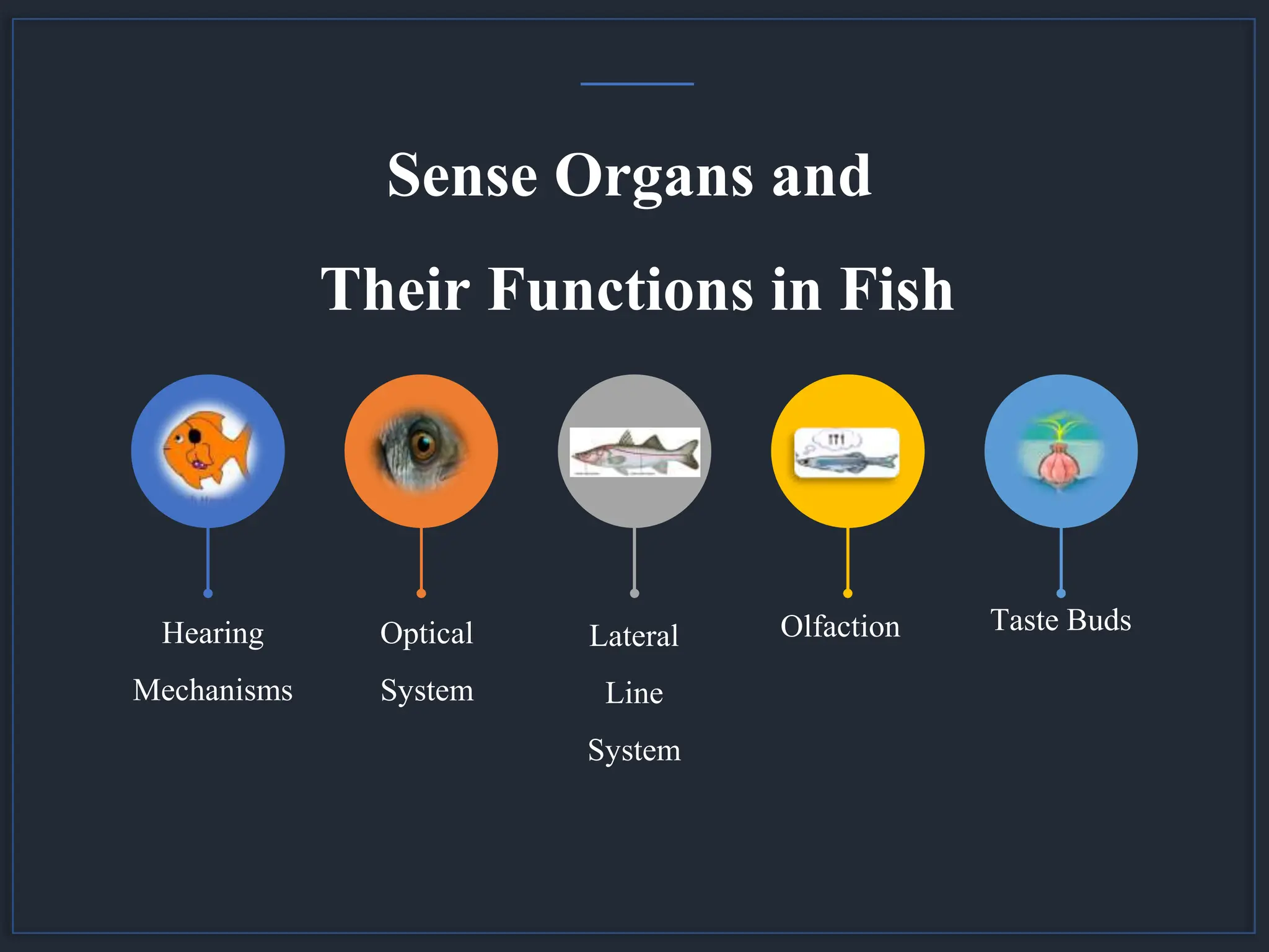
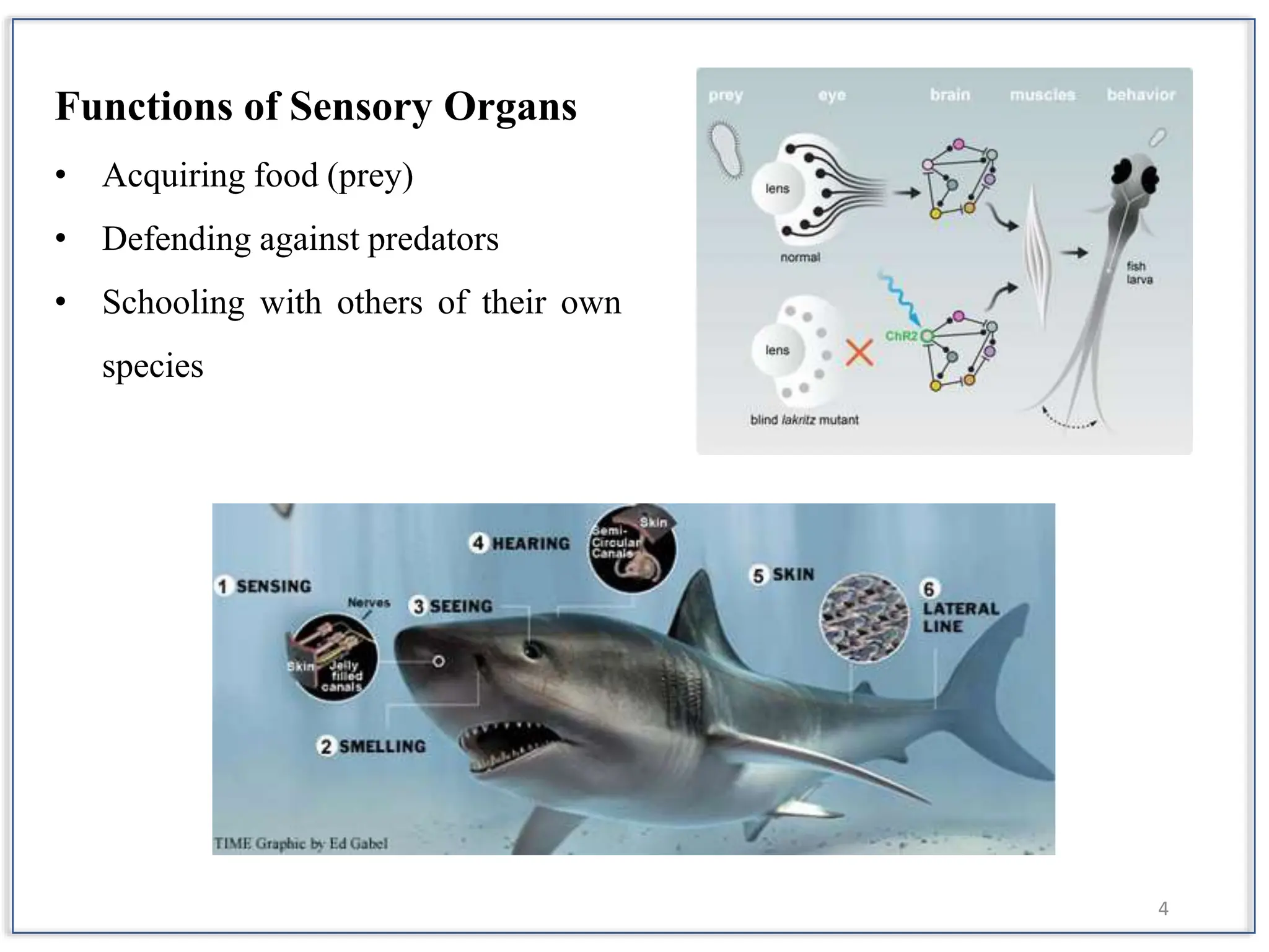
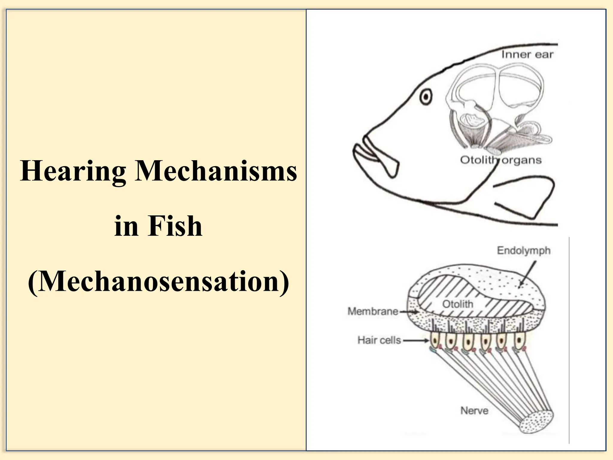
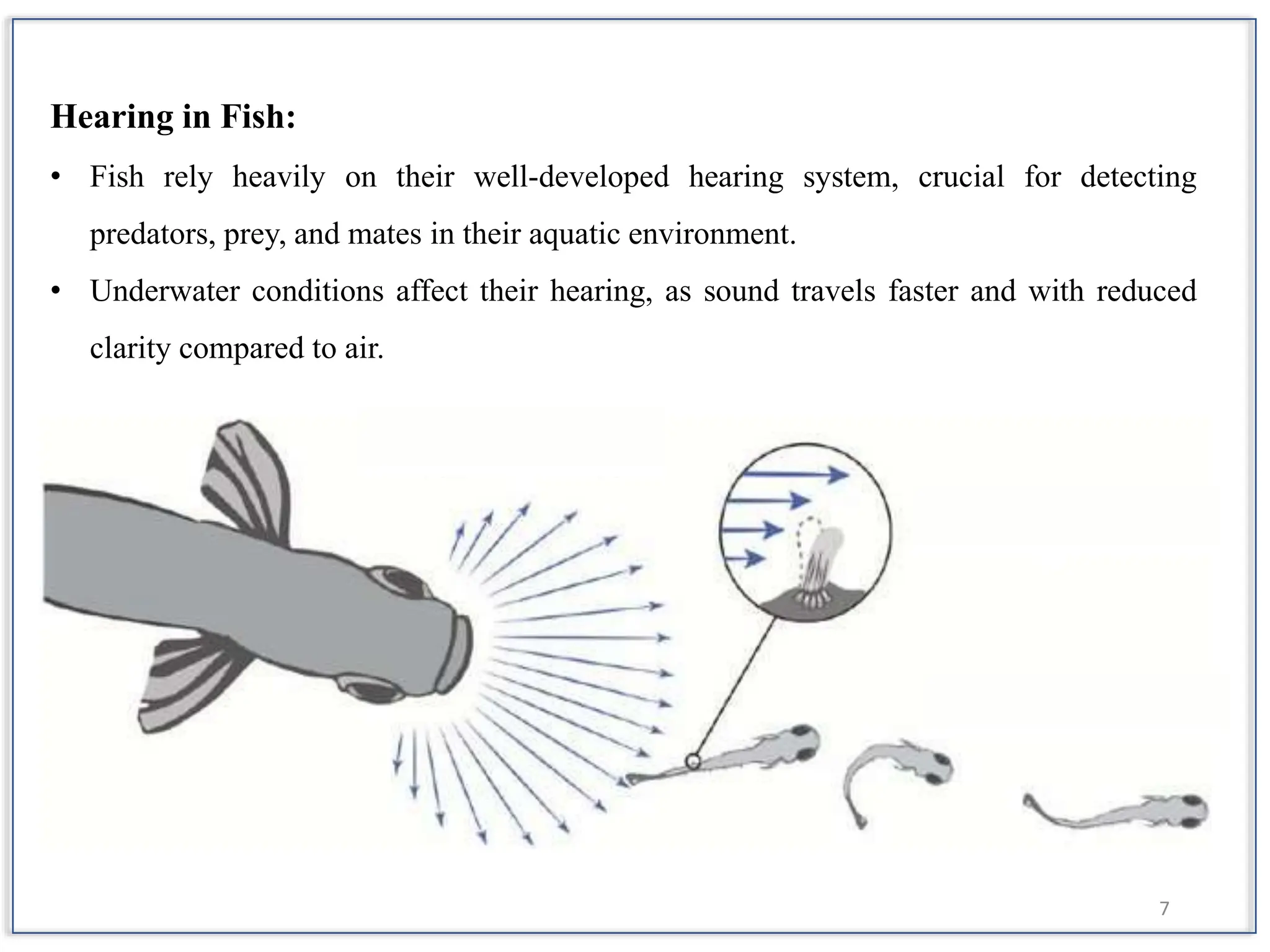
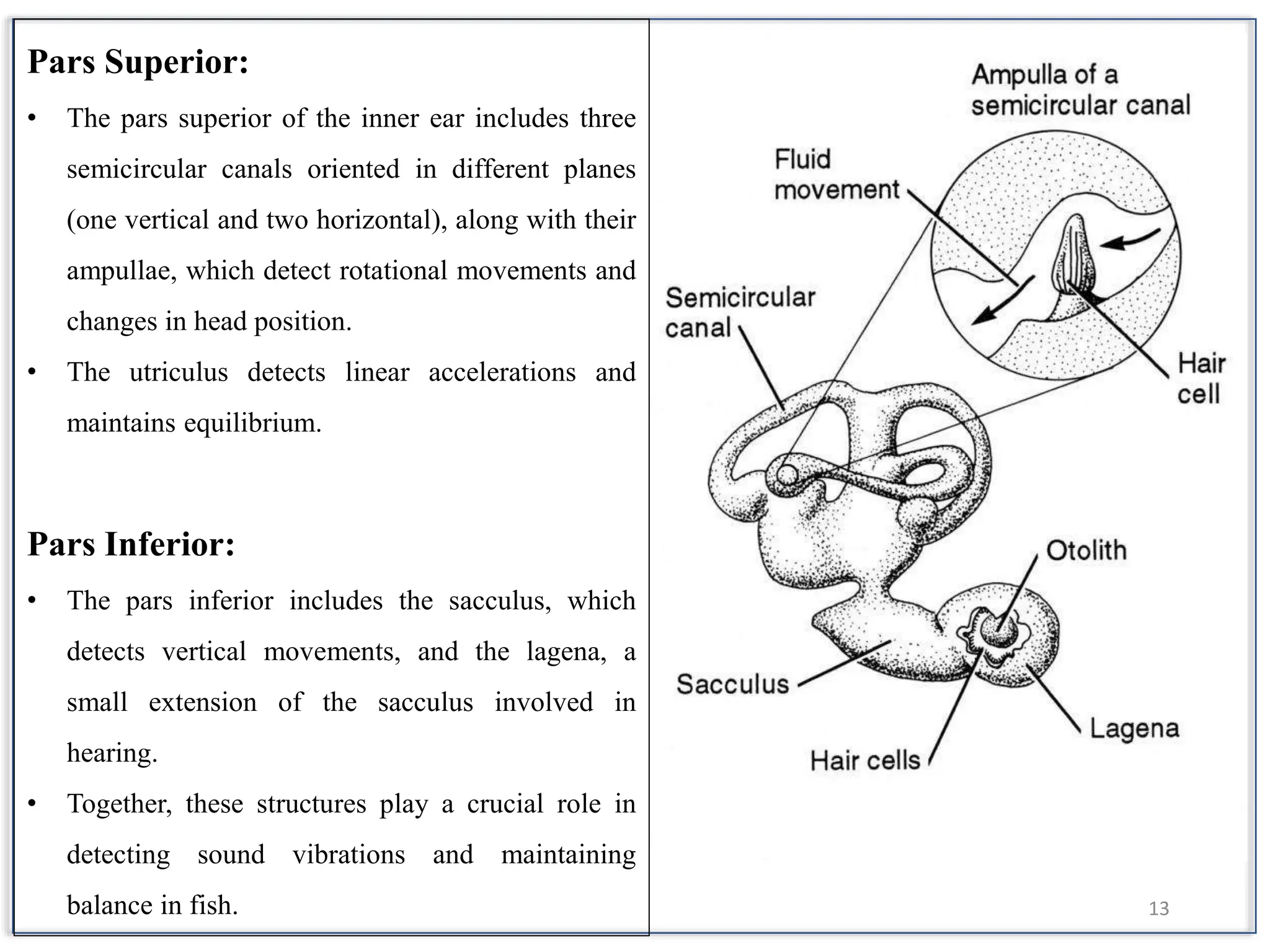
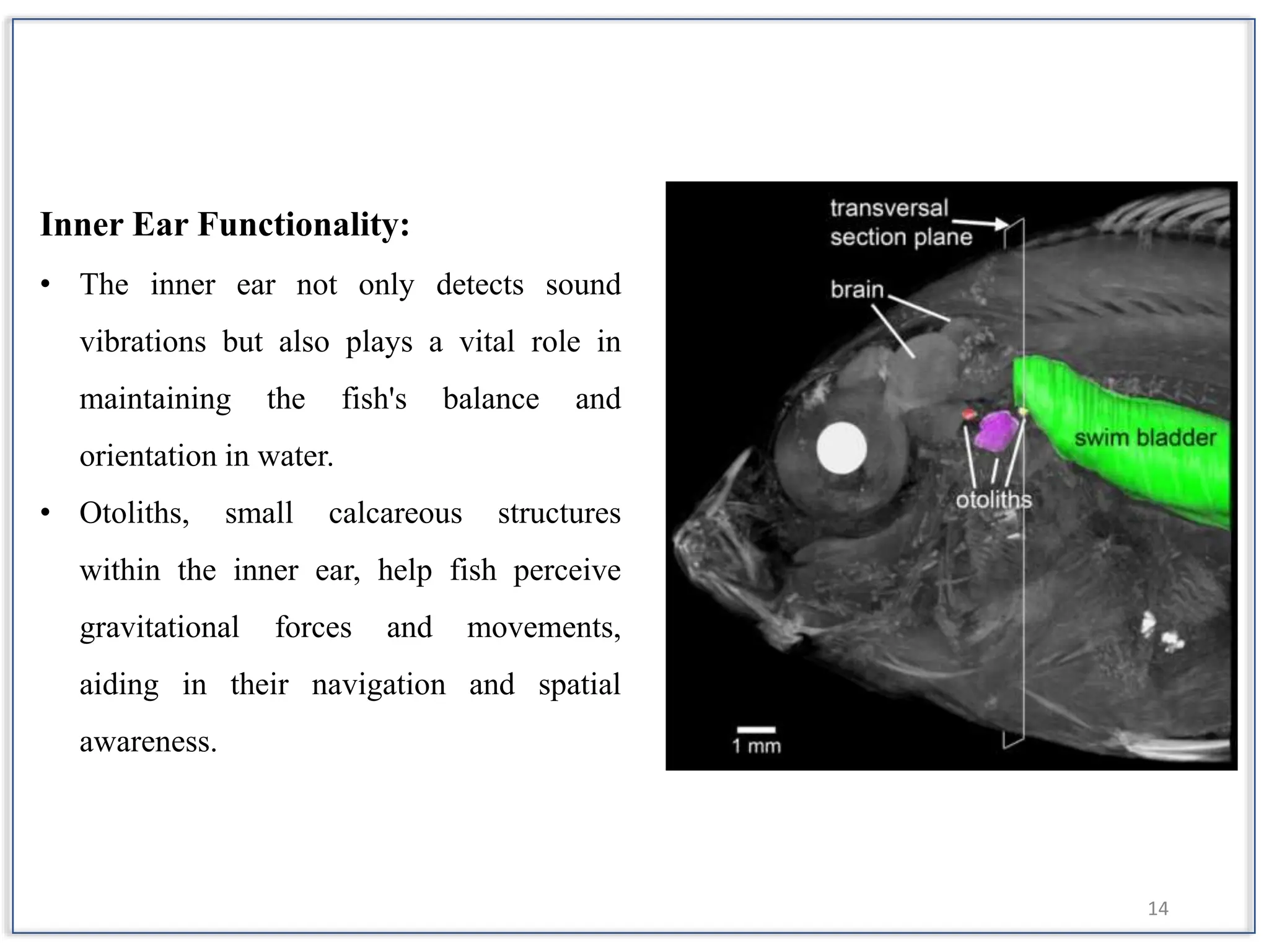
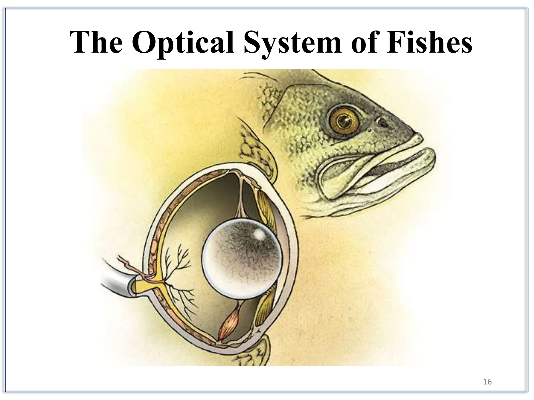
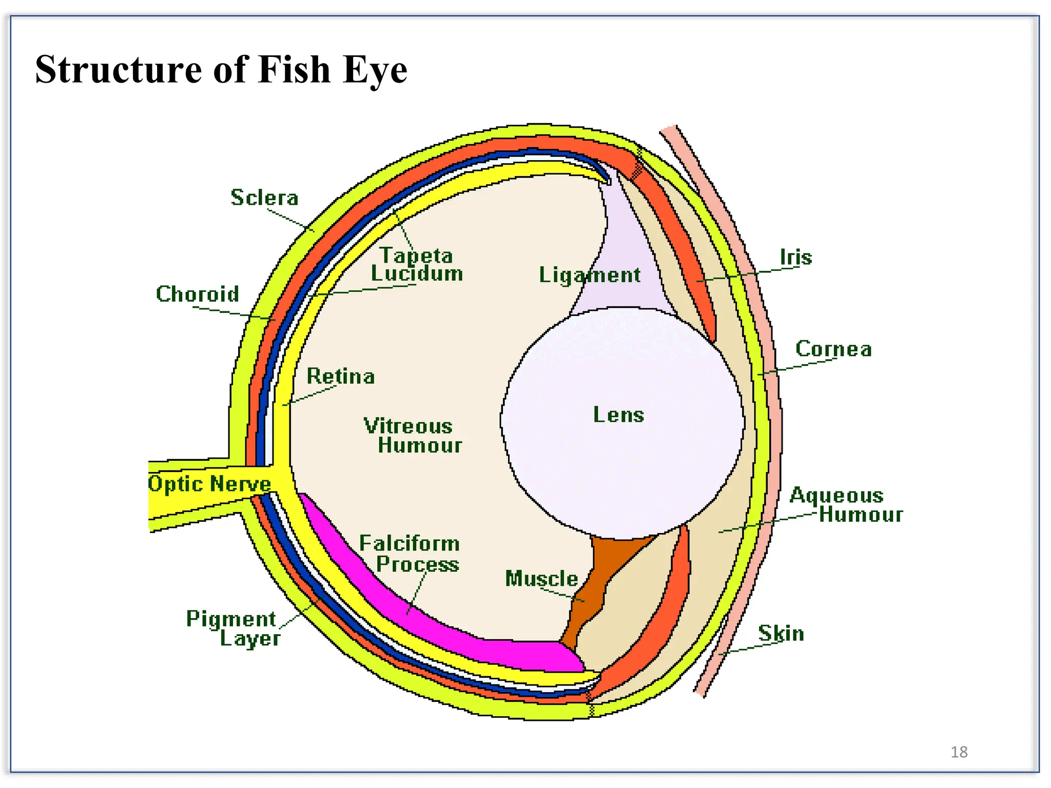
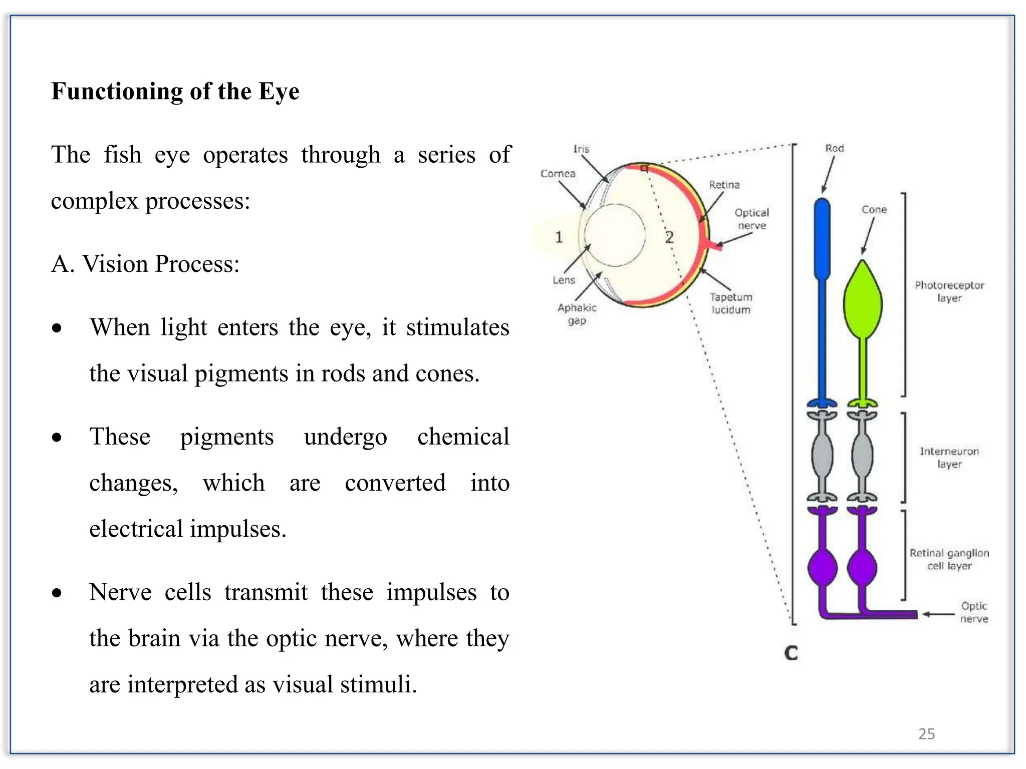
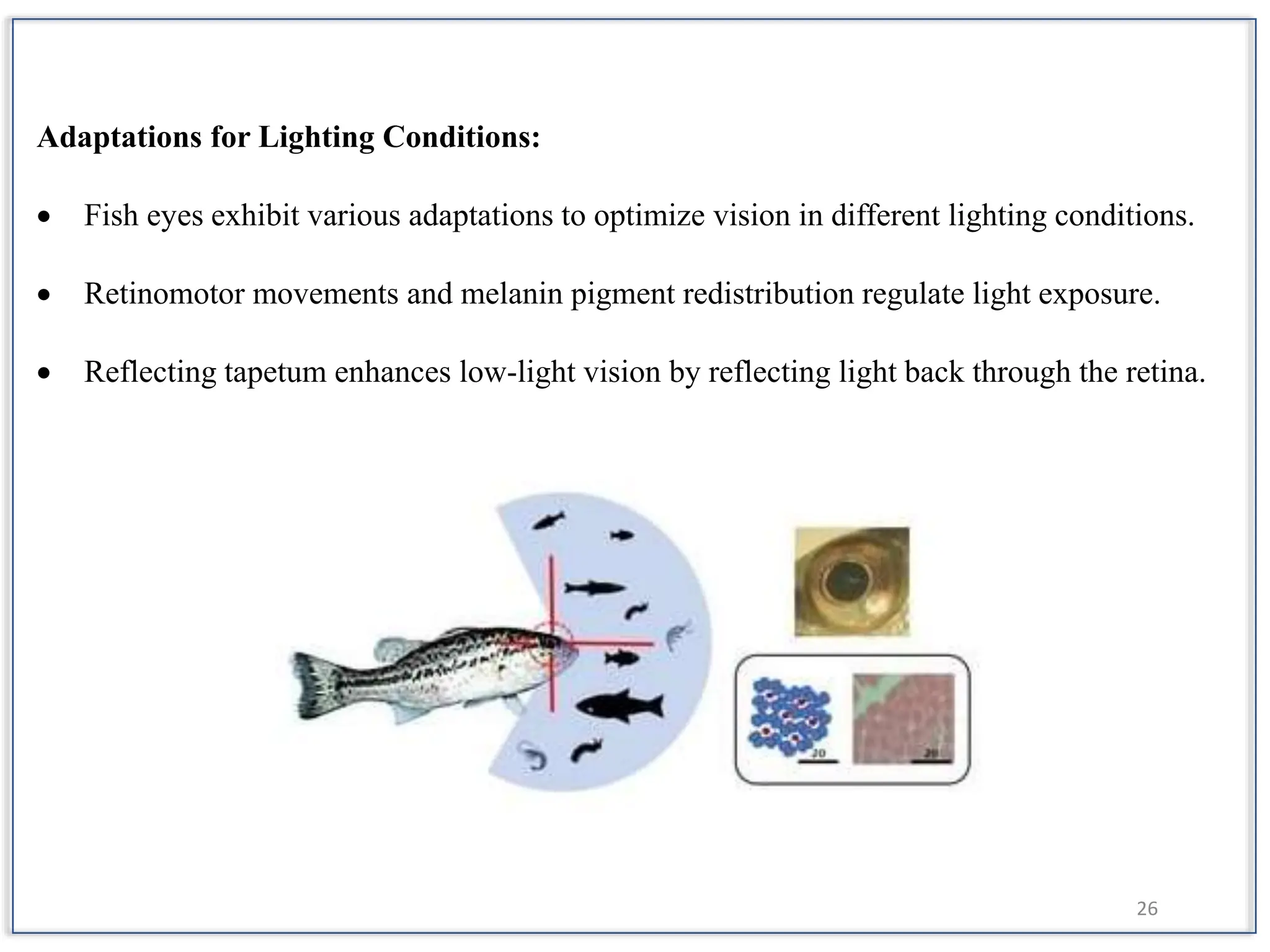
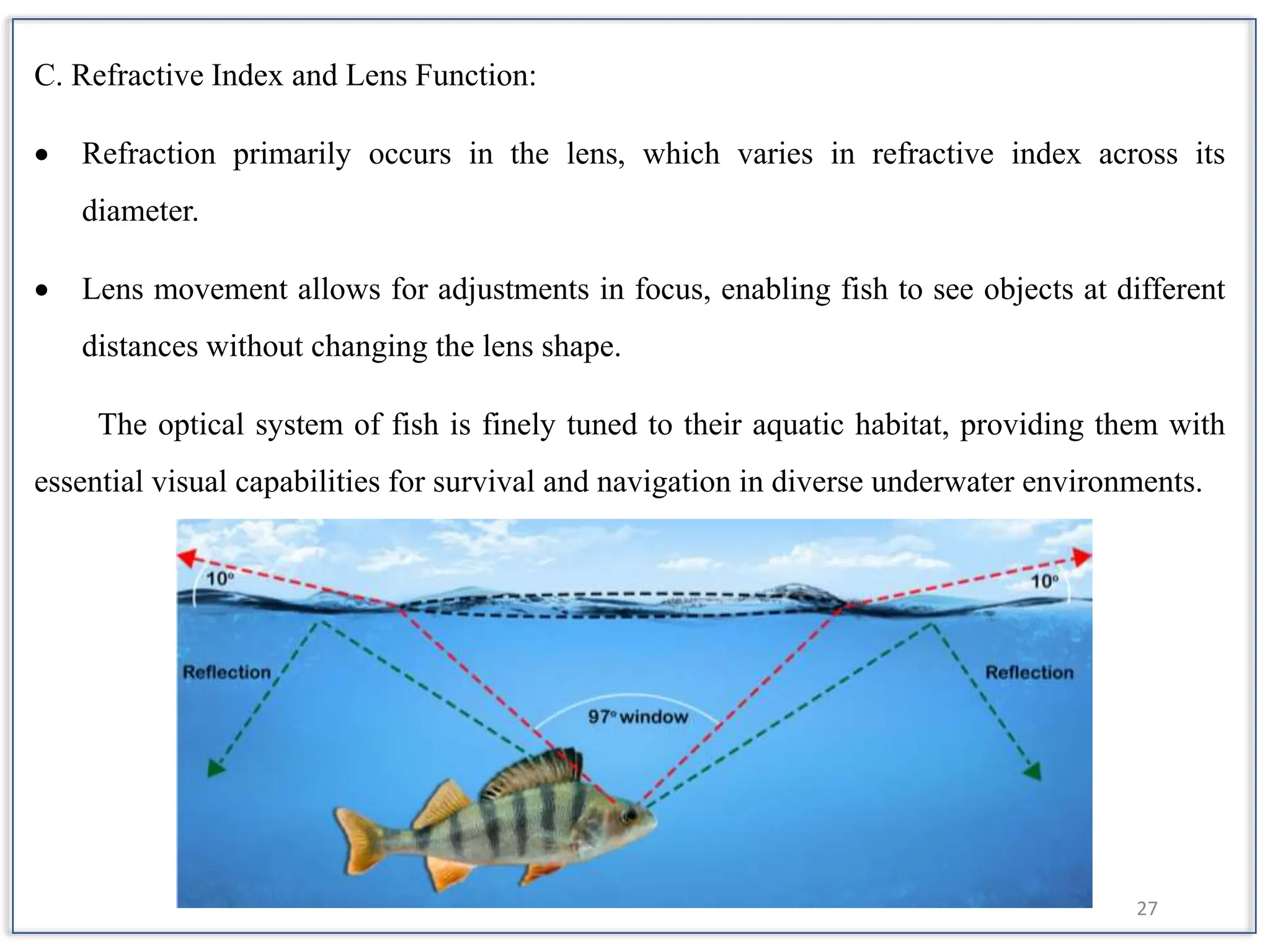
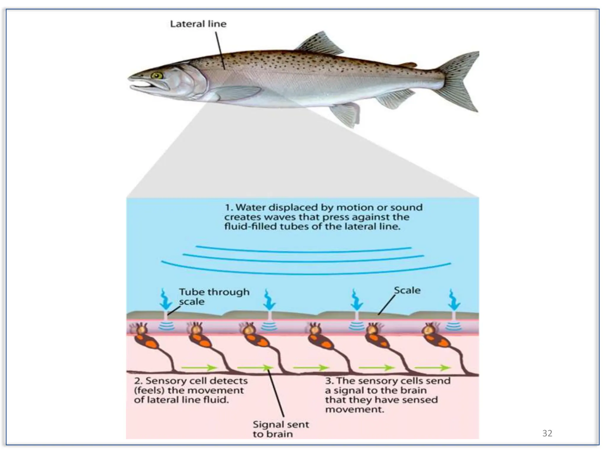
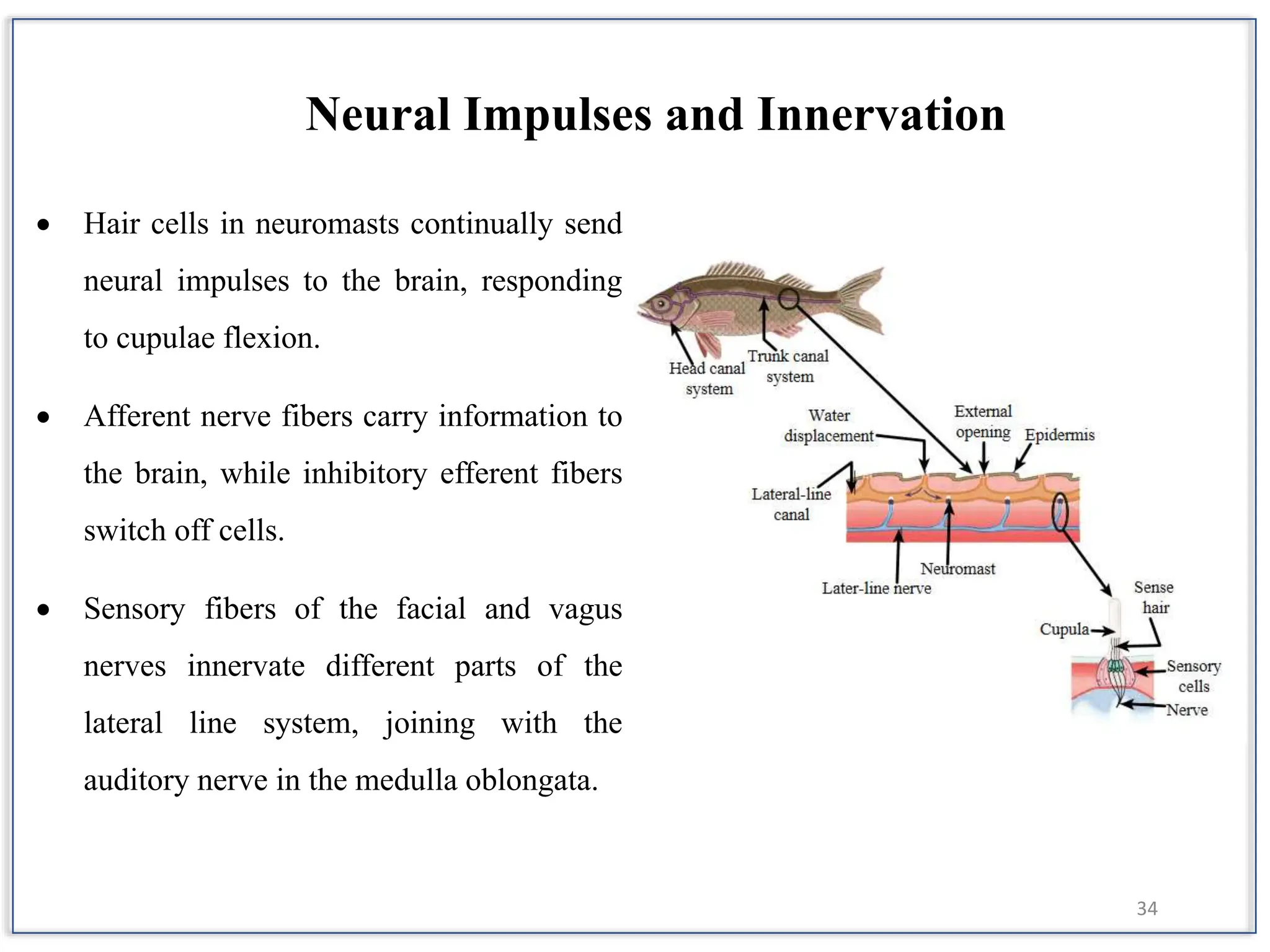
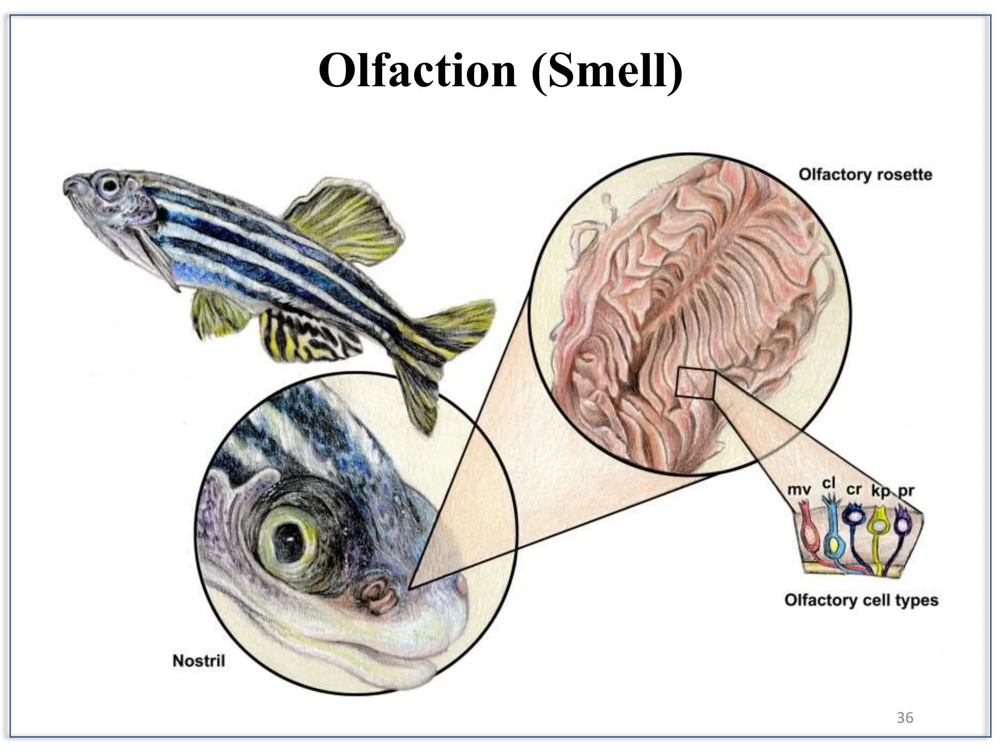
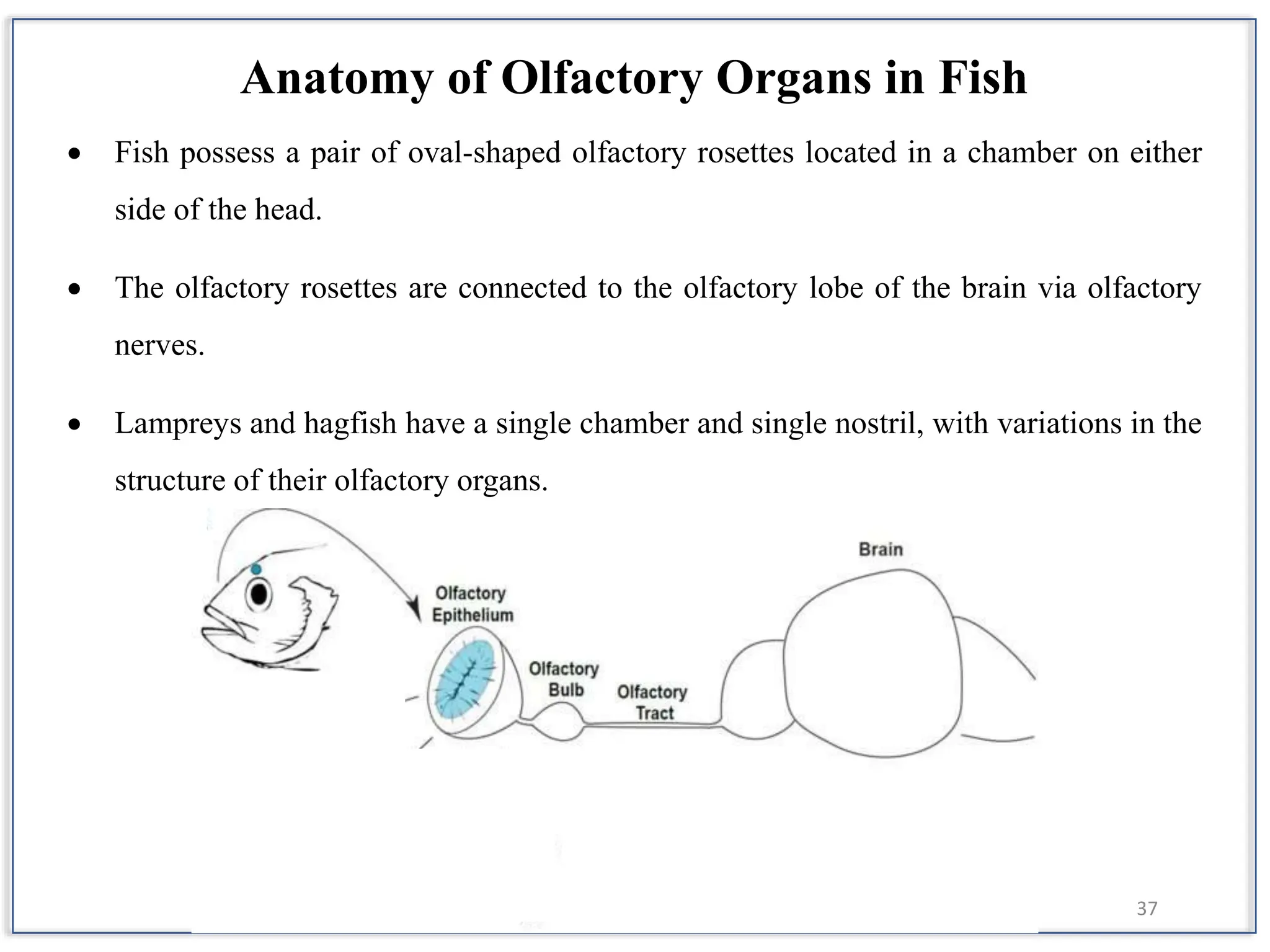
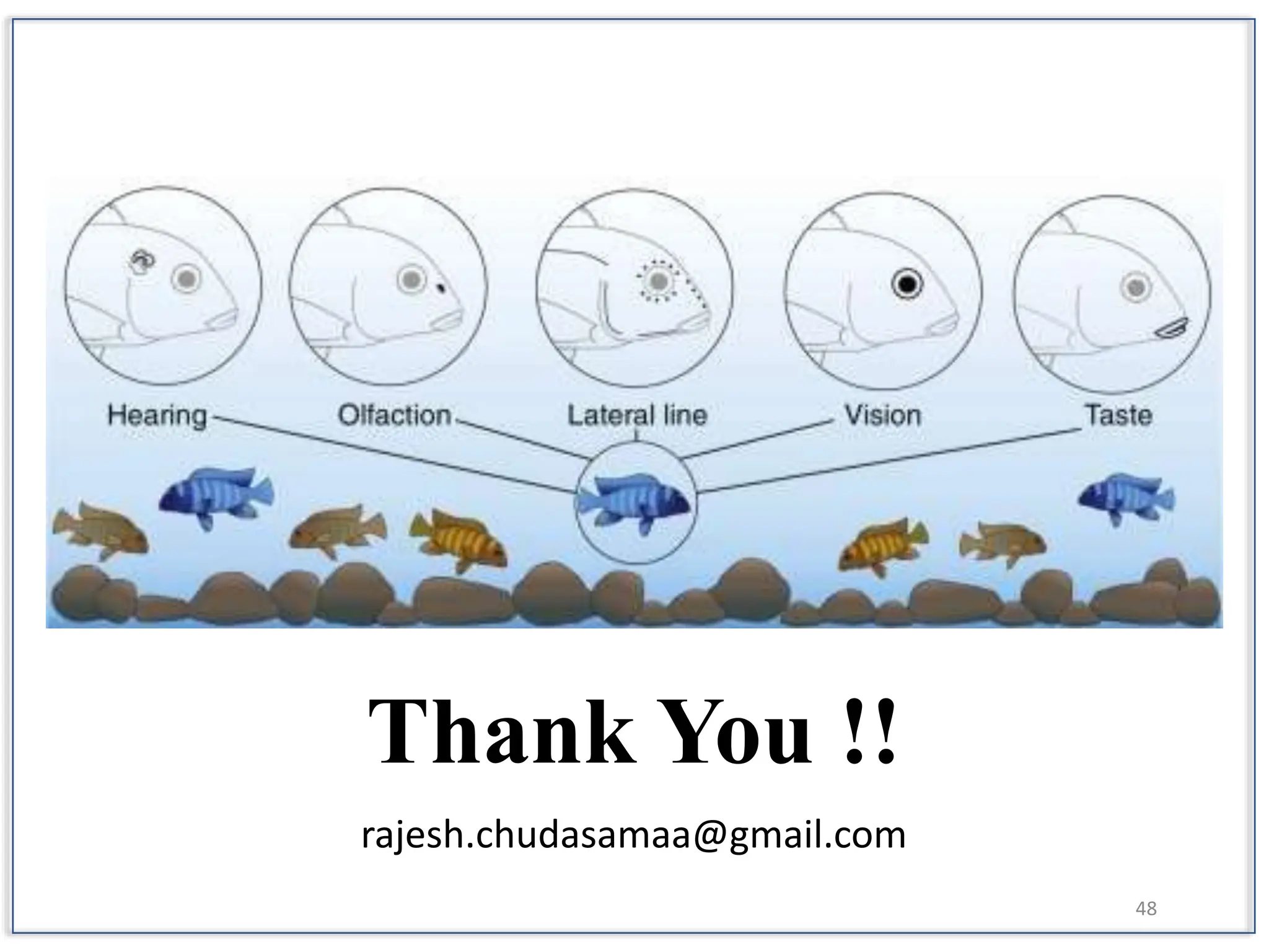
College of Fisheries Science
Kamdhenu University
Sub: Functional Physiology of Fishes (FRM 603)
Submitted By
Rajesh V. Chudasama,
Reg. No. 231303002, Ph.D. (AQC),
1st Sem. COF-VRL, KU.
Submitted To
Dr. H. L. Parmar,
Assistant Professor,
FRM Dept., COF-VRL, KU.
1
Sense Organs and
Their Functions in Fish
Hearing
Mechanisms
Optical
System
Lateral
Line
System
Olfaction Taste Buds
3
• The highly specialized organs that
receive physical and chemical
stimuli from the environment and
various body parts, and transform
them into stimuli are called sense
organs or sensory receptors.
• Closely associated with the nervous
system.
Physical Stimuli
• Change of heat
• Light intensity and quality
• Acoustical stimuli
4
Functions of Sensory Organs
• Acquiring food (prey)
• Defending against predators
• Schooling with others of their own
species
5
HUMAN FISH
Lungs Gills
Stomach Stomach
Liver Liver
Kidneys Kidneys
Ears Lateral Line, Otoliths
Skin Scales & Slime Layer
Nose Nares
Arms Pectoral Fins
Legs Pelvic Fins
6
Hearing Mechanisms
in Fish
(Mechanosensation)
7
Hearing in Fish:
• Fish rely heavily on their well-developed hearing system, crucial for detecting
predators, prey, and mates in their aquatic environment.
• Underwater conditions affect their hearing, as sound travels faster and with reduced
clarity compared to air.
8
Hearing Mechanisms:
• Fish detect sound through specialized sensory organs like lateral lines, which sense
pressure changes in the water, and otoliths (ear stones), which respond to sound
vibrations.
• Some fish species, such as carp and herring, enhance their hearing by using their swim
bladders to amplify sound waves.
9
Carp Hearing:
• Carp possess a unique adaptation called the Weberian organ, which consists of
specialized vertebral processes that transmit vibrations from the swim bladder to the
inner ear.
• This adaptation significantly enhances their ability to detect subtle underwater
vibrations and sounds.
10
11
Shark Hearing:
• While difficult to test directly, sharks are believed to have a keen sense of hearing.
• They have small openings on each side of their heads leading directly to their inner ears,
allowing them to potentially detect prey from considerable distances by picking up low-
frequency sounds.
12
Anatomy of Fish Inner Ear:
• The inner ear in fish is critical for both hearing and maintaining balance.
• It consists of membranous sacs housed within chambers on either side of the
fish's skull.
• These sacs include the pars superior with semicircular canals for balance and
the utriculus, and the pars inferior containing the sacculus and lagena primarily
responsible for hearing.
13
Pars Superior:
• The pars superior of the inner ear includes three
semicircular canals oriented in different planes
(one vertical and two horizontal), along with their
ampullae, which detect rotational movements and
changes in head position.
• The utriculus detects linear accelerations and
maintains equilibrium.
Pars Inferior:
• The pars inferior includes the sacculus, which
detects vertical movements, and the lagena, a
small extension of the sacculus involved in
hearing.
• Together, these structures play a crucial role in
detecting sound vibrations and maintaining
balance in fish.
14
Inner Ear Functionality:
• The inner ear not only detects sound
vibrations but also plays a vital role in
maintaining the fish's balance and
orientation in water.
• Otoliths, small calcareous structures
within the inner ear, help fish perceive
gravitational forces and movements,
aiding in their navigation and spatial
awareness.
15
Comparative Anatomy and Physiology:
• The Weberian ossicles in some fish, like Ostariophysi, connect the swim
bladder to the inner ear, enhancing their hearing sensitivity.
• Otoliths in bony fish are composed of calcium carbonate secreted by sensory
cells and play a crucial role in detecting sound and maintaining balance.
• Neural impulses from hair cells in the inner ear transmit sensory information to
the brain, triggering motor responses that help fish maintain stability and
navigate their environment effectively.
• Water's higher density compared to air makes it a more efficient conductor of
sound pressure waves, facilitating underwater communication and sensory
perception in fish.
16
The Optical System of Fishes
17
• Fish possess a complex optical
system that is essential for their
survival, aiding in navigation,
foraging, and predator
avoidance.
• This system consists of various
structures and adaptations
tailored to their underwater
environment.
18
Structure of Fish Eye
19
A. Cornea:
 The cornea, situated at the front of the eye,
is typically of constant thickness in teleosts.
 It consists of three layers: corneal
epithelium, stroma, and endothelium.
 In most species, the cornea is transparent,
allowing light to enter the eye.
B. Sclerotic Layer:
 Surrounding the eyeball, the sclerotic layer
provides structural support and toughness.
 In elasmobranches, this layer is supported
by fibrous tissue, while in some species like
Latimaria, it has a thick cartilage layer.
20
C. Choroid or Reflecting Layer:
 Situated beneath the sclerotic layer, the
choroid is highly vascularized.
 It contains a choroid gland that secretes
oxygen to meet the metabolic demands of
the retinal tissue.
D. Iris:
 The iris, positioned between the cornea and
lens, regulates the amount of light entering
the eye.
 While some species have a fixed pupil,
others, like elasmobranches, possess
muscles to adjust pupil size.
21
E. Lens:
 Located behind the iris, the lens focuses
incoming light onto the retina.
 It is filled with aqueous humor and is
transparent, composed primarily of non-
collagenous protein.
F. Adjustments in Lens Shape:
 Fish can adjust the shape of their lens to
focus on objects at different distances
without changing its shape.
22
Retina and Visual Pigments
The retina, lining the inner surface of the eye, is responsible for detecting light and
converting it into neural signals. It consists of two main layers:
A. Outer Granular Layer:
 Heavily pigmented layer containing rod and cone visual nerve cells.
 Rods are responsible for low-light vision, while cones contribute to color vision and
daylight vision.
23
B. Inner Less Pigmented Layer:
 Contains various types of nerve cells, including horizontal cells, bipolar cells, and
amacrine cells.
 These cells facilitate the transmission of visual information to the optic nerve.
 Visual pigments, located in the outer segments of rods and cones, are crucial for
light detection. These pigments consist of opsin proteins linked to vitamin A1 or A2
aldehyde and vary in their sensitivity to light.
24
25
Functioning of the Eye
The fish eye operates through a series of
complex processes:
A. Vision Process:
 When light enters the eye, it stimulates
the visual pigments in rods and cones.
 These pigments undergo chemical
changes, which are converted into
electrical impulses.
 Nerve cells transmit these impulses to
the brain via the optic nerve, where they
are interpreted as visual stimuli.
26
Adaptations for Lighting Conditions:
 Fish eyes exhibit various adaptations to optimize vision in different lighting conditions.
 Retinomotor movements and melanin pigment redistribution regulate light exposure.
 Reflecting tapetum enhances low-light vision by reflecting light back through the retina.
27
C. Refractive Index and Lens Function:
 Refraction primarily occurs in the lens, which varies in refractive index across its
diameter.
 Lens movement allows for adjustments in focus, enabling fish to see objects at different
distances without changing the lens shape.
The optical system of fish is finely tuned to their aquatic habitat, providing them with
essential visual capabilities for survival and navigation in diverse underwater environments.
28
Lateral Line System
29
The lateral line system is a unique sensory system found in fishes, integral to the
acoustico lateralis system. It involves sensory lines distributed on the head and body,
comprising the lateral line canal and neuromast organs.
Structure of the Lateral Line Canal
 The lateral line canal exists as a continuous
groove on the head and body, extending to
the base of the caudal fin.
 It contains sensory receptors arranged in
rows and follows the path of nerves.
 In some species, such as lung fishes, the
canal may be partially roofed over by
denticles, with pores opening on the skin
surface.
30
Development and Distribution of Lateral Line Canals
 During embryonic development, the lateral line canal differentiates as grooves along the
longitudinal axis.
 The dorsal and ventral canals disappear later, leaving only the lateral canals in adults.
 Canals may terminate into branches in the head region or lose connection with trunk
canals.
31
Functionality of the Lateral Line System
 The lateral line system helps fishes sense sounds, vibrations, gravity, and water
displacements.
 It responds to linear and angular accelerations of the fish's body, providing a sense
of direction in three-dimensional space.
 Fish can detect objects through echolocation using reflected waves.
 Components include sensory lines, pit organs, and ampullae of Lorenzini.
32
33
Neuromast Organs and Sensory Cells
 Neuromasts develop as thickenings on the head, extending into definite lines along the
body.
 Sensory areas develop from two types of cells: sensory and supporting cells.
 Neuromasts are receptors containing hair cells and cupulae, sensitive to movements of
the watery endolymph fluid through canals.
34
 Hair cells in neuromasts continually send
neural impulses to the brain, responding
to cupulae flexion.
 Afferent nerve fibers carry information to
the brain, while inhibitory efferent fibers
switch off cells.
 Sensory fibers of the facial and vagus
nerves innervate different parts of the
lateral line system, joining with the
auditory nerve in the medulla oblongata.
Neural Impulses and Innervation
35
 The lateral line system detects both
particle displacement and sound pressure.
 Near-field effects involve particle
displacement close to the source, while
far-field effects relate to sound pressure at
a distance.
 Canal-based receptors offer protection
from continuous water stimulation during
rapid swimming, allowing detection of
weak water displacements.
Detection of Sound
36
Olfaction (Smell)
37
Anatomy of Olfactory Organs in Fish
 Fish possess a pair of oval-shaped olfactory rosettes located in a chamber on either
side of the head.
 The olfactory rosettes are connected to the olfactory lobe of the brain via olfactory
nerves.
 Lampreys and hagfish have a single chamber and single nostril, with variations in the
structure of their olfactory organs.
38
Structure of Olfactory Receptors
 Olfactory receptors are typically located in ciliated olfactory pits, which have
incurrent and excurrent nostrils or channels divided by a flap of skin.
 Movement of cilia, muscular movement of the branchial pump, or swimming creates
water flow into the olfactory pit, creating a pressure difference between incurrent and
excurrent nostrils.
39
40
 The olfactory sensory epithelium consists
of four types of receptor cells: olfactory
(receptor), supporting (subtentacular),
basal, and mucous (secretory) cells.
 Sensory cells are uniformly distributed on
both sides, with primary and secondary
neurons distinguished.
 Basal cells give rise to other cell types and
replace degenerating cells.
Types of Olfactory Cells
41
 Olfactory receptor cell dendrites show intense alkaline phosphatase activity and
less acid protease activity.
 Lipids are widely found in the olfactory epithelium.
 Mucous secreted by mucous cells contains mucin protein and acid
mucopolysaccharides, forming a protective layer around sensory hair.
Biochemical Composition and Functionality
42
 Olfactory stimuli are communicated to
the olfactory lobe of the brain through
the first cranial nerve.
 Carnivorous and predatory fishes
generally possess a better olfactory
sense than herbivorous fishes.
 Olfactory cues are crucial for
migratory fish like salmon to locate
their native streams.
Olfactory Stimuli and Sensory Perception
43
Taste Buds in Fish
44
 Gustatory sensory cells, or taste receptor cells, occur in clusters called taste buds in
epidermal locations, including the oral cavity and lips.
 Taste buds are also found on the head, barbels, fins, and flanks in some species.
 Taste buds contain different cell types, such as tubular or light cells, based on
morphology and staining affinity.
45
Neuroanatomy and Neurotransmission in Taste Cells
 Taste buds are innervated by nerve endings, with neurotransmission likely
involving ATP in teleost, elasmobranch, and lamprey taste cells.
 Serotonin, glutamate, and GABA are localized in some teleost taste cells, serving
synaptic, paracrine, and autocrine functions.
 Cranial nerves innervating taste buds are multimodal, with sensory afferent and
motor efferent fibers playing different roles in food search, ingestion, and
palatability determination.
46
Conclusion
The sensory systems of fish are crucial for their survival, enabling them to perceive
and respond to their environment effectively. Fish rely on their senses of sight, smell, taste,
hearing, and the mechanosensation provided by the lateral line system to navigate, locate
prey, avoid predators, and communicate. Their eyes, with complex structures and visual
pigments, allow them to perceive light, shapes, colors, and movements underwater,
adapting to varying lighting conditions. The olfactory and gustatory systems detect
chemical cues, helping fish find food, identify mates, and navigate. Specialized receptors
and taste buds enhance their sensitivity to chemical stimuli. The lateral line system detects
water movements, vibrations, and pressure changes, aiding in spatial orientation, predator
avoidance, and prey detection. These integrated sensory systems enable fish to thrive in
diverse aquatic habitats. Understanding fish sensory perception is vital for conservation,
fisheries management, and aquaculture practices.
47
References
Bone, Q., & Moore, R. (2008). Biology of fishes. Taylor & Francis.
Diana, J. S., & Höök, T. O. (2023). Biology and ecology of fishes. John Wiley & Sons.
Dijkgraaf, S. (1960). Hearing in bony fishes. Proceedings of the Royal Society of London. Series B.
Biological Sciences, 152(946), 51-54.
Douglas, R., & Djamgoz, M. (2012). The visual system of fish. Springer Science & Business Media.
Hara, T. J. (Ed.). (2012). Fish chemoreception (Vol. 6). Springer Science & Business Media.
Kasturi Samantaray, K. S. (2015). Physiology of finfish and shellfish (pp. 250-pp).
Kasumyan, A. O. (2003). The lateral line in fish: structure, function, and role in behavior. Journal of
Ichthyology, 43(2), S175.
Sloman, K. A., Balshine, S., & Wilson, R. W. (Eds.). (2005). Fish physiology: Behaviour and
physiology of fish.
Smith, L. S. (1982). Introduction to fish physiology (p. 352pp).
48
Thank You !!
rajesh.chudasamaa@gmail.com

Sense organs and their functions in Fish

  • 1.
    College of FisheriesScience Kamdhenu University Sub: Functional Physiology of Fishes (FRM 603) Submitted By Rajesh V. Chudasama, Reg. No. 231303002, Ph.D. (AQC), 1st Sem. COF-VRL, KU. Submitted To Dr. H. L. Parmar, Assistant Professor, FRM Dept., COF-VRL, KU. 1
  • 2.
    Sense Organs and TheirFunctions in Fish Hearing Mechanisms Optical System Lateral Line System Olfaction Taste Buds
  • 3.
    3 • The highlyspecialized organs that receive physical and chemical stimuli from the environment and various body parts, and transform them into stimuli are called sense organs or sensory receptors. • Closely associated with the nervous system. Physical Stimuli • Change of heat • Light intensity and quality • Acoustical stimuli
  • 4.
    4 Functions of SensoryOrgans • Acquiring food (prey) • Defending against predators • Schooling with others of their own species
  • 5.
    5 HUMAN FISH Lungs Gills StomachStomach Liver Liver Kidneys Kidneys Ears Lateral Line, Otoliths Skin Scales & Slime Layer Nose Nares Arms Pectoral Fins Legs Pelvic Fins
  • 6.
  • 7.
    7 Hearing in Fish: •Fish rely heavily on their well-developed hearing system, crucial for detecting predators, prey, and mates in their aquatic environment. • Underwater conditions affect their hearing, as sound travels faster and with reduced clarity compared to air.
  • 8.
    8 Hearing Mechanisms: • Fishdetect sound through specialized sensory organs like lateral lines, which sense pressure changes in the water, and otoliths (ear stones), which respond to sound vibrations. • Some fish species, such as carp and herring, enhance their hearing by using their swim bladders to amplify sound waves.
  • 9.
    9 Carp Hearing: • Carppossess a unique adaptation called the Weberian organ, which consists of specialized vertebral processes that transmit vibrations from the swim bladder to the inner ear. • This adaptation significantly enhances their ability to detect subtle underwater vibrations and sounds.
  • 10.
  • 11.
    11 Shark Hearing: • Whiledifficult to test directly, sharks are believed to have a keen sense of hearing. • They have small openings on each side of their heads leading directly to their inner ears, allowing them to potentially detect prey from considerable distances by picking up low- frequency sounds.
  • 12.
    12 Anatomy of FishInner Ear: • The inner ear in fish is critical for both hearing and maintaining balance. • It consists of membranous sacs housed within chambers on either side of the fish's skull. • These sacs include the pars superior with semicircular canals for balance and the utriculus, and the pars inferior containing the sacculus and lagena primarily responsible for hearing.
  • 13.
    13 Pars Superior: • Thepars superior of the inner ear includes three semicircular canals oriented in different planes (one vertical and two horizontal), along with their ampullae, which detect rotational movements and changes in head position. • The utriculus detects linear accelerations and maintains equilibrium. Pars Inferior: • The pars inferior includes the sacculus, which detects vertical movements, and the lagena, a small extension of the sacculus involved in hearing. • Together, these structures play a crucial role in detecting sound vibrations and maintaining balance in fish.
  • 14.
    14 Inner Ear Functionality: •The inner ear not only detects sound vibrations but also plays a vital role in maintaining the fish's balance and orientation in water. • Otoliths, small calcareous structures within the inner ear, help fish perceive gravitational forces and movements, aiding in their navigation and spatial awareness.
  • 15.
    15 Comparative Anatomy andPhysiology: • The Weberian ossicles in some fish, like Ostariophysi, connect the swim bladder to the inner ear, enhancing their hearing sensitivity. • Otoliths in bony fish are composed of calcium carbonate secreted by sensory cells and play a crucial role in detecting sound and maintaining balance. • Neural impulses from hair cells in the inner ear transmit sensory information to the brain, triggering motor responses that help fish maintain stability and navigate their environment effectively. • Water's higher density compared to air makes it a more efficient conductor of sound pressure waves, facilitating underwater communication and sensory perception in fish.
  • 16.
  • 17.
    17 • Fish possessa complex optical system that is essential for their survival, aiding in navigation, foraging, and predator avoidance. • This system consists of various structures and adaptations tailored to their underwater environment.
  • 18.
  • 19.
    19 A. Cornea:  Thecornea, situated at the front of the eye, is typically of constant thickness in teleosts.  It consists of three layers: corneal epithelium, stroma, and endothelium.  In most species, the cornea is transparent, allowing light to enter the eye. B. Sclerotic Layer:  Surrounding the eyeball, the sclerotic layer provides structural support and toughness.  In elasmobranches, this layer is supported by fibrous tissue, while in some species like Latimaria, it has a thick cartilage layer.
  • 20.
    20 C. Choroid orReflecting Layer:  Situated beneath the sclerotic layer, the choroid is highly vascularized.  It contains a choroid gland that secretes oxygen to meet the metabolic demands of the retinal tissue. D. Iris:  The iris, positioned between the cornea and lens, regulates the amount of light entering the eye.  While some species have a fixed pupil, others, like elasmobranches, possess muscles to adjust pupil size.
  • 21.
    21 E. Lens:  Locatedbehind the iris, the lens focuses incoming light onto the retina.  It is filled with aqueous humor and is transparent, composed primarily of non- collagenous protein. F. Adjustments in Lens Shape:  Fish can adjust the shape of their lens to focus on objects at different distances without changing its shape.
  • 22.
    22 Retina and VisualPigments The retina, lining the inner surface of the eye, is responsible for detecting light and converting it into neural signals. It consists of two main layers: A. Outer Granular Layer:  Heavily pigmented layer containing rod and cone visual nerve cells.  Rods are responsible for low-light vision, while cones contribute to color vision and daylight vision.
  • 23.
    23 B. Inner LessPigmented Layer:  Contains various types of nerve cells, including horizontal cells, bipolar cells, and amacrine cells.  These cells facilitate the transmission of visual information to the optic nerve.  Visual pigments, located in the outer segments of rods and cones, are crucial for light detection. These pigments consist of opsin proteins linked to vitamin A1 or A2 aldehyde and vary in their sensitivity to light.
  • 24.
  • 25.
    25 Functioning of theEye The fish eye operates through a series of complex processes: A. Vision Process:  When light enters the eye, it stimulates the visual pigments in rods and cones.  These pigments undergo chemical changes, which are converted into electrical impulses.  Nerve cells transmit these impulses to the brain via the optic nerve, where they are interpreted as visual stimuli.
  • 26.
    26 Adaptations for LightingConditions:  Fish eyes exhibit various adaptations to optimize vision in different lighting conditions.  Retinomotor movements and melanin pigment redistribution regulate light exposure.  Reflecting tapetum enhances low-light vision by reflecting light back through the retina.
  • 27.
    27 C. Refractive Indexand Lens Function:  Refraction primarily occurs in the lens, which varies in refractive index across its diameter.  Lens movement allows for adjustments in focus, enabling fish to see objects at different distances without changing the lens shape. The optical system of fish is finely tuned to their aquatic habitat, providing them with essential visual capabilities for survival and navigation in diverse underwater environments.
  • 28.
  • 29.
    29 The lateral linesystem is a unique sensory system found in fishes, integral to the acoustico lateralis system. It involves sensory lines distributed on the head and body, comprising the lateral line canal and neuromast organs. Structure of the Lateral Line Canal  The lateral line canal exists as a continuous groove on the head and body, extending to the base of the caudal fin.  It contains sensory receptors arranged in rows and follows the path of nerves.  In some species, such as lung fishes, the canal may be partially roofed over by denticles, with pores opening on the skin surface.
  • 30.
    30 Development and Distributionof Lateral Line Canals  During embryonic development, the lateral line canal differentiates as grooves along the longitudinal axis.  The dorsal and ventral canals disappear later, leaving only the lateral canals in adults.  Canals may terminate into branches in the head region or lose connection with trunk canals.
  • 31.
    31 Functionality of theLateral Line System  The lateral line system helps fishes sense sounds, vibrations, gravity, and water displacements.  It responds to linear and angular accelerations of the fish's body, providing a sense of direction in three-dimensional space.  Fish can detect objects through echolocation using reflected waves.  Components include sensory lines, pit organs, and ampullae of Lorenzini.
  • 32.
  • 33.
    33 Neuromast Organs andSensory Cells  Neuromasts develop as thickenings on the head, extending into definite lines along the body.  Sensory areas develop from two types of cells: sensory and supporting cells.  Neuromasts are receptors containing hair cells and cupulae, sensitive to movements of the watery endolymph fluid through canals.
  • 34.
    34  Hair cellsin neuromasts continually send neural impulses to the brain, responding to cupulae flexion.  Afferent nerve fibers carry information to the brain, while inhibitory efferent fibers switch off cells.  Sensory fibers of the facial and vagus nerves innervate different parts of the lateral line system, joining with the auditory nerve in the medulla oblongata. Neural Impulses and Innervation
  • 35.
    35  The lateralline system detects both particle displacement and sound pressure.  Near-field effects involve particle displacement close to the source, while far-field effects relate to sound pressure at a distance.  Canal-based receptors offer protection from continuous water stimulation during rapid swimming, allowing detection of weak water displacements. Detection of Sound
  • 36.
  • 37.
    37 Anatomy of OlfactoryOrgans in Fish  Fish possess a pair of oval-shaped olfactory rosettes located in a chamber on either side of the head.  The olfactory rosettes are connected to the olfactory lobe of the brain via olfactory nerves.  Lampreys and hagfish have a single chamber and single nostril, with variations in the structure of their olfactory organs.
  • 38.
    38 Structure of OlfactoryReceptors  Olfactory receptors are typically located in ciliated olfactory pits, which have incurrent and excurrent nostrils or channels divided by a flap of skin.  Movement of cilia, muscular movement of the branchial pump, or swimming creates water flow into the olfactory pit, creating a pressure difference between incurrent and excurrent nostrils.
  • 39.
  • 40.
    40  The olfactorysensory epithelium consists of four types of receptor cells: olfactory (receptor), supporting (subtentacular), basal, and mucous (secretory) cells.  Sensory cells are uniformly distributed on both sides, with primary and secondary neurons distinguished.  Basal cells give rise to other cell types and replace degenerating cells. Types of Olfactory Cells
  • 41.
    41  Olfactory receptorcell dendrites show intense alkaline phosphatase activity and less acid protease activity.  Lipids are widely found in the olfactory epithelium.  Mucous secreted by mucous cells contains mucin protein and acid mucopolysaccharides, forming a protective layer around sensory hair. Biochemical Composition and Functionality
  • 42.
    42  Olfactory stimuliare communicated to the olfactory lobe of the brain through the first cranial nerve.  Carnivorous and predatory fishes generally possess a better olfactory sense than herbivorous fishes.  Olfactory cues are crucial for migratory fish like salmon to locate their native streams. Olfactory Stimuli and Sensory Perception
  • 43.
  • 44.
    44  Gustatory sensorycells, or taste receptor cells, occur in clusters called taste buds in epidermal locations, including the oral cavity and lips.  Taste buds are also found on the head, barbels, fins, and flanks in some species.  Taste buds contain different cell types, such as tubular or light cells, based on morphology and staining affinity.
  • 45.
    45 Neuroanatomy and Neurotransmissionin Taste Cells  Taste buds are innervated by nerve endings, with neurotransmission likely involving ATP in teleost, elasmobranch, and lamprey taste cells.  Serotonin, glutamate, and GABA are localized in some teleost taste cells, serving synaptic, paracrine, and autocrine functions.  Cranial nerves innervating taste buds are multimodal, with sensory afferent and motor efferent fibers playing different roles in food search, ingestion, and palatability determination.
  • 46.
    46 Conclusion The sensory systemsof fish are crucial for their survival, enabling them to perceive and respond to their environment effectively. Fish rely on their senses of sight, smell, taste, hearing, and the mechanosensation provided by the lateral line system to navigate, locate prey, avoid predators, and communicate. Their eyes, with complex structures and visual pigments, allow them to perceive light, shapes, colors, and movements underwater, adapting to varying lighting conditions. The olfactory and gustatory systems detect chemical cues, helping fish find food, identify mates, and navigate. Specialized receptors and taste buds enhance their sensitivity to chemical stimuli. The lateral line system detects water movements, vibrations, and pressure changes, aiding in spatial orientation, predator avoidance, and prey detection. These integrated sensory systems enable fish to thrive in diverse aquatic habitats. Understanding fish sensory perception is vital for conservation, fisheries management, and aquaculture practices.
  • 47.
    47 References Bone, Q., &Moore, R. (2008). Biology of fishes. Taylor & Francis. Diana, J. S., & Höök, T. O. (2023). Biology and ecology of fishes. John Wiley & Sons. Dijkgraaf, S. (1960). Hearing in bony fishes. Proceedings of the Royal Society of London. Series B. Biological Sciences, 152(946), 51-54. Douglas, R., & Djamgoz, M. (2012). The visual system of fish. Springer Science & Business Media. Hara, T. J. (Ed.). (2012). Fish chemoreception (Vol. 6). Springer Science & Business Media. Kasturi Samantaray, K. S. (2015). Physiology of finfish and shellfish (pp. 250-pp). Kasumyan, A. O. (2003). The lateral line in fish: structure, function, and role in behavior. Journal of Ichthyology, 43(2), S175. Sloman, K. A., Balshine, S., & Wilson, R. W. (Eds.). (2005). Fish physiology: Behaviour and physiology of fish. Smith, L. S. (1982). Introduction to fish physiology (p. 352pp).
  • 48.