Downloaded 15 times
![FRONTOETHMOID SUTURE
PPF
Int. maxillary art. & its br.
infratemporal fossa-pterygomaxillary fissure-pterygopalatine fossa
[termnl br of opht A.]](https://image.slidesharecdn.com/sellaranatomycopy-201122153119/85/Sellar-anatomy-copy-3-320.jpg)
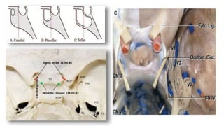
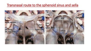

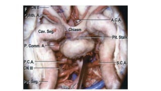
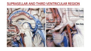
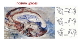
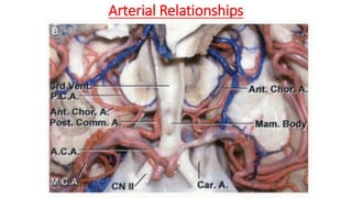
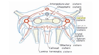
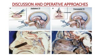
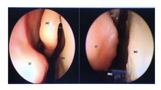
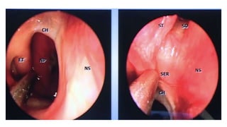
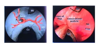
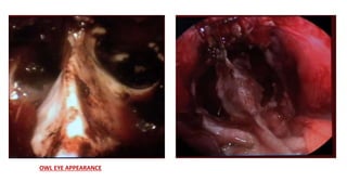
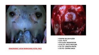
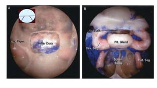


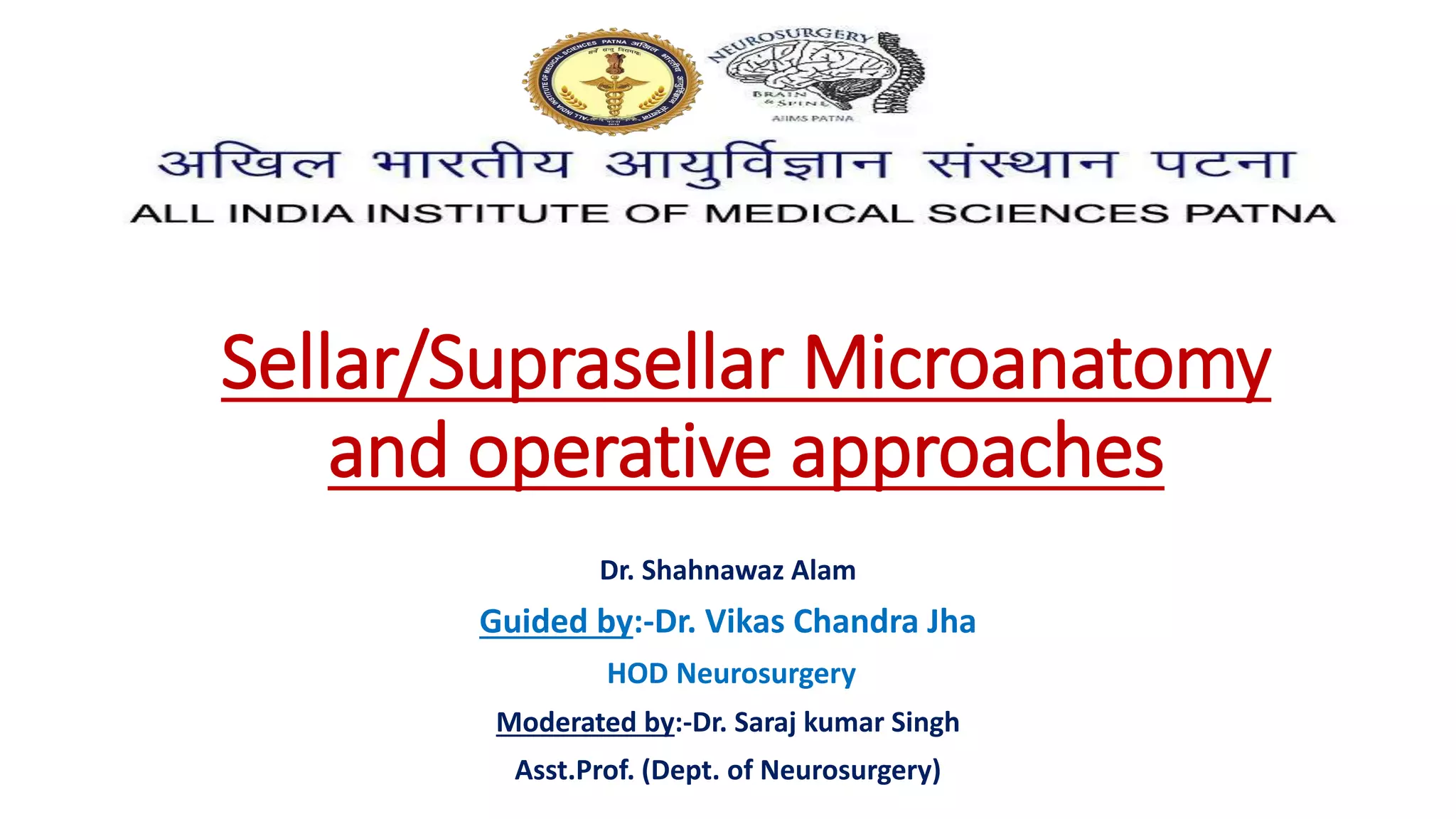
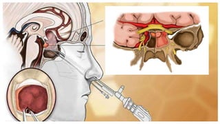
This document discusses the microanatomy of the sellar and suprasellar regions as well as operative approaches for accessing these areas. It describes the bones, venous connections, arterial relationships and incisura spaces in the regions. Several surgical approaches are mentioned, including sublabial, transseptal, endonasal, and subfrontal transfrontal transsphenoidal approaches. Key anatomical structures like the optic chiasm, carotid protuberance, cavernous sinus and clival indentation are located using an "owl eye" panoramic view of the fetal face mimicking centers.


![ONFH[AVN HIP] -TRIPLE REGIME -A NOVAL SURGICAL CONCEPT .pptx](https://cdn.slidesharecdn.com/ss_thumbnails/onfhavnhip2026koaconcalicutdrgokuldevdrmashraf-260210064517-213ec005-thumbnail.jpg?width=640&height=640&fit=bounds)


