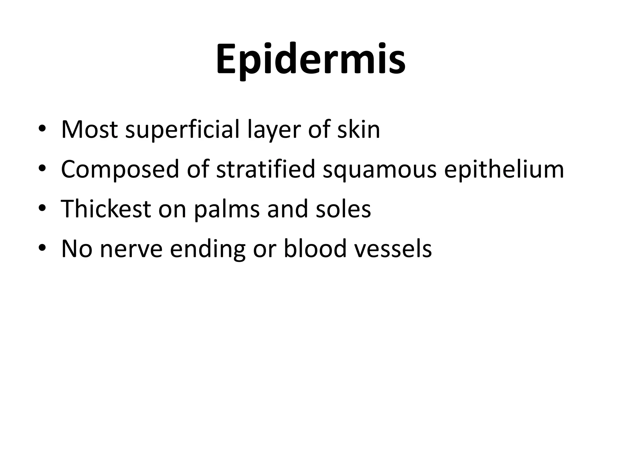
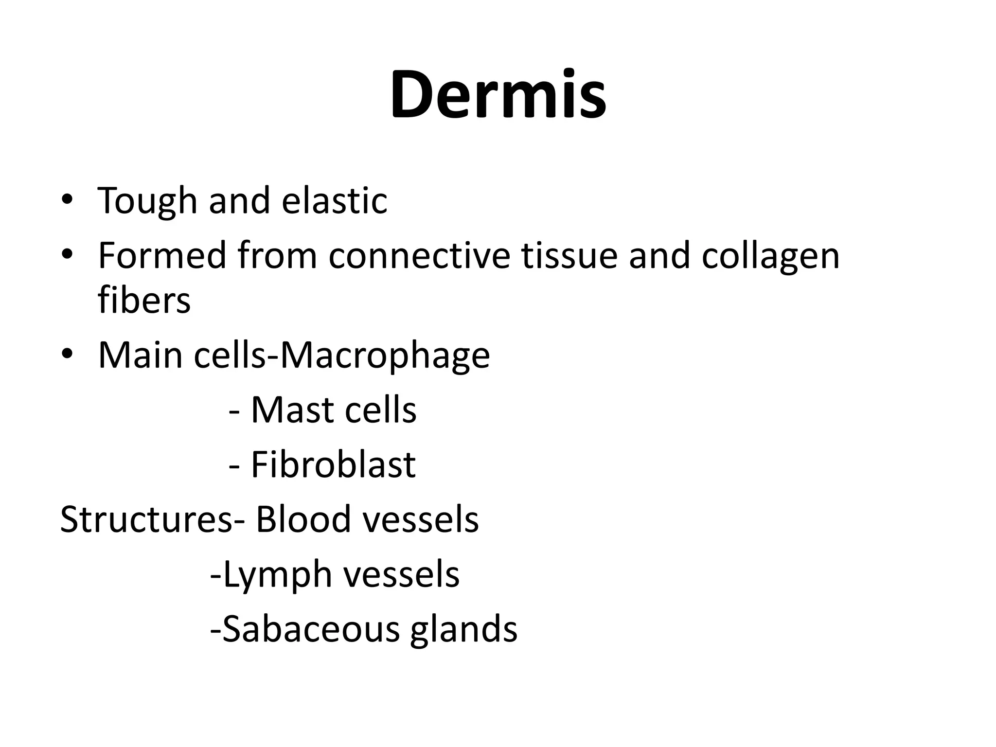
The document summarizes the examination of skin. It discusses the two main layers of skin - the epidermis and dermis. The epidermis is the most superficial layer, composed of stratified squamous epithelium. It contains no blood vessels or nerve endings. The dermis lies below the epidermis, formed from connective tissue and collagen fibers. It contains structures like blood vessels, lymph vessels, and sebaceous glands. The document also outlines important aspects to assess during a dermatological examination such as the patient's history, characteristics of the lesion, and common dermatological terms used to describe skin abnormalities.
2. Surface area of 1.5-2mm in adults
Layers- mainly two-1.Epidermis
2.Dermis
Skin
3. • Most superficial layer of skin
• Composed of stratified squamous epithelium
• Thickest on palms and soles
• No nerve ending or blood vessels
Epidermis
4. • Tough and elastic
• Formed from connective tissue and collagen
fibers
• Main cells-Macrophage
- Mast cells
- Fibroblast
Structures- Blood vessels
-Lymph vessels
-Sabaceous glands
Dermis
5.
6. • How long has the lesion been
present ?
• Has its distribution changes over
time?
• Has it been associated with sun
exposure / exposure to heat/cold?
Dermatological history
7. • Is their any associated Pruritis?
• Is the lesion painful?
• Does patient have any constitutional
symptoms?
• Does patient having any past h/o skin
disease or allergy ?
8. • Does patient having any past h/o
systemic disease e.g DM ?
• Does patient having any h/o
exposure to chemicals ?
• Does patient have family h/o
melanoma ?
• What medications patient is taking ?
9. • Lesions must be described according to
their color , shape, and using
dermatological terms
• Distribution and pattern must be
described
• Lesion must be palpated for consistency
,tenderness ,temperature ,depth etc.
Points to be assesed -
10. Macule
Flat lesion up to 1 cm in diameter of skin color
change
Circular , oval, or irregular
Well demarcated
E.g- flat nevus
Dermatological terms
12. Papule
• Elevated superficial solid lesion up to 1 cm
diameter
• Round or flat topped
• Irregularly distributed
• Sharply defined (superficial ) or indistinct
border (deep)
• E.g- acne
13. Plaque
• Flat elevated superficial lesion 1cm or more
• Often formed by coalescence of papules
• May be centrally depressed
• Usually well defined
• E.g psoriasis
14. Nodule
• Superficial solid “marble like” lesion greater
than 1 cm diameter
• Larger , often deeper and firmer than papule
• Hard or soft
• E.g. -acne or keloid
15. Wheal (“hives”)
• Irregular and oedematous like elevations
• Round ,or irregular
• Discrete or may be coalesce
• E.g. Urticaria
16. Vesicle
• Well defined superficial elevation containing
serous fluid up to 1cm
• Discrete irregularly scattered grouped or
linear
• E.g. varicella
17. Bulla
• Superficial elevation containing serous fluid
more than 1 cm
• Differ from vesicles only in size
• E.g contact dermatitis
18. Pustules
• Small superficial elevation containing turbid
purulent fluid (usually necrotic inflammatory
cells )
• Vary in size and shape
• Exudates may be white-yellow ,greenish-
yellow
• E.g. Pustular acne , Pustular psoriasis